, using NFAT2 Antibody. The lane on the left was treated with blocking peptide.")

.
Bands result from membrane strip incubation.")


 by IF/ICC. The samples were fixed with PFA and permeabilized in 0.1% Triton X-100, then blocked in 10% serum for 45 minutes at 25°C. Samples were then incubated with primary Ab(#DF6446) and mouse anti-beta tubulin Ab(#T0023) for 1 hour at 37°C. An AlexaFluor594 conjugated goat anti-rabbit IgG Ab(Red) and an AlexaFluor488 conjugated goat anti-mouse IgG Ab(Green) were used as the secondary antibody.
The nuclear counter stain is DAPI (blue).")
 TRAP staining, Bar= 50 μm; (B) IF staining of TLR4, Bar=100 μm; (C) Protein expression of RANKL, RANK, OC-STAMP, AP-1, NFATc1, TRAF6, NF-κB, TLR4, MyD88, p-IκBα, TNF-α, and MMP-12 in NR8383 cells treated with SiO2 and Ac-SDKP or not. Data are presented as the mean ± SD. n = 3 per group.")
 were combined using Photoshop software. The blots were cropped using Photoshop and are compliant with the digital image and integrity polices of Oncotarget. c-Fos and NFATc1 protein levels as well as IkBα protein expression and activity (phosphorylation) were detected using Western blot in RAW264.7 cells treated with PE, low-dose curcumin, and high-dose curcumin (a, c) and air pouch tissue samples (b, d) treated with PE, solvent, or PE+ curcumin. Non-treated (blank) samples were used as controls. The upper panels show typical results for Western blot images")
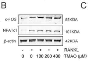
 difference from other corresponding groups. Insets are representative bands")
, CnA (B) as well as NFAT (C) in sham, and UUO mice with different treatments. GAPDH was used as an internal control. ##P < 0.01, UUO group vs. sham group; *P < 0.05, **P < 0.01, UUO +HKC group vs. UUO group. Bar graphs represent the mean ± SEM (n = 3).")
 and RANKL (30 ng/ml) and then treated with or without rSj‑Cys (0.3 µM) for different time periods (4 or 24 h). The mRNA and protein expression levels of NF‑κB‑associated signaling molecules IκBα and p65, and of downstream targets NFATc1 and c‑Fos, were assessed by (A) reverse transcription‑quantitative PCR and (B) western blot analysis.")
 in parathyroid hormone (PTH)-induced NFAT nuclear translocation in human umbilical vein endothelial cells (HUVECs).(G,H) Representative Western blot images showing fractionation assay results indicating the presence of p-NFATC1 in the cytoplasmic")
 The expression of miR-939 and miR-376a in UC patient tissues and normal human tissues. B) The expressions of NF-κB and NFAT in UC patient tissues and normal human tissues. Compared with normal group; *p<0.05.")
, PI3K inhibitor LY294002 (B), and NFAT inhibitor 11R-VIVIT (C). D-E Concentrations of IL-5 and IL-13 in the culture supernatants of pulmonary ILC2s were determined by ELISA in the presence of MEK inhibitor U0126-EtOH (D), PI3K inhibitor LY294002 (E), and NFAT inhibitor 11R-VIVIT (F). G-H The expression of signal proteins in the isolated ILC2s were determined by Western blot under the conditions of the presence or absence of U0126-EtOH (G) or LY294002 (H). Data are representative of at least two individual experiments, error bars represent SEM;")
 The (B) RNA and (C) protein levels of TRAP, c‐Fos, and NFATc1 in bone joint from mice of OVX group and sham group were checked by qPCR and western blotting assay. D The TRAP staining of bone joint from OVX mice and sham group. (E and F) The BMM and RAW264.7 cells were stimulated with RANKL, then (E) RNA and (F) protein levels of TRAP, c‐Fos, NFATc1, and KAT6A were measured by qPCR and western blotting assay. **p")
. B Ca2+ oscillation experiment was utilized to assess variations in Ca2+ concentration (n = 3). Original magnification: ×200. C Western blot was conducted to detect the expression of NFATc1 and PLCγ2 in OVX mice (n = 3). D IHC was employed to assess the expression of BTK and PLCγ2 in OVX mice. Original magnification: ×400. NSC697923: the specific inhibitor of UBE2N; PCI-32765: the BTK inhibitor; *P < 0.05, **P < 0.01, ***P < 0.001.")
 targets RANKL to inhibit osteoclast differentiation. a) and b) Expression and quantification of RANK proteins in each group. c) Fluorescence image of SiRANK in osteoclasts. Green fluorescence (SiRANK) (200×, scale bar = 50 μm). d) and e) Expression and quantification of osteoclast differentiation-related proteins in each group. f) Schematic diagram of the mechanism of XTS-targeted RANKL inhibition. g) Fluorescence expression of differentiation-related proteins in osteoclasts of each group (200×, scale bar = 100 μm).")
 RT-qPCR analysis of osteoclastogenic marker genes in BMMs treated with various concentrations of INS (0, 15, and 30 μM) for 5 days in the presence of RANKL and M-CSF. Relative mRNA expression levels of Acp5, c-Fos, Ctsk, and Nfatc1 were normalized to β-actin. (E, F) Representative Western blot images and quantification showing that INS dose-dependently upregulates the expression of antioxidant-related proteins (Nrf2, HO-1, SOD1, and catalase). (G, H) Representative Western blot images and quantification of osteoclastogenic marker proteins (CTSK, NFATc1, MMP9, and c-Fos) in RANKL-stimulated BMMs treated with INS (0, 15, and 30 μM) at days 1, 3, and 5 of differentiation. Data are presented as mean ± SD from three independent experiments performed in triplicate. *P < 0.05, **P < 0.01, ***P < 0.001 compared to the RANKL-stimulated control group (0 μM INS).")
| Product: | NFAT2 Antibody |
| Catalog: | DF6446 |
| Description: | Rabbit polyclonal antibody to NFAT2 |
| Application: | WB IHC IF/ICC |
| Cited expt.: | WB, IHC, IF/ICC |
| Reactivity: | Human, Mouse, Rat |
| Prediction: | Pig, Bovine, Horse, Rabbit |
| Mol.Wt.: | 80-120kD(Observed); 101kD(Calculated). |
| Uniprot: | O95644 |
| RRID: | AB_2838409 |
Control Products

Related Downloads
Protocols
Product Info
*The optimal dilutions should be determined by the end user. For optimal experimental results, antibody reuse is not recommended.
*Tips:
WB: For western blot detection of denatured protein samples. IHC: For immunohistochemical detection of paraffin sections (IHC-p) or frozen sections (IHC-f) of tissue samples. IF/ICC: For immunofluorescence detection of cell samples. ELISA(peptide): For ELISA detection of antigenic peptide.
Cite Format: Affinity Biosciences Cat# DF6446, RRID:AB_2838409.
Fold/Unfold
cytoplasmic 1; MGC138448; NF ATc; NF ATc1; NF-ATc; NF-ATc1; NF-ATc1.2; NFAC1_HUMAN; NFAT 2; NFAT transcription complex cytosolic component; NFATC 1; NFATc; NFATc1; Nuclear factor of activated T cells cytoplasmic 1; Nuclear factor of activated T cells cytoplasmic calcineurin dependent 1; Nuclear factor of activated T cells cytosolic component 1; nuclear factor of activated T-cells 'c'; Nuclear factor of activated T-cells;
Immunogens
A synthesized peptide derived from human NFAT2, corresponding to a region within the internal amino acids.
Expressed in thymus, peripheral leukocytes as T-cells and spleen. Isoforms A are preferentially expressed in effector T-cells (thymus and peripheral leukocytes) whereas isoforms B and isoforms C are preferentially expressed in naive T-cells (spleen). Isoforms B are expressed in naive T-cells after first antigen exposure and isoforms A are expressed in effector T-cells after second antigen exposure. Isoforms IA are widely expressed but not detected in liver nor pancreas, neural expression is strongest in corpus callosum. Isoforms IB are expressed mostly in muscle, cerebellum, placenta and thymus, neural expression in fetal and adult brain, strongest in corpus callosum.
- O95644 NFAC1_HUMAN:
- Protein BLAST With
- NCBI/
- ExPASy/
- Uniprot
MPSTSFPVPSKFPLGPAAAVFGRGETLGPAPRAGGTMKSAEEEHYGYASSNVSPALPLPTAHSTLPAPCHNLQTSTPGIIPPADHPSGYGAALDGGPAGYFLSSGHTRPDGAPALESPRIEITSCLGLYHNNNQFFHDVEVEDVLPSSKRSPSTATLSLPSLEAYRDPSCLSPASSLSSRSCNSEASSYESNYSYPYASPQTSPWQSPCVSPKTTDPEEGFPRGLGACTLLGSPRHSPSTSPRASVTEESWLGARSSRPASPCNKRKYSLNGRQPPYSPHHSPTPSPHGSPRVSVTDDSWLGNTTQYTSSAIVAAINALTTDSSLDLGDGVPVKSRKTTLEQPPSVALKVEPVGEDLGSPPPPADFAPEDYSSFQHIRKGGFCDQYLAVPQHPYQWAKPKPLSPTSYMSPTLPALDWQLPSHSGPYELRIEVQPKSHHRAHYETEGSRGAVKASAGGHPIVQLHGYLENEPLMLQLFIGTADDRLLRPHAFYQVHRITGKTVSTTSHEAILSNTKVLEIPLLPENSMRAVIDCAGILKLRNSDIELRKGETDIGRKNTRVRLVFRVHVPQPSGRTLSLQVASNPIECSQRSAQELPLVEKQSTDSYPVVGGKKMVLSGHNFLQDSKVIFVEKAPDGHHVWEMEAKTDRDLCKPNSLVVEIPPFRNQRITSPVHVSFYVCNGKRKRSQYQRFTYLPANVPIIKTEPTDDYEPAPTCGPVSQGLSPLPRPYYSQQLAMPPDPSSCLVAGFPPCPQRSTLMPAAPGVSPKLHDLSPAAYTKGVASPGHCHLGLPQPAGEAPAVQDVPRPVATHPGSPGQPPPALLPQQVSAPPSSSCPPGLEHSLCPSSPSPPLPPATQEPTCLQPCSPACPPATGRPQHLPSTVRRDESPTAGPRLLPEVHEDGSPNLAPIPVTVKREPEELDQLYLDDVNEIIRNDLSSTSTHS
Predictions
Score>80(red) has high confidence and is suggested to be used for WB detection. *The prediction model is mainly based on the alignment of immunogen sequences, the results are for reference only, not as the basis of quality assurance.
High(score>80) Medium(80>score>50) Low(score<50) No confidence
Research Backgrounds
Plays a role in the inducible expression of cytokine genes in T-cells, especially in the induction of the IL-2 or IL-4 gene transcription. Also controls gene expression in embryonic cardiac cells. Could regulate not only the activation and proliferation but also the differentiation and programmed death of T-lymphocytes as well as lymphoid and non-lymphoid cells. Required for osteoclastogenesis and regulates many genes important for osteoclast differentiation and function (By similarity).
Phosphorylated by NFATC-kinase and GSK3B; phosphorylation induces NFATC1 nuclear exit and dephosphorylation by calcineurin promotes nuclear import. Phosphorylation by PKA and DYRK2 negatively modulates nuclear accumulation, and promotes subsequent phosphorylation by GSK3B or casein kinase 1.
Cytoplasm. Nucleus.
Note: Cytoplasmic for the phosphorylated form and nuclear after activation that is controlled by calcineurin-mediated dephosphorylation. Rapid nuclear exit of NFATC is thought to be one mechanism by which cells distinguish between sustained and transient calcium signals. The subcellular localization of NFATC plays a key role in the regulation of gene transcription (PubMed:16511445). Nuclear translocation of NFATC1 is enhanced in the presence of TNFSF11. Nuclear translocation is decreased in the presence of FBN1 which can bind and sequester TNFSF11 (By similarity).
Expressed in thymus, peripheral leukocytes as T-cells and spleen. Isoforms A are preferentially expressed in effector T-cells (thymus and peripheral leukocytes) whereas isoforms B and isoforms C are preferentially expressed in naive T-cells (spleen). Isoforms B are expressed in naive T-cells after first antigen exposure and isoforms A are expressed in effector T-cells after second antigen exposure. Isoforms IA are widely expressed but not detected in liver nor pancreas, neural expression is strongest in corpus callosum. Isoforms IB are expressed mostly in muscle, cerebellum, placenta and thymus, neural expression in fetal and adult brain, strongest in corpus callosum.
Rel Similarity Domain (RSD) allows DNA-binding and cooperative interactions with AP1 factors.
The N-terminal transactivation domain (TAD-A) binds to and is activated by Cbp/p300. The dephosphorylated form contains two unmasked nuclear localization signals (NLS), which allow translocation of the protein to the nucleus.
Isoforms C have a C-terminal part with an additional trans-activation domain, TAD-B, which acts as a transcriptional activator. Isoforms B have a shorter C-terminal part without complete TAD-B which acts as a transcriptional repressor.
Research Fields
· Cellular Processes > Cell growth and death > Cellular senescence. (View pathway)
· Environmental Information Processing > Signal transduction > MAPK signaling pathway. (View pathway)
· Environmental Information Processing > Signal transduction > cGMP-PKG signaling pathway. (View pathway)
· Environmental Information Processing > Signal transduction > cAMP signaling pathway. (View pathway)
· Environmental Information Processing > Signal transduction > Wnt signaling pathway. (View pathway)
· Human Diseases > Infectious diseases: Viral > Hepatitis B.
· Human Diseases > Infectious diseases: Viral > HTLV-I infection.
· Human Diseases > Immune diseases > Inflammatory bowel disease (IBD).
· Organismal Systems > Development > Osteoclast differentiation. (View pathway)
· Organismal Systems > Immune system > Natural killer cell mediated cytotoxicity. (View pathway)
· Organismal Systems > Immune system > Th1 and Th2 cell differentiation. (View pathway)
· Organismal Systems > Immune system > Th17 cell differentiation. (View pathway)
· Organismal Systems > Immune system > T cell receptor signaling pathway. (View pathway)
· Organismal Systems > Immune system > B cell receptor signaling pathway. (View pathway)
· Organismal Systems > Endocrine system > Oxytocin signaling pathway.
References

Application: IF/ICC Species: Mouse Sample: BMMs

Application: WB Species: human Sample:

Application: IHC Species: Rat Sample:
Application: WB Species: Mouse Sample:
Restrictive clause
Affinity Biosciences tests all products strictly. Citations are provided as a resource for additional applications that have not been validated by Affinity Biosciences. Please choose the appropriate format for each application and consult Materials and Methods sections for additional details about the use of any product in these publications.
For Research Use Only.
Not for use in diagnostic or therapeutic procedures. Not for resale. Not for distribution without written consent. Affinity Biosciences will not be held responsible for patent infringement or other violations that may occur with the use of our products. Affinity Biosciences, Affinity Biosciences Logo and all other trademarks are the property of Affinity Biosciences LTD.