- WB
- IHC
- IF/ICC
- Cites


.
Bands result from membrane strip incubation.")


 and mouse anti-beta tubulin Ab(T0023) for 1 hour at 37°C. An AlexaFluor594 conjugated goat anti-rabbit IgG(H+L) Ab(Red) and an AlexaFluor488 conjugated goat anti-mouse IgG(H+L) Ab(Green) were used as the secondary antibody.
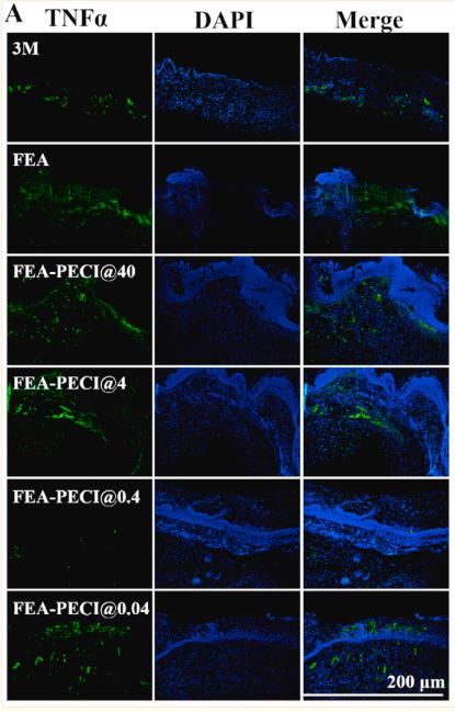
The nuclear counter stain is DAPI (blue).")

. b Densitometry of the TNF-a and IL-1b band correlated to the b-actin band. The bars represent the mean ± SE (n = 5/group). The results demonstrated that a significant increase of TNF-a and IL-1b expression in the TBI group (*p\0.01 vs. sham group), and treatment with TAK-242 significantly downregulated the level of TNF-a and IL-1b protein expression at 12, 24, and 48 h following TBI (# p\0.05 vs. TBI group). TNF-a, tumor necrosis factor-a; IL-1b, interleukin-1b")


- or vehicle-treated cystic kidneys. (A) TNF-α, MCP-1, CFB and SOD2 were analyzed by western blot in 9-week-old +/+ and Cy/+ kidneys. (B–E) Immunohistochemical staining for oxidative stress markers 8-OHdG and nitrotyrosine in the tubulointerstitial area. Computer-assisted morphometry was used to quantify changes of 8-OHdG and nitrotyrosine in each group. Scale bar = 50 µm. (F) NF-κB pathway ( p-p65, p65, p105 and p50) and mTOR pathway ( p-S6K and total S6K) were analyzed by western blot in 9-week-old +/+ and Cy/+ kidneys. Blots are representative of three independent experiments.")
 were used.(A-B)Immunohistochemistry assay of the DBM and surrounding tissues at 7 and 14 days for the secretion of (A) IL-10 and (B) TNF-α.")
Expression bands of claudin-4, occludin, ZO-1, IL-6, and tumor necrosis factor-α (TNF-α) in the kidneys of newborn rats exposed to normoxia or hyperoxia till 1st postnatal day (P1D), 3rd postnatal day (P3D), 5th postnatal day (P5D), 7th postnatal day (P7D), and 14th postnatal day (P14D) were detected by western blotting.")

Histomorphological and immunofluorescence evaluation for sterile gauze, WBMF-2VE−, and WBMF-2 on the 7th day. WBMF-2VE− and
WBMF-2 showed an attenuated inflammatory response and rapid keratinocyte proliferation and migration. The black, blue, and green arrows
represent inflammatory cell infiltration, neovessel, and fibroblasts, respectively. The green dotted rectangles point out the epidermal tongues and
magnify them (the open green arrows indicate keratinocytes). TNF-α presents red fluorescence, and VEGF presents green fluorescence. Black scale
bar: 200 μm, green scale bar: 20 μm, white scale bar: 50 μm. Quantitative analysis of the relative percentage of area coverage by (b) TNF-α and (c)
VEGF on the 7th day. For all data, the sterile gauze group was set as 100%. *P < 0.05, **P < 0.005.")
 Efficacies of NS, TA, DS and DS-TA NPs in treating OA, as shown by articular sections stained with H&E or safranin O, and their corresponding immunohistochemical staining
sections. Scale bar: 100 μm. (B, C, D) The levels of IL-1β, IL-6 and TNF-α in cartilage samples in each treatment group. Date are presented as mean ± SD (n = 3, *p b 0.05).")
 Representative figures of TNF-α, IL-1β and IL-10 protein. (D-F) Quantitative analyses of TNF-α, IL-1β and IL-10 protein. (mean ± S.D, n = 3).")
 The expressions of TNF-α, IL-6, IL-1β, and
ICAM-1 were evaluated by Western blotting at 3 days post-SCI in each group (n = 6). (B) Quantification of TNF-α, IL-6, IL-1β, and ICAM-1
expressions (n = 6, all the data are expressed as means ± SD, two-way ANOVA followed by Tukey's post hoc test was applied). (C–F)
Immunofluorescence staining was used to detect the level of TNF-α, IL-6, IL-1β, and ICAM-1 from each group (n = 6, scale bar = 50 µm). (G)
Statistical analysis of immunofluorescence staining for positive expression of TNF-α, IL-6, IL-1β, and ICAM-1 in nerve cells from each group
(n = 6, all the data are expressed as means ± SD, two-way ANOVA followed by Tukey's post hoc test was applied). * means p < 0.05; **means
p < 0.01; and *** means p < 0.001")
. In DM rats treated with osthole, levels of TNF-α protein were lower than those in the untreated DM group (p < 0.01). No difference was observed between the DM group and the DM+DMSO group (p > 0.05). Bar graphs show the ratio of TNF-α protein to β-actin protein in each group. Data are displayed as the means ±SEM (n = 8 per group). **p < 0.01 compared to the control group; ##p < 0.01 compared to the DM group.")

 Representative images of HE and TRAP staining, showing the reduced osteolytic legion and TRAP-positive OCs in the PF-treated groups. (D–G) Representative images of IHC staining of RANKL, OPG, OCN and TNF-α. (H) TRAPpositive OCs number. (H) Quantitative analysis of the expression of RANKL, OPG and RANKL/OPG ratio. The data are presented as the mean ± SD (*p < 0.05,**p < 0.01, ***p < 0.001).")
 for 24 h, the expression of OCTN1 (62 kDa), OCTN2 (63 kDa), IL6 (24 kDa), IL18 (26 kDa), IL1β (31 kDa),and TNF-α (26 kDa) was significantly regulated in human respiratory epithelial cells;β-actin (42 kDa) was used as a loading control. Bands were analysed using ImageJ 1.45s (National Institutes of Health, USA). NC indicates the normal control groups. Data are shown as the mean ± SD (n = 3).")
; metabolic pathway analysis in MICT versus SED (b);mRNA levels of PPAR-γ, HSL, ATGL, and TNF-α in adipose tissues (c);correlation analysis of 6 metabolites (d); metabolic pathway analysis in HIIT versus SED (e); and contents of PPAR-γ, HSL, ATGL, TNF-α, and P450scc protein (f).")
 Representative immunoblots and relative protein levels of TNF-α,IL-6, NF-κB, and HO-1")

 Optical density analysis of the TNF-α, IL-1β and IL-6 proteins.")
. b–e The inflammatory marker proteins immunofluorescence staining and histograms at 14 days, including CD86, GFAP, TNF-α, iNOS(magnification × 400). All data presented as mean ± SEM in each group. *p < 0.05 vs. SCI + saline group, **p < 0.01 vs. SCI + saline group. #p < 0.05 comparison between SCI + EPO group and SCI + EPO + PD98059, ##p < 0.01 comparison between SCI + EPO group and SCI + EPO + PD98059")
 Control group, (B) Sham group, (C) Aβ group, (D) Aβ + Dserine group, (E) Aβ + DAAO group and (F) Aβ + BE group.")
 The western blot results show the expression levels of GFAP, TNF-α, p-JNK, JNK, p-c-jun, c-jun, ATF2 and NMDAR1 proteins in all experimental groups.")

 Immunohistochemistry was used to detect the expression of interleukin (IL)-1 β, IL-6 and tumor necrosis factor (TNF)-α in the flaps of the three groups of
rats; (B) IL-1β, IL-6 and TNF-α contents in the flaps of three groups of rats were expressed by integral absorbance (IA). **P < 0.01, CDPC-L group vs. control group;
**P < 0.01, CDPC-L group vs. CDPC-H group.")
 The levels of mRNA expression in brain tissue 24 h after hypoxic–ischemic (HI) injury normalized to those of β-actin for each sample. *P < 0.05, **P < 0.01, and ***P < 0.001 vs. the HI group. n = 5. Values are presented as mean ± SEM. n = 5. Protein expression level (B) and quantification data (C) of tumor necrosis factor-alpha (TNF-α) 24 h after HI injury. *P < 0.05 vs. the sham group. #P < 0.05 vs. the HI group. Values are presented as mean ± SEM. n = 3. Representative images of immunofluorescence staining of TNF-α (green) in the cortex (D), hippocampus (E), and nucleus (blue) labeled with DAPI. Scale bar = 25 μm.")
 The levels of mRNA expression in brain tissue 24 h after hypoxic–ischemic (HI) injury normalized to those of β-actin for each sample. *P < 0.05, **P < 0.01, and ***P < 0.001 vs. the HI group. n = 5. Values are presented as mean ± SEM. n = 5. Protein expression level (B) and quantification data (C) of tumor necrosis factor-alpha (TNF-α) 24 h after HI injury. *P < 0.05 vs. the sham group. #P < 0.05 vs. the HI group. Values are presented as mean ± SEM. n = 3. Representative images of immunofluorescence staining of TNF-α (green) in the cortex (D), hippocampus (E), and nucleus (blue) labeled with DAPI. Scale bar = 25 μm.")
 Immunohistochemistry was used to detect the expression of interleukin (IL)-1 β, IL-6, and tumor necrosis factor (TNF)-α in the flaps of the three groups of
rats; (B) IL-1 β, IL-6, and TNF-α contents in the flaps of the three groups of rats were expressed by integral absorbance (IA). **P < 0.01, DEX-L group vs. control
group; **P < 0.01, DEX-L group vs. DEX-H group. n = 6 per group.")

 is the QT-PCR analysis results
of some key genes in the tumor tissues; (I) is the western bolting analysis results of some key proteins in the tumor tissues; (J) is the level of LPS in serum;
(K) is the plasma concentration of 5-Fu. Animals were randomly divided into nine groups: control (no treatments), xenograft-only (Model), HEP-only
(FP), pre-xenograft HEP (PN), pre-xenograft HEP+5-Fu (PF), HEP+5-Fu (NP), 5-Fu-only (NF), antibiotics+HEP+ 5-Fu (AP), and antibiotics+5-Fu (AF).
Only the control and HEP-only groups had no xenografts. Values are expressed as means ± SDs (n = 8), #P < 0.05 compared with control group, ∗P <
0.05, ∗∗P < 0.01 compared with model group, indicates significant differences compared to the model group.")
 eNOS, CD31 and α-SMA levels in mesenteric arteries of SHRs as measured using western blot. (B) eNOS expression in mesenteric arteries. (C) KD decreased NO
levels in serum of SHRs. (D-E) Quantification of CD31 and α-SMA of Western blots. All data are expressed as a mean ± SEM. n = 6 in each group. *P < 0.05;
**P < 0.01; KD, Ketogenic diet; ND, Normal diet; SHR, Spontaneously hypertensive rats; WT, Wistar rat.")
 The levels of mRNA expression in brain tissue 24 h after hypoxic–ischemic (HI) injury normalized to those of β-actin for each sample. *P < 0.05, **P < 0.01, and ***P < 0.001 vs. the HI group. n = 5. Values are presented as mean ± SEM. n = 5. Protein expression level (B) and quantification data (C) of tumor necrosis factor-alpha (TNF-α) 24 h after HI injury. *P < 0.05 vs. the sham group. #P < 0.05 vs. the HI group. Values are presented as mean ± SEM. n = 3. Representative images of immunofluorescence staining of TNF-α (green) in the cortex (D), hippocampus (E), and nucleus (blue) labeled with DAPI. Scale bar = 25 μm.")
 The levels of mRNA expression in brain tissue 24 h after hypoxic–ischemic (HI) injury normalized to those of β-actin for each sample. *P < 0.05, **P < 0.01, and ***P < 0.001 vs. the HI group. n = 5. Values are presented as mean ± SEM. n = 5. Protein expression level (B) and quantification data (C) of tumor necrosis factor-alpha (TNF-α) 24 h after HI injury. *P < 0.05 vs. the sham group. #P < 0.05 vs. the HI group. Values are presented as mean ± SEM. n = 3. Representative images of immunofluorescence staining of TNF-α (green) in the cortex (D), hippocampus (E), and nucleus (blue) labeled with DAPI. Scale bar = 25 μm.")

 Immunofluorescence staining of TNF-α in kidneys. (b) Serum
TNF-α level. (c) Renal TNF-α relative mRNA level. Date are represented as the mean ± SD. *p<0.05, **p<0.01, ***p<0.001, ****p<0.0001.")
 levels were analysed by western blotting. The data are presented as the mean ± SD. N = 4, ##P < 0.01 vs.
the normal group; **P < 0.01 vs. the CIA model group. Bonferroni’s test was used for multiple comparisons between groups.")

 Representative pictures of TNF-α (a–d) and
IL-6 (e–h) utilizing immunohistochemistry (400×); bars present the semi-quantification of the TNF-α and IL-6 respectively (n = 8). (B) Representative
bands of TNF-α and IL-6 by means of western blotting; β-actin proteins were used as loading controls; the bars represent semi-quantification of
TNF-α and IL-6 respectively. All data were expressed as mean ± SEM. (n = 3). **P < 0.01 vs. control group; #P < 0.05, ##P < 0.01 vs. DN group. TNF-α:
tumor necrosis factor-α; IL-6: interleukin-6. Black arrows, representative immunostaining areas of TNF-α and IL-6 respectively")
 and TNF-α IHC staining (f–j, TNF-α: × 400 magnification). Tissue sections of cerebellum were staind with TNF-α IHC staining (k–o, TNF-α: × 400 magnification)")
. All data are presented as mean ± SD. *P < 0.05 versus the NC group; #P < 0.05 versus the DM group; △P < 0.05 versus HA and CUR groups")
 Co-expression of TLR4/CD68 in rats exposed to silica, Bar=50 μm; (B) Co-expression of TNF-α/CD68 in rats exposed to silica, Bar=50 μm; (C) Levels of TLR4. MyD88, p-IκBα, and TNF-α in rats lungs measured by Western blotting. Data are presented as the mean ± SD. n = 3 per group. (D) Levels of TNF-α and RANKL in serum of rats measured by ELISA. Data are presented as the mean ± SD. n = 10 per group.")
. Quantitative results of TNF-α (b), IL-1β (c) and MCP-1 (d). IHC staining of oxidative stress indexes in three groups, including 8-OHdG and 4-HNE (e). Quantitative results of 8-OHdG (f) and 4-HNE (g) in IHC staining. Correlation of serum CN-1 expression with 8-OHdG (h) and 4-HNE (i). Representative microscopic images are shown (× 200 magnification). *p < 0.05; **p < 0.01; ***p < 0.001; ****p < 0.0001")
 Protein expression of TNFα, Caspase8 and RIP1/RIP3 pathway by western blot in HK-2 cells. (B) Representative micrographs showing IHC staining of TNFα, Caspase8 RIP1 and RIP3 proteins in mouse renal tissues. *P<0.05 compared with Sham/Saline, #P<0.05 compared with IRI/Saline. (C) Protein expression of TNFα, Caspase8 and RIP1/RIP3 pathway by western blot in HK-2 cells under ischemia-reperfusion injury. (D) Representative micrographs showing IHC staining of TNFα, Caspase8, RIP1 and RIP3 proteins in mouse renal ischemia-reperfusion injury tissues. *P<0.05 compared with IRI/veliparib/pre-NC, #P<0.05 compared with IRI/veliparib/anti-NC.")
 Protein expression of TNFα, Caspase8 and RIP1/RIP3 pathway by western blot in HK-2 cells. (B) Representative micrographs showing IHC staining of TNFα, Caspase8 RIP1 and RIP3 proteins in mouse renal tissues. *P<0.05 compared with Sham/Saline, #P<0.05 compared with IRI/Saline. (C) Protein expression of TNFα, Caspase8 and RIP1/RIP3 pathway by western blot in HK-2 cells under ischemia-reperfusion injury. (D) Representative micrographs showing IHC staining of TNFα, Caspase8, RIP1 and RIP3 proteins in mouse renal ischemia-reperfusion injury tissues. *P<0.05 compared with IRI/veliparib/pre-NC, #P<0.05 compared with IRI/veliparib/anti-NC.")
 Immunohistochemistry of inflammatory cytokines (TNF-α, IL-6, and IL-1β) (B) Immunofluorescent staining of macrophages (F4/80, iNOS) (C) Representative western blots of p-p38, p38, p-JNK, JNK, p-ERK, and ERK in colon tissue (D–F) Relative density of each signaling band was calculated. Data are expressed as the mean ± SEM, n = 3. Data were analyzed using one-way ANOVA. #p < 0.05, ##p < 0.01 compared to the control group. *p < 0.05, **p < 0.01 compared to the model group.")
 TNF-α and (B) IL-6 levels in serum. (C) Protein expression levels of TNF-α, IL-6 and HO1 in the liver tissues were detected via western blotting. Semi-quantitative analysis of (D) TNF-α, (E) IL-6 and (F) HO-1 was normalised to β-actin. Data are presented as the mean ± SD, n=3. ***P<0.001 vs. control group; #P<0.05, ##P<0.01, ###P<0.001 vs. model group. Con, Control; Mod, Model; AS-IV, Astragaloside IV (80 mg/kg); Met, Metformin (200 mg/kg); HO-1, heme oxygenase-1.")
 (B) qPCR and western blot analyses of IL-6, TNF-α, and MCP-1 in cardiac tissue of the WT Sham, TN-C KO Sham, WT MI, and TN-C KO MI groups on the fifth day
after MI. (C) Representative immunohistochemistry and quantitate of IL-6, TNF-α, and MCP-1 expression in cardiac tissues on the fifth day after MI (n = 6 per group).
(D) (E) Representative qPCR and western blot analyses of IL-6, TNF-α, and MCP-1 in cardiomyocytes infected with si-NC or si-TN-C before treated by H2O2 (150 μM
24 h) (n = 4 per group). Scale bar = 20 μm. Data are mean ± SD. *P < 0.05, **P < 0.01, ***P < 0.001, ****P < 0.0001. IL indicates interleukin; MCP, monocyte
chemoattractant protein; TN-C, tenascin-C; TNF, tumor necrosis factor; WT, wild type; TN-C KO, TN-C knockout mice; MI, myocardial infarction; qPCR, quantitative
real-time polymerase chain reaction.")
. The transfection efficiency of bovine miR-125b mimic or inhibitor in MAC-T cells as measured by fluorescence micrograph (A) and qPCR (B). At 48 h post-transfection, MAC-T cells were challenged with LPS for 3 h. Cells were then collected to measure mRNA expression and production of NKIRAS2 (C), NF-κB (D), IL-6 (E), and TNF-α (F). Expression levels of mRNA and protein were determined by qPCR and Western blot, respectively. *p < 0.05, **p < 0.01, ***p < 0.001.")
 Western blots and (c) quantification of IL-4, IL-6,TNFα, GFAP, and Iba1 proteins in each group after SCI; columns represent the mean ± SD (n = 5)")
 Representative iNOS, COX-2, TNF-α, IL-1β, IL-6, and
β-actin bands. (B), (C), (D), (E), (F) The relative ratios of grayscale values of iNOS, COX-2, TNF-α, IL-1β, and IL-6 to that of corresponding β-actin, n = 4. ###P <
0.001, CUMS + saline vs. sham; *P < 0.05; **P < 0.01; ***P < 0.001, CUMS + saline vs. CUMS + AAV-siNCALD. All data are shown as the mean ± SEM.")
 The mRNA expression levels of IL-6, TNF-α, and MCP-1 in renal tissue
were analyzed by RT-PCR. (B) The protein expression levels of IL-6, TNF-α, and MCP-1were detected by western blot. (C-E) The ratio of IL-6, TNF-α, and MCP-1 to
GAPDH was calculated. Data are expressed as mean ± SEM (n = 6). **** p < 0.0001 vs. control; #### p < 0.0001 vs. model. The ns. means no significance.")
, IL-10 (B), IL-1β (C), IL-6 (D), TNF-α (E) and MCP-1 (F)
secreted by RAW 264.7 cells on AH-Ti, AH-Ti-PDA and AH-Ti-PDA-DJK-5 under the inflammatory environment with or without RANKL stimulation for 1, 3 and 5
days. *p < 0.05 compared with AH-Ti, #p < 0.05 compared with AH-Ti-PDA and Жp < 0.05 compared with the addition of RANKL. G: Histological evaluation with IL6 and TNF-α staining. Representative histological photographs of AH-Ti, AH-Ti-PDA and AH-Ti-PDA-DJK-5 implanted into the infected skin of rats for 7 days.")
 RT-qPCR was used to detect the mRNA expression of IL-1β, IL-6, TNF-α, and MCP-1 in renal tissues of mice (10 mice in each group). (B) Western blotting was used to analyze the protein expression of COX-2, iNOS, TNF-α, HMGB1, IL-6, and IL-1β in renal tissues (10 mice in each group). (C) Protein expression of COX-2, iNOS, TNF-α, HMGB1, IL-6, and IL-1β in panel B was quantified by the optical density method (10 mice in each group). The measurement data are expressed as the mean ± standard deviation. *, P < 0.05, indicates data comparison between two groups.")
 Relative mRNA level of IL-1β, IL-6, and TNF-α in the colon was measured through qPCR. Expression of IL-1β, IL-6, and TNF-α in colon tissue lysates were determined by Western blot from DSS-challenged mice.")
 caveolin 1, (B) p-p38 MAPK, (C)MMP-13, (D) IL-1β, (E) TNF-α, (F) Bax, and (G) Bcl-2. *P < 0.05 indicates statistical differences between each treatment group and the model group.")
 protein expression. (a) Representative Western blotting results for IL-1β, IL-6,TNF-α, and GAPDH.")
. (a) Western blot analysis of the proteins MMP9, MAPK8, IL-6, and TNF-α in the blood.")
. B Quantitative analysis of intracellular lipid droplets. C The expressions of TNFα, IL-1, and Cleaved-caspase3 in control, PA, and C14 group (n = 6), as detected using western blotting. D The results of TNFα, IL-1, and Cleaved-caspase3 expression. E The free fatty acids of H9c2 cells in each group was measured (n = 6) and compared with the treated and control group. F The data revealed positive expression of TNFα and cleaved-caspase 3 in H9c2. (G-H) Immunofluorescence staining results showing the levels of TNF-α and Cleaved-caspase3 in H9c2 (n = 6, Scale bar = 100 μm). Data are represented as means ± SD, where two-way ANOVA followed by Tukey’s post hoc test employed. *P < 0.05, **P < 0.01, and ***P < 0.001")
. B Quantitative analysis of intracellular lipid droplets. C The expressions of TNFα, IL-1, and Cleaved-caspase3 in control, PA, and C14 group (n = 6), as detected using western blotting. D The results of TNFα, IL-1, and Cleaved-caspase3 expression. E The free fatty acids of H9c2 cells in each group was measured (n = 6) and compared with the treated and control group. F The data revealed positive expression of TNFα and cleaved-caspase 3 in H9c2. (G-H) Immunofluorescence staining results showing the levels of TNF-α and Cleaved-caspase3 in H9c2 (n = 6, Scale bar = 100 μm). Data are represented as means ± SD, where two-way ANOVA followed by Tukey’s post hoc test employed. *P < 0.05, **P < 0.01, and ***P < 0.001")
. B, C ELISA assays of TNF-α and IL-6 after various treatments. Data are presented as the means ± SD (n = 3), *P < 0.05, **P < 0.01")
 Western blotting for the indicated proteins in ventricular extracts from Tab2fl/fl, MerCreMer (MCM), and Tab2fl/fl-MCM mice 2 weeks after treatment with tamoxifen as described in Methods. TAB2 protein level was normalized for the internal control GAPDH and expressed as fold change. *P < 0.05 versus Tab2fl/fl or MCM. n = 4. (B and C) Masson’s trichrome–stained, paraffin-embedded cardiac sections from mice as described in A. Scale bars: 1 mm in B and 50 μm in C. (D) Myocardial fibrosis quantified with MetaMorph software. *P < 0.05 versus Tab2fl/fl or MCM. n = 5–7. (E) Western blot (left) and quantification (right) of the indicated proteins normalized to GAPDH in cardiac extracts from mice indicated in A. (F) Heart weight to body weight ratio (HW/BW) of mice of the indicated genotypes. *P < 0.05 versus Tab2fl/fl or MCM. (G) Lung weight to body weight ratio (LW/BW) of mice of the indicated genotypes. *P < 0.05 versus Tab2fl/fl or MCM. (H) Representative echocardiographic M-mode images from mice indicated in A. The vertical white arrowed lines indicate left ventricular dimension in end-diastole (LVED). (I–K) Echocardiographic assessment of fractional shortening (FS) and left ventricular dimension in end-diastole (LVED) and end-systole (LVES) in mice of the indicated genotypes. *P < 0.05 versus Tab2fl/fl or MCM. n = 5–7. Statistical analysis was performed using 1-way ANOVA with Tukey’s post hoc test.")
. On day 5 and 8, the staining intensity of IL-6 and TNF-α in the 0.75 μg/μL EVs group were weaker comparing with blank control group (*P<0.05 n=5). There is no statistical difference between blank control group and 0.75 μg/μL EVs group on day 5 and 8 in IL-1β. EVs, extracellular vesicles.")
 Calcitriol decreased myocyte apoptosis and death. Representative photographs (A) and quantitative analysis of TUNEL staining of the cardiomyocytes in mice with the indicated treatments (n = 5). Scale bar represents 50 μm. (C,D) Western blot analysis of protein expressions in the whole-cell lysates (VDR, IL-1β, IL-10, TNF-α, and NF-κB p65) or nuclear lysate (VDR and p-NF-κB p65) in mice with various treatments (n = 5). (E–G) Serum IL-1β, TNF-α, and IL-10 levels in mice with the indicated treatments (n ≥ 8). Data presented here are representative images (A,C) or quantitative analysis (B,D–G), respectively. (Scale bar represents 50 μm. Data represent means ± S.D. * p < 0.05, ** p < 0.01 versus sham group, # p < 0.05, ## p < 0.01 versus MI group; one-way ANOVA with a post hoc test of Tukey’s analysis).")
 were detected 5 days after isoflurane and sevoflurane anesthesia. A, B. The results revealed that isoflurane (p = 0.000, for Nestin; p = 0.000, for Sox2) and sevoflurane (p = 0.000, for Nestin; p = 0.000, for Sox2) inhibited the expression of Nestin and Sox2. Isoflurane inhibited the expression of Nestin or Sox2 compared to that of sevoflurane (p = 0.040, for Nestin; p = 0.010, for Sox2). Furthermore, isoflurane (p = 0.000, for IL-6; p = 0.000, for TNF-α; p = 0.000, for CD11b) and sevoflurane (p = 0.000, for IL-6; p = 0.000, for TNF-α; p = 0.000, for CD11b) increased the levels of IL-6, TNF-α and the microglial marker CD11b in hippocampus. C-F. Isoflurane (p = 0.000, for p-VEGFR2; p = 0.000, for p-VEGFR2/VEGFR2) and sevoflurane (p = 0.000, for p-VEGFR2; p = 0.000, for p-VEGFR2/VEGFR2) decreased the level of p-VEGFR2 and the ratio of p-VEGFR2/VEGFR2. Data are mean ± SEM. **p < 0.01, *p < 0.05")

 and
inflammatory biomarkers (TNF-α and IL-1β) as determined by Western blot analysis. G. Semi-quantification of cleaved Caspase-3/ Caspase-3, Bax/Bcl-2, TNF-α and
IL-1β). H. Concentration of cytokines TNF-α, IL-1β and IL-6, respectively, in HK-2 cells as detected by ELISA. The data in B, C, E, G and H are expressed as means ±
SD, *P < 0.05 vs. control group, #P < 0.05 vs. Cis + mimic NC group, &P < 0.05 vs. Cis + inhibitor NC group. ※P < 0.05 vs. Cis group.")
 Representative immunofluorescent staining and quantification of the ionized calcium-binding adaptor molecule 1 (Iba-1+) cells in the cornu ammonis 1 (CA1), cornu ammonis 3 (CA3), and dentate gyrus (DG) regions of the hippocampus (HIP) (n = 3, 2 images per mouse, scale bar 50 μm). The image captured from the box was marked with a solid line (scale bar 10 μm). (C, D) The mRNA expression levels of the cluster of differentiation 68 (CD68) and proinflammatory cytokines, including the tumor necrosis factor-alpha (TNF-α), interleukin-1 beta (IL-1β), and interleukin-6 (IL-6) in the HIP (n = 6). (E, F) Immunofluorescent staining and quantification of Iba-1+ cells in the prefrontal cortex (PFC) (n = 3, 2 images per mouse, scale bar 50 μm). (G, H) The mRNA expression levels of CD68 and proinflammatory cytokines, including TNF-α, IL-1β, and IL-6 in the PFC (n = 6). The relative mRNA levels were normalized with reference gene (β-actin). (I, J) The protein expression levels of TNF-α in the HIP and PFC (n = 8). Values are represented as mean ± SEM. * P < 0.05, ** P < 0.01, *** P < 0.001. Tukey-Kramer test.")
-induced S-AKI mice. (A) The serum levels of TNF-α, IL-6, and IL-1β were determined using assay kits in each group of mice. (B) Gene expression of TNF-α and IL-6 in renal tissues was quantitated using qRT-PCR. (C) The expressions of TNF-α, IL-6, and MCP-1 in renal tissues were analyzed using Western blot. (D) The protein levels of TNF-α, IL-6, and MCP-1 were quantified by densitometry and normalized with GAPDH. (E) TUNEL staining and immunofluorescence staining images of cleaved caspase-3 (C Casp-3) expression in renal sections were collected (scale bars = 50 μm). (F) TUNEL staining positive cells were counted in each group. (G) The gene expression ratio of BAX/BCL-2 in renal tissues was quantitated using qRT-PCR. (H) The expressions of BAX, BCL-2, and C Casp-3 in renal tissues were analyzed using Western blot. (I) The protein levels of BAX, BCL-2, and C Casp-3 were quantified by densitometry and normalized with GAPDH (time point: 12 h). Data are presented as mean ± SD, n = 5. *p < 0.05, **p < 0.01, ***p < 0.001, ****p < 0.0001 vs. control group; #p < 0.05, ##p < 0.01, ###p < 0.001, ####p < 0.0001 vs. LPS group.")
 FACS analysis of AF cells treated with AOPPs (50–400 µg/mL, 24 h) induced G1 phase arrest. (B) SA-β-gal staining of AF cells treated with AOPPs (50–400 µg/mL, 24 h). AOPPs increased the proportion of senescent AF cells. Scale bar: 100 µm. (C) AOPPs significantly increased the expression of NOX4, p53, p21, p16, IL-1β and TNF-α in AF cells. *P < 0.05 compared to the control. **P < 0.01 compared to the control. Error bars represent standard errors.")
 MRI showed that blocking NOX4 expression by AAV reduced the Pfirrmann grade of disc degeneration caused by RSA and AOPPs. *P < 0.05. **P < 0.01. Error bars represent the interquartile range. (B, C) HE staining and saffron O/fast green staining showed less disc destruction caused by AOPPs and RSA in discs with blocked NOX4 expression and correspondingly decreased disc injury scores. **P < 0.01 and #P < 0.05 compared with the other four groups. (D) Western blot analysis showed that blocking NOX4 expression reduced the expression of p53, p21, p16, IL-1β and TNF-α in disc tissue injected with AAV. (E, F) Immunofluorescence assay showed that blocking NOX4 expression reduced the expression of p53, p21, p16, p-p38, p-ERK, p-JNK, IL-1β and TNF-α. (G) Diagram of AOPPs-induced NOX4, p53, p21, p16, IL-1β and TNF-α expression. #P < 0.05 compared to the AAV-NC+RSA and AAV-NC+AOPP groups. *P < 0.05. **P < 0.01. Error bars represent standard errors.")
, as well as p65, ID1, p-SMAD1/5, and osteogenic genes, concluding OCN and SOX9 (b). Scale bar = 25 μm. (c) Immunofluorescence and quantitative analysis against Smad5. Scale bar = 25 μm. Data are means ± SD (n = 3) ∗p < 0.05 and ∗∗p < 0.01. #p < 0.05 and ##p < 0.01. (d) The mRNA expression, Western blot analysis of tendon tissues, and relevant quantitative analysis of p65, ID1, OCN, and SOX9. Data are means ± SD (n = 3). ∗p < 0.05 and ∗∗p < 0.01. #p < 0.05 and ##p < 0.01.")
. B Lung injury score was evaluated according to the degree and scope of lung injury, including the degree of interstitial edema, septal wall thickness, and inflammatory cell infiltration. C Representative images of immunohistochemical staining of fibrin were obtained in the lungs from three groups of mice (400 ×). D The western blot results showed IL-1β, TNF-α, and IL-6 protein levels in lung tissues, and the relative expression levels of IL-1β, TNF-α, and IL-6 normalized to tubulin. Values are shown as mean ± SD. **P")
. Representative blots are shown. *P")
 Oil red O was used to measure the level of lipid accumulation (magnification 100x, scale bar = 250 μm). (b) The oil red O-positive area was analyzed and quantified. (c–j) The relative mRNA expression levels of FABP1, SCD1, CD36, HMGCR, ACACA, CCL5, IL-1β, and TNF-α were determined by qRT-PCR. (k) The proinflammatory factor protein levels of IL-1β, IL-6, and TNF-α. (l) The protein levels related to lipid metabolism of SREBP-1, ACOX1, CD36, CPT1α, and HMGCR were analyzed by western blotting, and the relative ratios were calculated and expressed as the mean ± SD; n = 3. #P < 0.05 means that the difference between the NC group and the PA group is significant. ∗P < 0.05 means that the difference between the PA group and the FV (30 mg/mL) group or the FV (15 mg/mL) group is significant.")
 The relative protein levels of PGC-1α, Nrf2, and SIRT1. (B–D) Values of quantitative analysis (n = 4). (E) Relative mRNA levels of SIRT1, PGC-1α, Nrf1, Nrf2, TNF-α, and IL-1β. (F) Percentage of immunostaining for Sirt1. (G) Immunohistochemistry shows the expression of hepatic SIRT1 (400× magnifications). (H) The relative protein levels of TFAM and (I) the relative protein levels of TNF-α. Data are presented as mean ± SEM. * p < 0.05, ** p < 0.01, *** p < 0.01 vs. control group. ns: no significance.")
 and preserved ΔΨm (× 20 magnification). c The protein levels of cyc were determined. d, e L-carnitine administration significantly reduced the content of MDA and increased the antioxidant enzymes (SOD activity). f Immunoblot analysis of TRPA1, IL-1β and TNF-α. Representative blots were from three independent experiments. g Representative photographs of Ca.2+ fluorescence intensity in different FLS treatment groups at 0, 30, 60, and 90 s (× 630 magnification). Data are presented as the mean ± SD (n = 3). ns, not significant. *P")
 Expression of STING, p-IRF3, TNF-α, and IL-1β, measured by western blotting. (B, C) Greater expression of STING and p-IRF3 in the D-I/R group than the I/R group. (D) Expression of p-IRF3, measured by immunofluorescence assays. (E, F) Greater expression of TNF-α and IL-1β in the D-I/R group than the I/R group. (G) Greater mRNA expression of IFNβ in the D-I/R group than the I/R group. Data are expressed as means ± SD,")

 thoracic aorta vascular and (B) myocardial tissue. Magnification, ×400. LBP, Lycium barbarum polysaccharide; EX, exhaustive exercise; Keap1, Kelch-like ECH-associated protein 1; Nrf2, NF-E2-related factor 2; NQO1, quinone oxidoreductase 1; GCLC, glutamyl-cysteine synthetase catalytic subunit; GCLM, glutamate-cysteine ligase modified subunit; AE, aerobic exercise.")
 Immunohistochemistry images of IL-1β, IL-6 and TNF-α. All images were obtained at identical mag-nification, ×200, scale bar = 50 μm. (B) Quantitative analysis of IL-1β, IL-6 and TNF-α content (n = 3). Data are represented as mean ± SEM. **P")
 Calcein-AM imaging of intracellular iron levels in H9C2 cells. (B) Western blot images and protein expression levels of DMT1, TfR1, FPN1 and FtH in LPS- and CVB-D-treated H9C2 cells. (C) hamp mRNA expression levels in LPS- and CVB-D-treated H9C2 cells. (D) Protein expression levels of pro-IL-1β, TNF-α, IL-6, p-STAT3 and STAT3 in LPS- and CVB-D-treated H9C2 cells. Data are presented as the mean ± SD (n=6). **P")
 area. (A) IHC-stained tissue images of the enthesis FC areas according to week age. TNF-α and IL-6 are used as primary antibodies. The black dot line indicates the tidemark. (B) Comparison results of TNF-α expression in the FC area. (C) Comparison results of IL-6 expression in the FC area. Statistical significance: * p < 0.05, ** p < 0.01, *** p < 0.001. † p < 0.05 (8-week-old mice in the Sedentary group vs. 12-week-old mice in the Sedentary group). TNF, tumor necrosis factor; IL, interleukin; IHC, immunohistochemical; UFC, uncalcified fibrocartilage; CFC, calcified fibrocartilage. Scale bar; 50 µm.")
 and spinal cord (D, E, F) of rats of each group. Values are presented as the means ± standard error of the mean. n = 6, **P")
 Magnification after immunohistochemical staining of hypoxia‐inducible factor‐1α (HIF‐1α) and vascular endothelial growth factor (VEGF) on day 7. (B) Expression of HIF‐1α on day 7. Roxadustat (RXD) improved the expression of HIF‐1α. (C) Expression of VEGF on day 7. RXD improved the expression of VEGF. **P")
 Relative mRNA levels of TNF-α, IL-1β, NF-κB p65, and GAPDH. (B) Relative protein expressions of NF-κB p65, TNF-α, IL-1β, and GADPH. (C) Representative western blots TNF-α, IL-1β, NF-κB p65, and GADPH. Data are presented as mean ± SEM (n = 3).")
 IL-6 immunofluorescence staining in the gastric tissue of patients (Scale bar = 100 or 50 μm, n = 3). (B) The mean density of IL-6-positive cells in Panel (A). (C) p-STAT3 immunofluorescence staining of the gastric tissue of patients (Scale bar = 100 or 50 μm, n = 3). (D) The mean density of p-STAT3-positive cells in Panel (C). The bottom images were the larger images of the frame in the top images, and the arrows indicate the location of IL-6-positive cells (A) or p-STAT3-positive cells (C). (E) IL-6, TNF-α, and IL-1β immunofluorescence staining in the gastric tissue of CAG rats (n = 3). (F) The expression of IL-6 and the ratio of p-STAT3/STAT3 in the gastric tissue of CAG rats, as determined by Western blotting (n = 4). The data are presented as the mean ± SEM. * p < 0.05, ** p < 0.01 vs. NAG or Con groups. NAG: non-atrophic gastritis patients; CAG: chronic atrophic gastritis patients or rats; Con: the control group of rats.")
 IL-1β, IL-6, and (B) TNF-α levels in serum. (C) TLR4, IKKα, IKKβ, (D) NF-κB (p65), TNF-α, IL-1β, and (E) IKKγ, IL-6 mRNA transcription levels in aorta. (F) p-IKKβ, IKKβ, p-IκBα, IκBα, (G) TLR4, p-p65, p65, (H) p-IKKβ/IKKβ, p-IκBα/IκBα, p-p65/p65, and (I) TNF-α, IL-1β protein expression levels in aorta. (J) p-IKKβ, IKKβ, p65, p-IκBα, IκBα, TNF-α, IL-1β and (K) TLR4, p-p65 protein expression levels in aorta. (L) Double fluorescence staining of p-IKKβ and MOMA2 in the aortic root plaque (400 ×, scale bar: 50 µm), the area enclosed by the white circle represents the plaque area, and white arrows indicate the expression of p-IKKβ in macrophages. (M) Fluorescence intensity of p-IKKβ and MOMA2. (N) Fluorescence colocalization curve. (O) Fluorescence colocalization scatterplot. GAPDH served as an internal control, and data are presented as the mean ± SD.")
 operating at 400× magnification (a). The integral absorbances of flap VEGF, TLR4, NF-kB, IL-1β, IL-6, and TNF-α levels in the control and treatment groups (b). **P")
 Expression of PTGS2, FOS, JUN, EDN1 and TNF proteins in NPCs treated with TBHP, with or without PT. (B–F) Expression of PTGS2, FOS, JUN, EDN1 and TNF proteins relative to β-actin in NPCs treated with TBHP, with or without PT. Data are the mean ± standard deviation. # p < 0.05, ### p < 0.001, #### p < 0.0001 vs. the CTL group; * p < 0.05, ** p < 0.01 vs. the TBHP group treated with 100 μM TBHP.")
. Representative western blots (A, C, E, G, I) and quantification of nuclear factor kappa-B (NF-κB) (B), phosphorylated NF-κB (p-NF-κB; p65) (D), interleukin-6 (IL-6) (J, K), tumor necrosis factor-α (TNF-α) (F) and interleukin-1β (IL-1β) (H). Representative immunostaining and quantification of IL-1β (L, M), TNF-α (N, O), and IL-6 (P, Q) in IMATs. Magnification: ×200; scale bar = 50 μm. CON control, EMA combined therapies + compound-c, EMC combined therapies, EXE moderate exercise, MET metformin; PRE prediabetes. The data shown represent the mean ± standard error of the mean (n = 5–6/group). ap")
. Representative western blots (A, C, E, G, I) and quantification of nuclear factor kappa-B (NF-κB) (B), phosphorylated NF-κB (p-NF-κB; p65) (D), interleukin-6 (IL-6) (J, K), tumor necrosis factor-α (TNF-α) (F) and interleukin-1β (IL-1β) (H). Representative immunostaining and quantification of IL-1β (L, M), TNF-α (N, O), and IL-6 (P, Q) in IMATs. Magnification: ×200; scale bar = 50 μm. CON control, EMA combined therapies + compound-c, EMC combined therapies, EXE moderate exercise, MET metformin; PRE prediabetes. The data shown represent the mean ± standard error of the mean (n = 5–6/group). ap")
 for 24 h. CX3CL1, TNF-α, and IL-6 expression profiles were then measured by western blotting. F, G Levels of IL-6 and TNF-α in podocyte cell supernatant. H, I Representative micrographs of CX3CL1 and TNF-α staining in podocytes. Scale bar = 10 μm. (IL-6, interleukin 6; TNF-α, tumor necrosis factor-α; P value was calculated by one-way analysis of variance and Tukey’s test.")
 for 24 h. CX3CL1, TNF-α, and IL-6 expression profiles were then measured by western blotting. F, G Levels of IL-6 and TNF-α in podocyte cell supernatant. H, I Representative micrographs of CX3CL1 and TNF-α staining in podocytes. Scale bar = 10 μm. (IL-6, interleukin 6; TNF-α, tumor necrosis factor-α; P value was calculated by one-way analysis of variance and Tukey’s test.")
 for 24 h. CX3CL1, TNF-α, and IL-6 expression profiles were then measured by western blotting. F, G Levels of IL-6 and TNF-α in podocyte cell supernatant. H, I Representative micrographs of CX3CL1 and TNF-α staining in podocytes. Scale bar = 10 μm. (IL-6, interleukin 6; TNF-α, tumor necrosis factor-α; P value was calculated by one-way analysis of variance and Tukey’s test.")
 and TNF-α, MCP-1 (red) in lung SpC+ (green) alveolar epithelial cells. B Quantitative analysis of fluorescence intensities for IL-6, TNF-α, IL-8 and MCP-1 in SpC+ alveolar epithelial cells. C Levels of pro-inflammatory mediators (IL-6, MCP-1, RANTES and TNF-α) in the lung homogenates were determined by Luminex assay. D Levels of pro-inflammatory mediators (IL-6, IL-8, IP-10, MCP-1, RANTES and TNF-α) were determined by Luminex assay.")
 Representative bands of NF-κB, p-NF-κB, p-IKK-α, p-IKK-β, IκB, p-IκB, TNF and IL-6wereshown.")
 bright field examination, α-SMA expression, and Phalloidin staining. Scale bar: 200 and 100 μm. (b, n = 6) VSMC viability was evaluated in different doses of HLJDD using the cell counting kit-8. The effect of HLJDD on foam cell formation was tested in (c) ORO, (d) Nile Red, (e) Annexin V/PI staining, and (f) ROS generation. n = 3, 3, 5, and 5, respectively. (g) Phalloidin and Giemsa staining on VSMCs and A7r5 cell line, respectively. Scale bar: 100 μm. Foam cell formation was also evaluated via (h) Western blot for (i, n = 4) α-SMA, (j, n = 5) SM22-α, (k, n = 4) TNF-α, (l, n = 3) IL6, (m, n = 8) OPN, (n, n = 7) CD36, and (o, n = 5) Caspase 3. Data were presented as mean ± standard deviation in one-way ANOVA with Dunnett post hoc tests. ns: not significant")
 upregulates MKP-1 and pro-inflammatory cytokines in the hippocampus, increases hippocampal microglial number, and promotes depression-like behaviors in rats. A Timeline of the experimental procedure. B Representative fluorescence image of MKP-1 and microglial marker Iba-1 immunoexpression in the hippocampus of sham control (CON) and CUMS-exposed rats (N = 3 rats per treatment group). C Representative immunoblots of MKP-1, ERK, p-ERK, p-38, p-p38, JNK, p-JNK, [IL]-6, [IL]-1β, and [TNF]-ɑ protein expression in the hippocampus after CUMS. D CUMS increased the expression levels of MKP-1, [IL]-6, [IL]-1β, and [TNF]-ɑ but decreased p-ERK/ERK and p-p38/p38 in the hippocampus (C and D: Data presented as mean ± SEM of N = 6 rats per group. **P")
 activated the CB2 receptor on microglia to inhibit AngII-induced hypertension by suppressing glycolytic enzymes and proinflammatory cytokines in the PVN. (a) The expression of the CB2 receptor in microglia was upregulated in the PVN of AngII-induced hypertension mice. Iba1 (blue) and CB2 receptor (red). Scale bar = 10 μm. JWH133 activation of the CB2 receptor inhibited MAP (b), plasma norepinephrine levels (c), proinflammatory factors TNF-α, IL-1β, and IL-6 production (d), glycolytic enzymes PFK and LDHa expression (e) in AngII-induced hypertension mice. The results are expressed as mean ±SEM (n = 5 mice in each group, * p < 0.05, ** p < 0.01, *** p < 0.001). Original blot images can be found in Supplementary File S1.")
 Tunel immunofluorescence staining in 28 dpi. Scale bar = 50 μm. (B) Quantitative analysis of the cell apoptosis ratio (n = 3). (C) Nissl staining in 28 dpi. Scale bar = 50 μm. (D) Mean number of surviving neurons (n = 3). (E) TNF-α immunofluorescence staining in 28 dpi. Scale bar = 50 μm. (F) IL-1β immunofluorescence staining in 28 dpi. Scale bar = 50 μm. (G) ELISA assay of inflammatory cytokines collected at tissue supernatants for TNF-α (n = 3). (H) ELISA assay of inflammatory cytokines collected at tissue supernatants for IL-1β (n = 3). Data are expressed as the mean ± SEM, *p")
 (*P < 0.05, **P < 0.01, ***P < 0.001 and ****P < 0.0001). f, g Quantification of the levels of the antioxidant protein Nrf2 (red) and ROS (DHE, red) in each group of OPCs. Scale bar = 50 μm. The data are presented as the means ± SEMs (n = 6). ***P < 0.001 and ****P < 0.0001 versus the indicated groups.")
 Western blot was used to test the expressions of TNF-α, inducible nitric oxide synthase (iNOS), nuclear factor-κB (NF-κB), BAX, Bcl-2, toll-like receptor 4 (TLR4), and nucleotide-binding oligomerization domain 2 (NOD2) of intestinal tissues in rats, n = 3 in each group; ▲P < 0.05, ▲▲P < 0.01 vs. control group. ★P < 0.05, ★★P < 0.01 vs. NEC group.")
. B. The Western blotting results showed that amiloride attenuated the increase in p38 MAPK phosphorylation caused by morphine but did not affect the total p38 protein (n = 3). C. Amiloride reduced the increase in p-p65 induced by morphine (n = 3). D-E. The Western blotting results revealed that amiloride treatment attenuated the increases in the levels of IL-1β (D) and TNF-α (E) induced by morphine (n = 3). F. Immunofluorescence images showed that amiloride inhibited morphine-induced upregulation of p-p38, p-p65, IL-1β, and TNF-α in the spinal cord (n = 3). ***P < 0.001 compared to the saline group; #P < 0.05, ##P < 0.01, and ###P < 0.001 compared to the morphine-treated group. Scale bar 50 μm. Abbreviations: Mor = morphine and Ami = amiloride.")
 A: Western blot was used to detect protein levels; B: protein expression statistics. c P")
 Effect of TNF-α on mechanical withdrawal threshold in control group rats. Decreased mechanical withdraw threshold in control rats (n = 5) at 1, 2, 4, 8, 12 h after intramuscular injection of TNF-α (0.1 mg/ml). (B). TNF-α (0.1 mg/ml) could induce the high expressions of p-P65, MLCK, MEF2C, and MyoC. Expression levels were detected 2 h after injection of TNF-α or DMSO and the differences were statistically significant. (C). The mRNA expression levels of MLCK, MEF2C, and MyoC were higher in the MTrPs group and TNF-α group than in the control group, with GAPDH as the internal reference mRNA. (D). Using GAPDH as cytoplasmic protein internal reference and Lamin B as cytosolic protein internal reference, the nuclear protein expression of MEF2C was higher in both MTrPs and TNF-α groups than in the control group. *p < 0.05, **p < 0.01.")
 BMDMs were treated with the FYN activation inhibitor (AZD0530) for 24 hours after incubation with ox-LDL. Expression of inflammation (TNF-α, IL-1β, iNOS), oxidative stress (NRF2, NOX2, SOD2, NQO1, and AC-SOD2), and apoptosis (BAX and BCL2) was measured by immunoblotting and presented as bar plots together with group mean ± SEM (n = 6 per group). (E-H) BMDMs were treated with a KDM2A overexpression plasmid (OE-KDM2A) and rescued by AZD0530. The expression levels of inflammatory markers (TNF-α, IL-1β, iNOS), oxidative stress markers (NOX2, SOD2, and AC-SOD2), and apoptotic markers (BAX and BCL2) were measured by immunoblotting and presented as bar plots together with group mean ± SEM (n = 7 per group). (L) Oil-red O staining determined the lipid accumulation in RAW264.7 cells treated with OE-KDM2A and reversed by AZD0530, quantified by (M) bar plot (n=5 per group). The expressions were plotted together with the group mean ± SEM. data were compared by one-way ANOVA test.")
 The relative (A) The relative mRNA expression level of TNF-α (A1), α-SMA (A2), Collagen-I (A3). (B - C) Western blot analysis of p-P38MAPK (C1), TNF-α (C2). Data are presented as means±SD, from 3 independent experiments. (*P < 0.05, **P < 0.01 compared with the control group; #P < 0.05, ##P < 0.01 compared with the DCM group, n = 3).")
 qPCR was used to detect the expression of inflammatory cytokines and NLRP3 inflammasome mRNA levels. (B–E) Western blot was used to detect the expression of inflammatory cytokines and NLRP3 inflammasome protein levels. *, p < 0.05; **, p < 0.01.")

 HIF-1α, LAT1 and GLUT1 expression following various treatments for 24 h. Quantification of (C) LAT1, (D) GLUT1, (E) HIF-1α in Western Blot assay (n = 3). (B) Inflammatory cytokine expression following various treatments for 24 h. The mRNA levels of (F) LAT1, (G) GLUT1, and (H) HIF-1α following various treatments for 24 h. Data are presented as mean ± SD. ∗P < 0.05, ∗∗P < 0.01, ∗∗∗P < 0.001, indicating significant difference between groups or compared to the Blank group.")
 IF staining images of TNF-α (green), TGF-β1 (green), IL-6 (red), IL-10 (red), and DAPI (blue) in heart and adhesion tissues from different groups two weeks after surgery. B–E) Fluorescent area ratios of TNF-α/DAPI, TGF-β1/DAPI, IL-6/DAPI, and IL-10/DAPI (n = 3). F) IF staining images of CD68 (red), CD206 (green), and DAPI (blue) in heart and adhesion tissues from different groups two weeks after surgery. The expression of CD68 represents total macrophages, and that of CD206 represents M2 macrophages. G) The number of total macrophages obtained by counting the number of CD68-positive cells (n = 3). H) The ratio of M2 macrophages labeled with CD206 to total macrophages (n = 3). The ns indicates no significant difference. *p < 0.05, **p < 0.01 and ***p < 0.001, ****p < 0.0001.")
 Myeloperoxidase staining of liver tissues of mice in the control, UW, and UW+ACT1 groups at 200× magnification. Myeloperoxidase-positive cells were quantified. (B) The protein levels of CD62E, CD68, and TNF-α were detected using Western blot analysis. GAPDH served as the loading control. The experiments were repeated at least thrice. (C) Representative electron microscopy pictures of the livers at 6 hours after orthotopic liver transplantation in the control, UW, and UW+ACT1 groups at 2000× magnification and 6000× magnification. Arrows in (C) denote tight junctions. (D) CX43 expression in liver tissues from control, UW, and UW+ACT1 groups was examined using immunohistochemical staining. The data represent mean ± standard error of the mean (n = 3-4 mice per group). ⁎⁎P < .01 vs the control group. ##P < .01 vs the UW group. ACT1, α-Connexin carboxyl terminal peptide 1; UW, University of Wisconsin solution; TNF-α, tumor necrosis factor alpha; GAPDH, Glyceraldehyde 3-phosphate dehydrogenase; CX43, connexin 43.")

 promoted macrophages to M1 polarization. A, Representative image of colocalization between HCC-1 and TNF-α (tumor necrosis factor-α), IL (interleukin)-6, IL-1β, NOS2 (nitric oxide synthase), and CD86 using a confocal microscope. Protein expression of IL-1β, TNF-α, IL-6, TLR4 (toll-like receptor 4), NOS2, CD86, CD206, and Arg1 (arginase 1) in M0 macrophages cocultured with human umbilical vein endothelial cells (HUVECs) overexpressing HCC-1 (B) or treated with recombinant HCC-1 protein (20 ng/mL; C) was quantitatively analyzed, and β-tubulin was used as an internal loading control (D and E; n=6 per group, unpaired t test, Shapiro-Wilk test [SW] showed normal distribution). Data were exhibited as mean±SD. Scale bar=100 µm for left and 50 µm for middle.")
 Schematic representation of the RNA-seq assay. (B, C) Differential gene KEGG enrichment analysis categorized histograms as well as enrichment bubble plots. (D, E, F) GSEAs of related signaling pathways. (G, H, I) Expression levels of relevant protein bands. (J, K, L, M) Quantitative analysis of the expression levels for relevant proteins. (NS, no significant difference")
 Representative Western blotting images of inflammation-related proteins of TNF-α, IL-1β, p-NF-κB p65, and NF-κB p65 in the endometrium of control, adenomyosis model and melatonin treated groups during the implantation window. (B) Bar graph showing the quantification of TNF-α/GAPDH, IL-1β/GAPDH, p-NF-κB p65/GAPDH and the ratio of p-NF-κB p65/NF-κB p65 respectively. (C) Western blot detection of phospho-p44/42 MAPK, p44/42 MAPK, phospho-p38 MAPK, and p38 MAPK, in the endometrium during the state of implantation from mice of the control, adenomyosis model, and melatonin treated groups. (D) Quantitative analysis of phospho-p44/42 MAPK, p44/42 MAPK and the phospho-p44/42 MAPK/p44/42 MAPK ratio, phospho-p38 MAPK, p38 MAPK and the phospho-p38 MAPK/phospho-p38 MAPK ratio normalized against GAPDH. ****P")
 WB images of α7nAChR, Nrf2 and HO-1. (B) α7nAChR/β-tubulin. (C) Nrf2/β-tubulin. (D) HO-1/β-tubulin. (E) IF images of α7nAChR in different groups. 400×, Scale bar = 20 μm. (F) α7nAChR intensity. (G) WB images of iNOS, Arg1, IL1β, and TNFα. (H) iNOS/β-tubulin. (I) Arg1/β-tubulin. (J) IL1β/β-tubulin. (K) TNFα/β-tubulin. (L) IF images of Arg1. 400×, Scale bar = 20 μm. (M) Arg1 intensity. The data were presented as mean ± SD (n = 6/group).")
. (A) Representative H&E-stained intestinal sections. (B) Western blot analysis of p-NF-κB/NF-κB, TNF-α, occludin, and Notch1 protein expression with quantitative results. (C) Serum levels of IFN-γ, IL-1β, IL-6, and TNF-α measured by ELISA. *p < 0.05, **p < 0.01, ***p < 0.001 compared with CG. #p < 0.05, ##p < 0.01, ###p < 0.001 compared with MG.")
 Hematoxylin and Eosin (H&E) staining and immunohistochemistry (IHC) assay were applied to assess the CBD's effects on osteosarcoma lung metastasis and proliferation inhibition of tumor tissues. (B) IHC histogram analysis. Scale bars = 50 μm. (C) The concentration of TNF-α, CCL5, IL-1, IL-6 and GM-CSF in mouse serum was quantified using enzyme-linked immunosorbent assay (ELISA). (D) Serum levels of alanine aminotransferase (ALT), aspartate aminotransferase (AST), creatinine (Cr), and creatine kinase (CK) in mice were quantified by ELISA. Data are presented as the mean ± SD. Statistical difference is presented by *p < 0.05, **p < 0.01, ***p < 0.001 compared to the vehicle (Veh) group; n = 6 animals per group.")
 Results of western blot analysis experiments. (B) NF-κB statistical graph. (C) TNF-α statistical graph. (D) IL-1β statistical graph. (E) IHC of mouse colon. (F) NF-κB IHC statistical graph. (G) TNF-α IHC statistical graph. (H) IL-1β IHC statistical graph. *P < 0.05, **P < 0.01, *** P < 0.001 compared to the model group; #P < 0.05, ##P < 0.01 compared to the control group.")
 Results of western blot analysis experiments. (B) NF-κB statistical graph. (C) TNF-α statistical graph. (D) IL-1β statistical graph. (E) IHC of mouse colon. (F) NF-κB IHC statistical graph. (G) TNF-α IHC statistical graph. (H) IL-1β IHC statistical graph. *P < 0.05, **P < 0.01, *** P < 0.001 compared to the model group; #P < 0.05, ##P < 0.01 compared to the control group.")
 without any acupuncture intervention for 14 d; Sham group: inject the Artificial cerebrospinal fluid (10 mmol/L, 100 nL) and fixed with a rat sleeve (as per rats in the EA and NRA + EA groups) without any acupuncture intervention for 14 d; EA group: electro-acupuncture at Taichong (LR3) and Quchi (LI11) acupoints for 14 d; NRA + EA group: inject the N-methyl-D-aspartic acid receptor inhibitor (10 mmol/L, 100 nL) and electro-acupuncture at Taichong (LR3) and Quchi (LI11) acupoints for 14 d. EA: electroacupuncture; NRA + EA:N-methyl-D-aspartate receptor antagonist and electroacupuncture; TNF-α: tumor necrosis factor; PVN: paraventricular nucleus. One-way analysis of variance was adopted, and the least significant difference t-test was used for the comparison among groups. Data were presented as mean ± standard deviation (n = 16). aP < 0.01 and dP < 0.05, vs control group; b P < 0.05, vs model group; cP < 0.05, vs Sham group.")
, pro-IL-1β/GAPDH and cleaved-IL-1β/GAPDH (B), and pro-IL-18/GAPDH and cleaved-IL-18/GAPDH (C). All data were expressed as mean ± SD, n = 4 in each group,")
 Representative immunohistochemistry images showing the expression of inflammatory molecules CD68, TNF-α, and VEGF in kidney tissue from different group rats. (b-d) Quantitative analysis of IHC staining showing the positive areas of CD68, TNF-α, and VEGF. (e, f) Serum levels of IL-6 and TNF-α detected by ELISA. (g, h) qPCR analysis of the mRNA expression levels of IL-6 and TNF-α in kidney tissues from different group rats.")
. C, Expression of apoptosis-related proteins (Bcl2, BAX, and cleaved caspase-3). n = 4 independent samples.")
, TNF-α (B), CD206 (C) and Arg1 (D) (red) as well as CD68 (green) in different groups. Statistical analysis of iNOS (E), TNF-α (F), CD206 (G) and Arg1 (H) expressions in each group. I Tube formation of HUVECs after 8 h. J Semi-quantitative analysis depicting the tube length. K Semi-quantitative analysis depicting the number of tube. In all experiments, data were presented as mean ± SD for n = 3 biological replicates.")
. C Quantitative results of TNF-α, IL-1β and IL-18 assays in serum by ELISA (TNF-α: t6 = 3.757, p = 0.0094; IL-1β: t6 = 3.507, p = 0.0127; IL-18: t6 = 2.663, p = 0.0374; n = 4 mice per group). D Representative immunoblotting bands of Atg7, p62, Atg5 and Beclin1. E Quantitative results of Atg7, p62, Atg5 and Beclin1 immunoblotting assays (Atg7: t10 = 2.460, p = 0.0337; p62: t10 = 7.415, p")
 qRT-PCR of TNF-α, IL-17A, IL-1β, and HIF-1α mRNA expression. (B) Western Blot detection of TNF-α, IL-17A, IL-1β, and HIF-1α protein expression. (C) Immunofluorescence staining shows the fluorescent expression of TNF-α and IL-17A (Scale bar: 50 μM, n = 3).")
; B TLR9 protein expression level in aortic valve tissue (n = 3 one-way analysis of variance); C the expression level of RUNX2 protein in aortic valve tissue (n = 3 one-way analysis of variance); D the expression level of BMP2 protein in aortic valve tissue (n = 3 one-way analysis of variance); E the expression level of TNF-α protein in aortic valve tissue (n = 3 one-way analysis of variance); F the expression level of IL-10 protein in aortic valve tissue (n = 3 one-way analysis of variance); G RT-qPCR line chart of aortic valve tissue in each group (n = 3 one-way analysis of variance); H BMP2 mRNA expression level in aortic valve tissue (n = 3 one-way analysis of variance); I RUNX2 mRNA expression level in aortic valve tissue (n = 3 one-way analysis of variance); J TLR9 mRNA expression level in aortic valve tissue (n = 3 one-way analysis of variance); K the expression level of IL-1β mRNA in aortic valve tissue (n = 3 one-way analysis of variance); L The expression level of IL-10 mRNA in aortic valve tissue (n = 3 one-way analysis of variance)")
 Detection of ADAMTS1 and TNF-α levels using Western blotting and selection of the optimal concentration of TGZ and the optimal time point for ischemia–reperfusion injury. (F and G) Evaluation of Scr and BUN levels in all mice. (H–L) Representative images of mouse kidney histology using HE and PAS staining; evaluation of TNF-α, F4/80, NGAL, and Kim-1 protein levels in mouse kidneys using IHC staining. Scale Bar = 50 μm. *p-value")
 Representative Western blotting analyses of CD206, CD86, TNFα, IL-6, IL-10 and IL-1β expression in the lung tissues of CLP model mice. (Bsingle bondF) Quantification of the Western blotting data in (A). All the data are presented as the means ± SDs; n = 4; *P < 0.05 vs. the indicated group. (G) Representative fluorescence images of CD206 (red), F4/80 (green) and DAPI-stained nuclei (blue). Scale bar = 20 μm.")


 The TCPP@PPy-PPy/PVA gel’s impact on NPC viability within a 3D culture system was evaluated through live/dead fluorescence staining on days 1, 3, and 7. (B) The MTT assay was used to determine the impact of varying US intensity on cell viability, with the culture time plotted as a line graph. Data shown are means ± SD of n = 3 biological replicates. (C) NPCs from Control, TBHP, TBHP + US, TBHP + Gel, and TBHP + Gel + US groups were performed live/dead and phalloidin fluorescence staining. (D) The PCR assay results from the Control-, TBHP-, TBHP + US–, TBHP + Gel–, and TBHP + Gel + US–treated NPCs were normalized and subsequently displayed using a heatmap. Data shown are means ± SD of n = 3 biological replicates. (E) WB analysis of inflammatory factor expression in NPCs following treatment with Control, TBHP, TBHP + US, TBHP + Gel, and TBHP + Gel + US experimental conditions. (F) The WB assay band images of ECM metabolism–related protein in NPCs following treatment with Control, TBHP, TBHP + US, TBHP + Gel, and TBHP + Gel + US. (G) Control-, TBHP-, TBHP + US–, TBHP + Gel–, and TBHP + Gel + US–treated NPCs were stained by Alcian. The darker the blue staining represents the better the catabolism of proteoglycan in NPCs.")
. C Measurement of TNF-alpha concentration in serum using the Mouse TNF-alpha ELISA Kit on days 28 and 56. D Changes in HYP content in lung tissues of each group on days 28 and 56. Data are presented as mean ± SD, n = 5 per group. (C: control group; M: model group; E: exosome group; H: stem cell group. *P")
 Representative images from the wound healing assay (n = 3). (B) Percentage of MH7A cell coverage area (n = 3). (C) CCK-8 assay results demonstrating the cytotoxic effects of IL-17A on MH7A cells (n = 3). (D) Western blot analysis of MH7A cells (n = 3). (E) Protein expression levels of TNF-α (n = 3). (F) Protein expression levels of IL-6 (n = 3). All experiments were performed with three technical replicates. Data are presented as mean ± SD. One-way ANOVA with Dunnett's post hoc test was used to compare differences among groups. Statistical significance was determined as follows: ∗p < 0.05, ∗∗p < 0.01.")
 mRNA expression levels of IL-1β, TNF-α, IL-6, IL-18, NF-κB inflammatory cytokines and (B) protein levels of p-AKT, p-IKKα/β, and p-P65 at different time points. (C) Fluorescence images comparing the injury group and the control group, with IL-6 labeling neurons (green) and DAPI staining nuclei (blue). (D) Fluorescence images comparing the injury group and the control group, with TNF-α labeling neurons (red) and DAPI staining nuclei (blue). (* p")
 (scale bar: 100 μm). F The optical density values of HIF-1α, HSP90 and VEGF in the flaps of the three groups. G, H The intergral absorbance of HIF-1α and VEGF in the flaps of the three groups. I Western blotting of HO-1 and SOD1 expressions in the flaps of the three groups. J The optical density values of HO-1 and SOD1 in the flaps of the three groups. K Mean SOD activity of the flaps in the three groups. L Mean MDA content of the flaps in the three groups. M The optical density values of IL-6 in the flaps of the three groups. N, O The intergral absorbance of IL-6 and TNF-α in the flaps of the three groups. P Western blotting of IL-6 expressions in the flaps of the three groups. Q Immunohistochemistry for IL-6 and TNF-α expression of the flaps in the three groups (original magnification × 200) (scale bar: 100 μm). Data are presented as mean ± SD, n = 6 per group. ns P > 0.05, **P")
 Relative fluorescence intensity of Iba-1 in representative IF staining. (C) Representative blots of the NF-κB/NLRP3 signaling pathway showing protein expression (Relative amounts quantified by densitometric quantification of changes in gray values). (D) Relative expression changes in protein imprinting of IL-6. (E) Relative expression changes in protein imprinting of TNF-α. (F) Relative expression changes in protein imprinting of NF-κB. (G) Relative expression changes in protein imprinting of NLRP3. ***p")
WesternblotanalysisofTLR7,TLR8,TLR9, IL-1b,TNF-a,andIL 10proteinexpressionlevelsintheControl,SFTSV,andMENK–SFTSVgroups. (B)Histogramsofthegrayscaleanalysisofthecorrespondingproteins. Controlgroupvs.SFTSVgroupandSFTSVgroupvs.MENK-SFTSVgroup,*P")
 Western blot was used to detect the protein levels of iNOS, IL-6 and TNF-α in RAW264.7 treated with rmHMGB1. Relative quantification of the protein levels of iNOS (B), IL-6 (C) and TNF-α (D) (n=4). (E) The iNOS was detected by immunofluorescence combined with DAPI staining for nuclei. (F) Protein-protein interaction diagram was performed by STRING website (http://string-db.org/). (G) Western blot was used to detect the protein levels of TRAF6, NF-κB, and pNF-κB in RAW264.7 treated with rmHMGB1. Relative quantification of the protein levels of TRAF6 (H), and pNF-κB (I) (n=4). (J) Western blot was used to detect the protein levels of pNF-κB and NF-κB in RAW264.7 pretreatment with the TRAF6-specific inhibitor C25-140 (10 μM) prior to rmHMGB1 and LPS treatment. (K) Relative quantification of the protein levels of pNF-κB (n=3). Data are shown as mean ± SD, and analyzed by unpaired, two-tailed Student’s t-test analysis (K) or one‐way ANOVA analysis (B, C, D, H and I). **P < 0.01; ***P < 0.001; ****P < 0.0001.")
| Product: | TNF alpha Antibody |
| Catalog: | AF7014 |
| Description: | Rabbit polyclonal antibody to TNF alpha |
| Application: | WB IHC IF/ICC |
| Cited expt.: | WB, IHC, IF/ICC |
| Reactivity: | Human, Mouse, Rat |
| Prediction: | Pig, Bovine, Sheep, Dog |
| Mol.Wt.: | 17kD(soluble),25-35kD(membrane)(Observed); 26kD(Calculated). |
| Uniprot: | P01375 |
| RRID: | AB_2835319 |
Control Products

Product Info
*The optimal dilutions should be determined by the end user. For optimal experimental results, antibody reuse is not recommended.
*Tips:
WB: For western blot detection of denatured protein samples. IHC: For immunohistochemical detection of paraffin sections (IHC-p) or frozen sections (IHC-f) of tissue samples. IF/ICC: For immunofluorescence detection of cell samples. ELISA(peptide): For ELISA detection of antigenic peptide.
Cite Format: Affinity Biosciences Cat# AF7014, RRID:AB_2835319.
Fold/Unfold
APC1; APC1 protein; Cachectin; DIF; Differentiation inducing factor; Macrophage cytotoxic factor; Tnf; TNF superfamily, member 2; TNF, macrophage derived; TNF, monocyte derived; TNF-a; TNF-alpha; TNFA; TNFA_HUMAN; TNFSF2; Tumor necrosis factor alpha; Tumor necrosis factor; Tumor necrosis factor ligand superfamily member 2; Tumor Necrosis Factor, Membrane Form; Tumor necrosis factor, soluble form;
Immunogens
A synthesized peptide derived from human TNF alpha, corresponding to a region within the internal amino acids.
- P01375 TNFA_HUMAN:
- Protein BLAST With
- NCBI/
- ExPASy/
- Uniprot
MSTESMIRDVELAEEALPKKTGGPQGSRRCLFLSLFSFLIVAGATTLFCLLHFGVIGPQREEFPRDLSLISPLAQAVRSSSRTPSDKPVAHVVANPQAEGQLQWLNRRANALLANGVELRDNQLVVPSEGLYLIYSQVLFKGQGCPSTHVLLTHTISRIAVSYQTKVNLLSAIKSPCQRETPEGAEAKPWYEPIYLGGVFQLEKGDRLSAEINRPDYLDFAESGQVYFGIIAL
Predictions
Score>80(red) has high confidence and is suggested to be used for WB detection. *The prediction model is mainly based on the alignment of immunogen sequences, the results are for reference only, not as the basis of quality assurance.
High(score>80) Medium(80>score>50) Low(score<50) No confidence
Research Backgrounds
Cytokine that binds to TNFRSF1A/TNFR1 and TNFRSF1B/TNFBR. It is mainly secreted by macrophages and can induce cell death of certain tumor cell lines. It is potent pyrogen causing fever by direct action or by stimulation of interleukin-1 secretion and is implicated in the induction of cachexia, Under certain conditions it can stimulate cell proliferation and induce cell differentiation. Impairs regulatory T-cells (Treg) function in individuals with rheumatoid arthritis via FOXP3 dephosphorylation. Upregulates the expression of protein phosphatase 1 (PP1), which dephosphorylates the key 'Ser-418' residue of FOXP3, thereby inactivating FOXP3 and rendering Treg cells functionally defective. Key mediator of cell death in the anticancer action of BCG-stimulated neutrophils in combination with DIABLO/SMAC mimetic in the RT4v6 bladder cancer cell line. Induces insulin resistance in adipocytes via inhibition of insulin-induced IRS1 tyrosine phosphorylation and insulin-induced glucose uptake. Induces GKAP42 protein degradation in adipocytes which is partially responsible for TNF-induced insulin resistance (By similarity).
The TNF intracellular domain (ICD) form induces IL12 production in dendritic cells.
The soluble form derives from the membrane form by proteolytic processing. The membrane-bound form is further proteolytically processed by SPPL2A or SPPL2B through regulated intramembrane proteolysis producing TNF intracellular domains (ICD1 and ICD2) released in the cytosol and TNF C-domain 1 and C-domain 2 secreted into the extracellular space.
The membrane form, but not the soluble form, is phosphorylated on serine residues. Dephosphorylation of the membrane form occurs by binding to soluble TNFRSF1A/TNFR1.
O-glycosylated; glycans contain galactose, N-acetylgalactosamine and N-acetylneuraminic acid.
Cell membrane>Single-pass type II membrane protein.
Membrane>Single-pass type II membrane protein.
Secreted.
Secreted.
Secreted.
Belongs to the tumor necrosis factor family.
Research Fields
· Cellular Processes > Cell growth and death > Apoptosis. (View pathway)
· Cellular Processes > Cell growth and death > Necroptosis. (View pathway)
· Environmental Information Processing > Signal transduction > MAPK signaling pathway. (View pathway)
· Environmental Information Processing > Signaling molecules and interaction > Cytokine-cytokine receptor interaction. (View pathway)
· Environmental Information Processing > Signal transduction > NF-kappa B signaling pathway. (View pathway)
· Environmental Information Processing > Signal transduction > Sphingolipid signaling pathway. (View pathway)
· Environmental Information Processing > Signal transduction > mTOR signaling pathway. (View pathway)
· Environmental Information Processing > Signal transduction > TGF-beta signaling pathway. (View pathway)
· Environmental Information Processing > Signal transduction > TNF signaling pathway. (View pathway)
· Human Diseases > Drug resistance: Antineoplastic > Antifolate resistance.
· Human Diseases > Endocrine and metabolic diseases > Type II diabetes mellitus.
· Human Diseases > Endocrine and metabolic diseases > Insulin resistance.
· Human Diseases > Endocrine and metabolic diseases > Non-alcoholic fatty liver disease (NAFLD).
· Human Diseases > Endocrine and metabolic diseases > Type I diabetes mellitus.
· Human Diseases > Neurodegenerative diseases > Alzheimer's disease.
· Human Diseases > Neurodegenerative diseases > Amyotrophic lateral sclerosis (ALS).
· Human Diseases > Infectious diseases: Bacterial > Pertussis.
· Human Diseases > Infectious diseases: Bacterial > Legionellosis.
· Human Diseases > Infectious diseases: Parasitic > Leishmaniasis.
· Human Diseases > Infectious diseases: Parasitic > Chagas disease (American trypanosomiasis).
· Human Diseases > Infectious diseases: Parasitic > African trypanosomiasis.
· Human Diseases > Infectious diseases: Parasitic > Malaria.
· Human Diseases > Infectious diseases: Parasitic > Toxoplasmosis.
· Human Diseases > Infectious diseases: Parasitic > Amoebiasis.
· Human Diseases > Infectious diseases: Bacterial > Tuberculosis.
· Human Diseases > Infectious diseases: Viral > Hepatitis C.
· Human Diseases > Infectious diseases: Viral > Hepatitis B.
· Human Diseases > Infectious diseases: Viral > Influenza A.
· Human Diseases > Infectious diseases: Viral > Human papillomavirus infection.
· Human Diseases > Infectious diseases: Viral > HTLV-I infection.
· Human Diseases > Infectious diseases: Viral > Herpes simplex infection.
· Human Diseases > Cancers: Overview > Proteoglycans in cancer.
· Human Diseases > Immune diseases > Asthma.
· Human Diseases > Immune diseases > Inflammatory bowel disease (IBD).
· Human Diseases > Immune diseases > Systemic lupus erythematosus.
· Human Diseases > Immune diseases > Rheumatoid arthritis.
· Human Diseases > Immune diseases > Allograft rejection.
· Human Diseases > Immune diseases > Graft-versus-host disease.
· Human Diseases > Cardiovascular diseases > Hypertrophic cardiomyopathy (HCM).
· Human Diseases > Cardiovascular diseases > Dilated cardiomyopathy (DCM).
· Organismal Systems > Development > Osteoclast differentiation. (View pathway)
· Organismal Systems > Immune system > Antigen processing and presentation. (View pathway)
· Organismal Systems > Immune system > Toll-like receptor signaling pathway. (View pathway)
· Organismal Systems > Immune system > NOD-like receptor signaling pathway. (View pathway)
· Organismal Systems > Immune system > RIG-I-like receptor signaling pathway. (View pathway)
· Organismal Systems > Immune system > Hematopoietic cell lineage. (View pathway)
· Organismal Systems > Immune system > Natural killer cell mediated cytotoxicity. (View pathway)
· Organismal Systems > Immune system > IL-17 signaling pathway. (View pathway)
· Organismal Systems > Immune system > T cell receptor signaling pathway. (View pathway)
· Organismal Systems > Immune system > Fc epsilon RI signaling pathway. (View pathway)
· Organismal Systems > Endocrine system > Adipocytokine signaling pathway.
References
Application: IF/ICC Species: Mice Sample: wound tissues

Application: WB Species: mouse Sample: liver
Restrictive clause
Affinity Biosciences tests all products strictly. Citations are provided as a resource for additional applications that have not been validated by Affinity Biosciences. Please choose the appropriate format for each application and consult Materials and Methods sections for additional details about the use of any product in these publications.
For Research Use Only.
Not for use in diagnostic or therapeutic procedures. Not for resale. Not for distribution without written consent. Affinity Biosciences will not be held responsible for patent infringement or other violations that may occur with the use of our products. Affinity Biosciences, Affinity Biosciences Logo and all other trademarks are the property of Affinity Biosciences LTD.