


.
Bands result from membrane strip incubation.")
, using STAT3 Antibody at 1/1000 dilution.
5ug/NC membrane strip.
Exposure for 10s with Affinity™ ECL Kit(#KF8001).
Bands result from membrane strip incubation.")

 and mouse anti-beta tubulin Ab(T0023 1:200) for 1 hour at 37°C. An AlexaFluor594 conjugated goat anti-rabbit IgG(H+L) Ab(Red) and an AlexaFluor488 conjugated goat anti-mouse IgG(H+L) Ab(Green) were used as the secondary antibody.
The nuclear counter stain is DAPI(blue).")
 and mouse anti-beta tubulin Ab(#T0023) for 1 hour at 37°C. An AlexaFluor594 conjugated goat anti-rabbit IgG Ab(Red) and an AlexaFluor488 conjugated goat anti-mouse IgG Ab(Green) were used as the secondary antibody.
The nuclear counter stain is DAPI (blue).")

 Expression bands of STAT3 in the kidneys of newborn rats exposed to normoxia or hyperoxia from birth to 1st postnatal day(P1D), 3rd postnatal day (P3D), 5th postnatal day (P5D),7th postnatal day (P7D), and 14th postnatal day (P14D) were detected by western blotting. Relative expression adjusted to the reference gene β-actin and then standardized to the value of normoxia group on P1D is shown by box and whisker plots.The whiskers represent the minimal or the maximal gray value, and the boxes span the interquartile range of measurements for 10 rats with the mean value of 3 replicates (n = 10). ∗P < 0:05,one-way ANOVA, Bonferroni post hoc test.")

 Compound P-3 down-regulated the levels of IL-6 and TNF-α in RAW 264.7. The cell
viability of RAW 264.7 is higher than 80% under the treatment of compound P-3 at indicated
concentrations. (B) Compound P-3 suppressed the expression of STAT3, phosphorylated STAT3, and
Bcl-2 in both HCT-116 and MGC-803 cells. (C) Compound P-3 inhibited the nuclear translocation and
the total amount of STAT3 in both HCT-116 and MGC-803 cells. Data are presented as mean ± SD from
three independent experiments. * P < 0.05, ** P<0.01, *** P<0.001 versus the control group.")
 Representative images of Western blot. (B–I) quantification of the Western blot data by densitometric analysis and normalization to GAPDH (n = 3 independent experiments). **P < 0.01 vs model group.")

 on April 8, 2020.10Gy-irradiation and suppressed secretion after adding 0.8μM JAK1 inhibitor following irradiation. D-G: Representative images and quantitative analysis of ALP activity and mineralized nodule area of osteoblasts co-cultured with different types of CM from irradiated BMSCs (Con CM, Con/JAKi CM, IRIS CM and IRIS/JAKi CM).Magnification, ×100. H-I: Relative mRNA and protein expression levels of ALP and OC of osteoblasts co-cultured with four types of CM from irradiated BMSCs. All data were analyzed from three independent experiments. P values were calculated by Student’s t-test and one-way ANOVA analysis. Results are presented as mean ± SD.*p < 0.01, **p < 0.01, ***p < 0.001. ALP: alkaline phosphatase; OC: osteocalcin; Con CM: CM from control BMSCs; Con/JAKi CM: CM from control BMSCs with JAKi intervention; IRIS CM: CM from irradiation-induced senescent BMSCs; IRIS/JAKi CM: CM from irradiation-induced senescent BMSCs with JAKi intervention. SD:standard deviation. Scale bar: 100μm (D, F).")

 or a hyperuricemia (HUM)-inducing diet for 4 weeks. SREBP1c mRNA levels were determined by real-time PCR and protein levels were
detected by western blotting in mouse liver tissues. B and C, Relative mRNA expression of genes encoding metabolic enzymes associated with
lipid metabolism were detected in the mouse livers of normal control and HUM groups. The expression levels of all mRNAs were normalized to
GAPDH gene levels as an internal reference. D, Western blotting was performed to detect p-JAK2 (Tyr1007/1008) and p-STAT3 (Tyr705) protein
levels in the livers of mice fed a HUM-inducing diet or standard chow for 4 weeks. Representative western blots are shown from at least three
independent experiments. The significant differences were statistically analyzed by a two-sample t test between the two groups, and these are
marked. *P < .05 and **P < .01 represent model group compared to normal control group, n = 8. All displayed values are the mean ± SD")
 Overexpress USP5 in Panc1 cells activate STAT3 signaling. (B) Knockdown USP5 in AsPC1 cells inhibit STAT3 signaling. Data were quantified by densitometric analysis with ImageJ software. *P < 0.05; **P < 0.01; ***P < 0.001.")
, STAT3 (B). Values are expressed as means ± SD.
*p < 0.05 versus VEH.
#p < 0.05 versus CORT.
CORT, corticosterone group; CORT+EM, CORT + empagliflozin group; STAT3, Signal transducer and activator of transcription 3; T-STAT3, total STAT3; P-STAT3, phosphorylated STAT3; GAPDH, glyceraldehyde 3-phosphate dehydrogenase; SD, standard deviation; TLR4, Toll-like receptor 4; VEH, vehicle group.")
 inhibitory concentration; p-STAT3, phosopho-STAT3Tyr705")
 The original bands of BDNF, JAK2, p‐JAK2, STAT3, p‐STAT2, TERT and β‐actin. β‐actin was considered as loading controls. (b) The ratio of different proteins to β‐actin was calculated by the band density. *p < .05, **p < .01 vs control group; # p < .05 and ## p < .01 vs D‐gal group")
, 12i (B) and avitinib (C) in HBE, A549, NCIeH1975, HCC827 cell lines.")

 and mean values (b–g) of c-Fos, GFAP, Iba-1, PKCε, STAT3, and IL-6 proteins in the spinal cord. Intrathecally injected T-5224 (c-Fos/AP-1 inhibitor, 500 μg/50 μL), minocycline (Mino, 100 μg/50 μL), and L-2-aminoadipic acid (LAA, 1 mg/50 μL) reduced c-Fos, GFAP, Iba-1, PKCε, and IL-6 protein levels in FCA-treated rats, whereas STAT3 expression did not change between groups (P > 0.05). Data were normalized against GAPDH and are expressed as ratios (%) of control. Data are shown as means ± SD (n = 4–5). ∗P < 0.05, ∗∗P < 0.01; ∗∗∗P < 0.001, one-way ANOVA with Bonferroni tests.")
 hirudin and 10 μM losartan for 24 h, respectively. (A) The expression of STAT3, MAPK1 and IL-6 were analyzed by qRT-PCR, n = 6. (B) The IL-6 in cell supernatant was assayed by ELISA, n = 5. (C) Western blot for the expression of proteins including STAT3, p-STAT3, MAPK1, p-MAPK1 and IL-6. (D) Quantified by western blot analysis and normalized to control, n = 3. Bars represent the mean ± SD, *p < 0.05, **p < 0.01.")
 Expression of MMP9, p-STAT3, STAT3, Bcl-2, and Bax proteins in colon tissue (n = 4 per group). (C) Immunofluorescence staining of colon tissue, green: p-STAT3, red: MMP9, blue: DAPI. Values are expressed as mean ± SEM. # p < 0.05, ## p < 0.01, compared with normal mice; * p < 0.05, ** p < 0.01, compared with AOM/DSS-treated mice.")
 = 6). (A-E) The levels of TLR4, TRAF6, IL-6, ROR-γt, and STAT3 mRNA were measured by quantitative real-time PCR. (F-M) The levels of TLR4, TRAF6, p-IκBα/IκBα, NF-κB, ROR-γt, and p-STAT3/STAT3 proteins were measured by using the western blot. All error bars represent the mean ± standard deviation. ##P < 0.01, compared to the control group; ∗P < 0.05, ∗∗P < 0.01, compared to the AR group. SD: Saposhnikovia divaricata, AR: allergic rhinitis, TLR4: toll-like receptors 4, TRAF6: TNF receptor-associated factor 6, IL-6: interleukins-6, IκBα: inhibitor of NF-κb, ROR-γt: retinoid-related orphan nuclear receptor γt, and STAT3: signal transducer and activator of transcription 3.")
 staining and immunohistochemical (IHC) staining of transplanted tumor tissue; F: Statistical graph of IHC staining results. All analyses were repeated three times. The data is represented as the mean ± SD. aP < 0.05; bP < 0.01. NC: Negative control; Vector: The blank vector for lentivirus transfection.")
 Protein levels of p-NF-κB, NF-κB, IL-6, TNF-α, NLRP3, pro-caspase1, cleaved-caspase1, ASC, pro-IL-1β, and IL-1β were determined using Western blot method (n = 6). (E–G) mRNA levels of Nlrp3, Caspase1, and Asc were determined by qPCR (n = 5). (H–J) Serum levels of IL-1β, IL-6, and TNF-α (n = 5). ns, not significant; * p < 0.05; ** p < 0.01 vs. “Con” group; # p < 0.05, ## p < 0.01 vs. “EtOH” group.")
 pathway- related markers in HCT-116 cells was detected by Western blot. We used β-actin as the internal control. All experiments have been performed in triplicate and data were expressed as mean ±SD. ▲p")
 Immunoprecipitation of TNIK in C4-2 cells followed by immunoblot analysis of EGFR or TNIK. (B) Immunoprecipitation of EGFR in C4-2 cells followed by immunoblot analysis of EGFR or TNIK. (C) Co-immunofluorescence (co-IF) analysis of TNIK and EGFR proteins in C4-2 cells after transfected TNIK wild-type vector. Scale bars: 50 μm. (D) Schematic diagram of the Myc-EGFR constructs. C4-2 cells were transfected with the indicated plasmids for 48 h. The cell lysates were immunoprecipitated with anti-TNIK immunomagnetic beads and immunoblotted with the indicated antibodies. (E) The levels of EGFR/p-EGFR and EGFR-related protein were measured in C4-2 cells after transfected TNIK wild-type vector. (F) The levels of EGFR/p-EGFR and EGFR-related protein were measured in C4-2 cells after transfected siTNIK. (G) Expression of a TNIK kinase mutant (D171A) reduces phosphorylation of EGFR. (H) The levels of p-EGFR and NRF2 protein were measured in C4-2 cells after knocking down the ECD domain. (I) Levels of cell death in C4-2 cells treated with 4 μm erastin (up). Levels of lipid peroxidation in C4-2 cells following the same treatment (down). Data are shown as mean ± SD.")

 miRTarBase, miRDB and TargetScan were used to predict the potential targets of miR-124 and found the potential target genes (NEUROD1, LAMC1, ITGB1, KLF15, NR3C1, STAT3, CAV1); (B) The mRNAs expression levels of miR-124 potential targets; (C, D) The predicted binding sites between miR-124 and STAT3 according to the TargetScan; The neurons were randomly divided into 6 groups, i.e., vector+ mimics NC, vector +miR-124 mimics, STAT3-WT + mimics NC, STAT3-WT + miR-124 mimics, STAT3-Mut + mimics NC, and STAT3-Mut + miR-124 mimics. The relative luciferase activity was measured using a dual-luciferase reporter system; (E, F) The STAT3 mRNA was detected by qRT-PCR; (G) The protein levels of STAT3 was detected by Western blot; (H) Quantitative analysis of the protein levels of STAT3; Data were expressed as mean ± SD (derived from three independent experiments for each sample). NS, not significant for p > 0.05, * p < 0.05, ** p < 0.01, and *** p < 0.001 (Student’s t-test (B, D), or one-way analysis of variance with Tukey’s post hoc test (E, F, H)).")
 mRNA expression levels of inflammatory factors; (B,D) protein expression levels and phosphorylation of JAK, represented by grayscale values; (C,E) protein expression levels and phosphorylation of STAT3, represented by grayscale values. n = 8. * represents a significant difference, with p < 0.05. ** represents a significant difference, with p < 0.01.")

 Multiple immunofluorescence staining of intervertebral discs. (B) Comparison of the expression levels of serum IL-1β, IL-6, TNF-α, PEG2, IL-4, and IL-10. (C, D) The relative expression levels of p-JAK2/JAK2, p-STAT3/STAT3, iNOS/β-actin in the interdisc tissues of rats in each group. The unedited images are referenced in Fig. S2. *: P < 0.05, **: P < 0.01, ***: P < 0.001, ****: P < 0.0001, ns: P>0.05.")
 cells. (a, b) Bavachin induces GSH depletion in a dose-related manner over 24 h. (c, d) Bavachin greatly induces ROS generation in OS cells over 24 h. (e, f) Bavachin increases MDA accumulation in a dose-related manner. (g, h) Western blotting analysis shows the expression of p-STAT3, STAT3, P53, SLC7A11, and GPX4 in OS cells treated with bavachin (10 μM, 20 μM, and 40 μM) for 24 h. &p < 0.01 vs. the control group, #p < 0.001 vs. the control group, ∗p < 0.05, ∗∗p < 0.01, and ∗∗∗p < 0.001.")
, JAK2, PD-L1, and SOCS3 between parental (CT26) and acquired radioresistance (CT26-R) cells. *p")
 KEGG enrichment analysis of differential genes related to S100A8 in glioma. (B) Western blot was used to detect the target proteins of NF-κB and JAK-STAT in glioma and microglia cell lines. (C) Western blot was used to detect the target proteins of NF-kB in glioma. (D and E) Intracranial xenograft tumor growths with different treatment as indicated respectively were examined by bioluminescent images (left panels) and quantified by the fluorescence intensity (right panels). (F) Immunofluorescence staining verified the difference of CD206 expression. Scale bar, 50 µm. *, P < 0.05; **, P < 0.01; ***, P < 0.001. Con, control; NC, negative control; sh1, shS100A8-1.")
 Sequence analysis revealed that the STAT3 protein contains six structural domains, namely, NTD, CCD, DBD, LD, SH2 and TAD. (B) The binding profile of T52 (yellow) to the SH2 domain of STAT3 (gray) was investigated using AutoDock Vina, red depicts the docking sites of the T52 and SH2 domain. (C) Interaction mode of STAT3 SH2 domain with T52. The aforementioned figures were generated by PyMOL. The key residues marking the interaction between STAT3 and T52, and the hydrogen bonds are indicated by yellow dashed lines. (D) Western Blot detection of key proteins of the STAT3 signaling pathway. These experiments were repeated three times.")
 Western blot detection of the STAT3 signaling pathway protein expressions in T52-treated Mg63 and Hos cells with or without Colivelin TFA treatment. (B) Mg63 were incubated with 2 μM T52 for 2 h, respectively, and the distribution of STAT3 was analyzed via immunocytochemistry (scale bar = 20 μm). (C) CCK8 assay was used to determine the proliferation ability of T52-treated Mg63 and HOS cells with or without Colivelin TFA treatment (n = 3). (D) The proliferation of T52-treated Mg63 and Hos cells with or without Colivelin TFA treatment was analyzed by colony formation assay (n = 3). (E) A wound healing assay was used to determine the migratory ability of T52-treated Mg63 and Hos cells with or without Colivelin TFA treatment (n = 3). (F) The transwell chamber assay analyzed the migratory and invasion ability of T52-treated Mg63 and Hos cells with or without Colivelin TFA treatment (n = 3). *p < 0.05, ** p")
-induced Janus kinase 2/signal transducer and activator of transcription 3 (JAK2/STAT3) pathway. (a-c) Western blot was used to detect protein expression of JAK2 and STAT3 phosphorylation. Immunohistochemistry was used to detect the expression of JAK2 (d) and STAT3 (e) phosphorylation. ×200, #P < 0.05 (vs. sham group), *P < 0.05 (vs. CLP group). CLP: Cecal ligation and puncture, SFI: Shenfu injection, GAPDH: Glyceraldehyde-3-phosphate dehydrogenase, JAK2/STAT3: Janus kinase 2/signal transducer and activator of transcription 3")
 Western blot analysis on proliferation and apoptosis pathway-related protein changes in HCC38 cells. The densitometric analysis of phospho-AKT (Ser473)/AKT ratio (B,F), phospho-ERK (Thr202/Tyr204) /ERK ratio (C,G), phospho-STAT3 (Tyr705)/STAT3 ratio (D,H) and BAX/Bcl-2 ratio (E,I) in HCC38 cells (J) Western blot analysis on proliferation and apoptosis pathway-related protein changes in HCC1937 cells. The densitometric analysis of phospho-AKT (Ser473)/AKT ratio (K,O), phospho-ERK (Thr202/Tyr204)/ERK ratio (L,P), phospho-STAT3 (Tyr705)/STAT3 ratio (M,Q) and BAX/Bcl-2 ratio (N,R) in HCC1937 cells. Graphs (A,J) are representatives of three independent experiments. * p < 0.05; ** p < 0.01; *** p < 0.001. (original western blot images see Supplementary Materials)")
 Protein levels of SLC7A11 and GPX4 were detected by Western blot (n = 3). (b) The contents of MDA and GSH were evaluated using commercial kits (n = 3). (c) A tissue iron assay kit was used to determine the iron content (n = 3). (d) Protein levels of p-STAT3 and NLRP3 were detected by Western blot (n = 3).")
 Flow cytometry assay was used for CD4+ T-cell identification, n = 3. (B) Flow cytometry was used for Th17 (CD4+ IL-17+ cells) identification and CD4+ IL-17+ cells proportion analysis, n = 3. (D-F) The contents of (D) IL-6, (E) IL-17, and (F) IL-23 were detected using ELISA. n = 6. (G-I) The relative expressions of (G) STAT3, (H) p-STAT3, and (I) IL-17 were measured using Western blot. n = 3. (J) Representative image of protein bands. n = 3. The “n” means number of experimental replicates. Mean ± standard deviation, one-way analysis of variance was used for statistical analysis. **p")
 Dosing regimen in ISO-induced NRCFs. NRCFs were starved for 24 h, incubated with ZB (0.5 μmol L−1 or 1 μmol L−1), Met (10 μmol L−1), or equal volumes of DMSO for 1 h and then treated with ISO (10 μmol L−1) for 5 min or 15 min. Met was used as a positive control; (b) Western Blot analysis of BTK, STAT3, and PI3K/Akt signal pathways in NRCFs; (c) Quantification of BTK signal pathways in NRCFs (n = 4); (d) Quantification of STAT3 signal pathways in NRCFs (n = 4); (e) Quantification of PI3K signal pathways in NRCFs (n = 4); (f) Quantification of Akt signal pathways in NRCFs (n = 4). BTK, STAT3, PI3K, and Akt signal pathways were normalized to GAPDH. The ratio of phosphorylated protein to total protein reflects the activation of signaling pathways. The data are shown as mean ± SEM (one-way ANOVA with Tukey’s post-hoc multiple comparison tests). *, p < 0.05, **, p < 0.01, ***, p < 0.001, ****, p < 0.0001 vs. CON; #, p < 0.05, ##, p < 0.01, ####, p < 0.0001 vs. ISO. NRCFs, Primary neonatal rat cardiac fibroblasts; CON, control; ISO, isoproterenol; Met, metoprolol; ZB, Zanubrutinib.")
 GSEA for CN-AML patients with high ME1 expression in TCGA database and GSE6891 dataset. (b) IL-6/JAK/STAT3 signaling is enriched in the TCGA database and GSE6891 dataset. (c) Western blotting to investigate the effect of ME1 silencing or overexpression on the IL-6/JAK2/STAT3 pathway in AML cells. (d) THP-1 cells with or without ME1 knockdown treated with exogenous IL-6 and the IL-6/JAK2/STAT3 pathway were detected by Western blotting. AML, acute myeloid leukaemia; CN-AML, cytogenetically normal acute myeloid leukaemia; FDR, false discovery rate; GSEA, gene set enrichment analysis; ME1, malic enzyme 1; NC, negative control; NES, normalised enrichment score; OE, overexpression.")
 Pearson correlation analysis showed the correlation heat map between samples. n = 3. (B) RNA-seq was then performed and differentially expressed genes were subjected to cluster analysis. (C) Differential gene volcano map. (D) GSEA enrichment score curve. (E) Western blot detection of PI3K/Akt and JAK/STAT3 signal pathway-related protein expression. n = 3.")
 Protein expression levels of hepatic KLF15, total STAT3, and p-STAT3 (n = 3). (B) Hepatic IL-6 levels measured by ELISA (n = 5). (C) Schematic diagram illustrating the proposed mechanism by which YGYZ modulated bile acid metabolism and improved the immune microenvironment through gut microbiota and KLF15 regulation. Data are presented as mean ± SD. Statistical analysis was performed using one-way ANOVA and Tukey’s multiple comparisons test. *p")
 and spleen index (B) of HSF-treated tumor-bearing C57BL/6 mice (n=8). (C) Histological analysis for tumor tissues by H&E staining (n=8). (D) Effect of HSF on the levels of IFN-γ, IL-2, IL-10, and IL-4 cytokine in the serum detected by ELISA (n=8). (E) Effect of HSF on the mRNA expression levels of IFN-γ, IL-2, IL-4, IL-10, and IL-10R detected by qRT-PCR assay (n=8). (F) Effect of HSF on the protein expressions of IL-10R, p-STAT3, STAT3, p-STAT1, and STAT1 in tumor tissues detected by Western blot. Data were showed as each dot for one animal and the exact p shown on plot.")
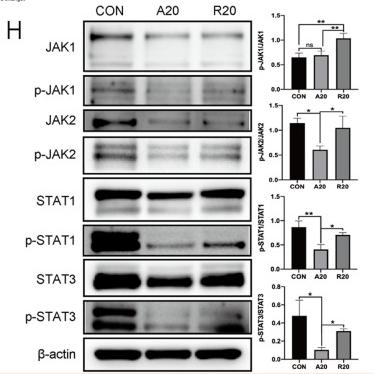
. The Western blot bands of MSGA regulates JAK-STAT signaling pathway, n = 3. (B-E). Grayscale analysis of p-JAK1/JAK1, p-JAK3/JAK3, p-STAT1/STAT1, and p-STAT3/STAT3 expression. Immunofluorescence (IF) images of STAT1 (F) and STAT3 (G) were photoed by laser scanning confocal microscope (Scale bar, 20 μm; Scale bar in representative images, 100 nm), n = 3. MSGA-L (100 μg/mL); MSGA-H (400 μg/mL). One-way ANOVA with Tukey’s post hoc test was used for the statistical analyses. Compared with M0 macrophages, statistically significant differences are indicated.")
. C Expression of TNF-α in different treatment groups by ELISA (n = 5). D The expressions of IL-22, IFN-γ, IL-1β and p-STAT3 proteins in different treatment groups were detected by Western blot. Quantitative data are presented as mean ± standard deviation (n = 5). Statistical significance is indicated as follows: ****P")
 Western blot experiments confirmed the activation of the CXCR2-mediated JAK–STAT signaling pathway around the haematoma (n = 6). (C, D) Western blot experiments detected the effects of Vinorine on the expression of JAK–STAT signaling pathway-related proteins in the perihaematoma region. (E) Western blot experiments detected the effects of Vinorine on the expression of MMPs in the perihaematoma region. (F) Western blot experiments detected the effects of Vinorine on the expression of apoptosis-related proteins such as BCL-2, Cleaved-Caspase 3, and Cleaved-Caspase 9 in the perihaematoma region. (G) Western blot experiments were conducted to assess the effects of Vinorine on the expression of blood–brain barrier proteins ZO-1 and CLDN5. (H) Western blot experiments were conducted to assess the effects of Vinorine on the expression of neuronal axonal proteins NF200 and PSD95. All data were expressed as mean ± standard deviation (SD). Statistical significance was determined by two-way analysis of variance (ANOVA) and Tukey's multiple comparisons test, *p")
 Western blot images of p-SMAD2, SMAD2, p-STAT3, STAT3, and GAPDH expression in response to vehicle, QBY, S1P, and W146 treatments. (B–G) Densitometric analysis of the corresponding band intensity ratios: (B) p-SMAD2/GAPDH, (C) p-SMAD2/SMAD2, (D) SMAD2/GAPDH, (E) p-STAT3/GAPDH, (F) p-STAT3/STAT3, and (G) STAT3/GAPDH. Data are expressed as mean ± SD (n=3). *P")
 Overexpressing and knocking down tRF-23 respectively led to higher and lower levels of p-JAK2/STAT3. JAK2/STAT3 inhibitor treatment also significantly reduced levels of p-JAK2/STAT3 relative to tRF-23 overexpression in the absence of such inhibition (p-JAK2, normalized to total JAK2; p-STAT3, normalized to total STAT3). Data are presented as the mean ± SD (n = 3 independent experiments with two technical replicates per independent sample). (C) JAK2/STAT3 signaling inhibitor treatment suppressed the osteogenic differentiation of hBMSCs on day 14 relative to those in which tRF-23 was overexpressed without inhibitor treatment. Scale bars: 50 μm. Matrix mineralization and ALP activity were quantified based on absorbance. Data are presented as the mean ± SD (n = 3 independent experiments with two technical replicates per independent sample). (D and E) RUNX2, OCN, and ALP levels on day 14 of hBMSC osteogenesis were detected via qPCR and western blotting. Data are presented as the mean ± SD (n = 3 independent experiments with two technical replicates per independent sample). Data were analyzed using paired two-tailed Student’s t tests. ∗∗p < 0.01 was considered significant. OE, overexpression; shRNA, short hairpin RNA; T-JAK2, total JAK2; T-STAT3, total STAT3.")
Heat map of differentially expressed mRNA. (B) Enrichment entries of DEGs GO analysis. (C) Enrichment entries of DEGs KEGG pathway analysis(top30). (D) qPCR validation of transcriptome sequence data. (E, F) Effects of different concentrations of CMSP(6, 8, 10)μg/mL on expression levels of JAK2/STAT3/c-Myc signal axis related proteins in CAL27 and SCC15 cells treated for 48h. β-Actin to control the load. n=3, compared with the control group")
 group (×400) (B) STAT3 of IL-17 A (mild to moderate, + to ++) group (×400) (C) AR of IL-17 A (+++) group (×400) (D) AR of IL-17 A (mild to moderate, + to ++) group (×400) (E) STAT3 was significantly higher in the IL-17 A (+++) group than in the IL-17 A (mild to moderate, + to ++) group (P = 0.046) (F) There was no significant difference in AR between the IL-17 A (+++) group and the IL-17 A (mild to moderate, + to ++) group (P = 0.455). *P")
 Heatmap of the top 30 genes in the DEGs of RELT (C) Clustered module maps of DEGs or WGCNA analysis: (D) Dendrograms of sample traits with scores for WGCNA analysis:(E) Screening of thresholds for WGCNA module analysis .F Correlation and P-value of genes with scores for each module of WGCNA analysis: (G) Scatter plots demonstrating the gene modules with strong correlation: (H) PPL network maps of pivotal genes: W Pivotal genes with GO and KEGG enrichment analyses: O. K Western blot validation of the JAK/STAT pathway. () LASSO coefficients pathway plot and cross-validation curve: . K Wester blot validation of the JAK2/STAT3 pathway in7860 and 769P cell lines (L) ELISA validation of lL-6 secretion in 786O and 769P cell lines (M) Western blot validation of lL-6 recovery experiments in 769P cell lines: N Monoclonal assay validation of cell proliferation: (O) Transwell migration assay validation of cell migration: N Flow cytometry analysis of apoptosis.")
| Product: | STAT3 Antibody |
| Catalog: | AF6294 |
| Description: | Rabbit polyclonal antibody to STAT3 |
| Application: | WB IHC IF/ICC |
| Cited expt.: | WB, IHC, IF/ICC |
| Reactivity: | Human, Mouse, Rat |
| Prediction: | Pig, Bovine, Horse, Sheep, Rabbit, Chicken |
| Mol.Wt.: | 86kDa(Observed); 88kD(Calculated). |
| Uniprot: | P40763 |
| RRID: | AB_2835144 |
Control Products

Related Downloads
Protocols
Product Info
*The optimal dilutions should be determined by the end user. For optimal experimental results, antibody reuse is not recommended.
*Tips:
WB: For western blot detection of denatured protein samples. IHC: For immunohistochemical detection of paraffin sections (IHC-p) or frozen sections (IHC-f) of tissue samples. IF/ICC: For immunofluorescence detection of cell samples. ELISA(peptide): For ELISA detection of antigenic peptide.
Cite Format: Affinity Biosciences Cat# AF6294, RRID:AB_2835144.
Fold/Unfold
1110034C02Rik; Acute Phase Response Factor; Acute-phase response factor; ADMIO; APRF; AW109958; DNA binding protein APRF; FLJ20882; HIES; MGC16063; Signal transducer and activator of transcription 3 (acute phase response factor); Signal transducer and activator of transcription 3; STAT 3; Stat3; STAT3_HUMAN;
Immunogens
A synthesized peptide derived from human STAT3, corresponding to a region within C-terminal amino acids.
- P40763 STAT3_HUMAN:
- Protein BLAST With
- NCBI/
- ExPASy/
- Uniprot
MAQWNQLQQLDTRYLEQLHQLYSDSFPMELRQFLAPWIESQDWAYAASKESHATLVFHNLLGEIDQQYSRFLQESNVLYQHNLRRIKQFLQSRYLEKPMEIARIVARCLWEESRLLQTAATAAQQGGQANHPTAAVVTEKQQMLEQHLQDVRKRVQDLEQKMKVVENLQDDFDFNYKTLKSQGDMQDLNGNNQSVTRQKMQQLEQMLTALDQMRRSIVSELAGLLSAMEYVQKTLTDEELADWKRRQQIACIGGPPNICLDRLENWITSLAESQLQTRQQIKKLEELQQKVSYKGDPIVQHRPMLEERIVELFRNLMKSAFVVERQPCMPMHPDRPLVIKTGVQFTTKVRLLVKFPELNYQLKIKVCIDKDSGDVAALRGSRKFNILGTNTKVMNMEESNNGSLSAEFKHLTLREQRCGNGGRANCDASLIVTEELHLITFETEVYHQGLKIDLETHSLPVVVISNICQMPNAWASILWYNMLTNNPKNVNFFTKPPIGTWDQVAEVLSWQFSSTTKRGLSIEQLTTLAEKLLGPGVNYSGCQITWAKFCKENMAGKGFSFWVWLDNIIDLVKKYILALWNEGYIMGFISKERERAILSTKPPGTFLLRFSESSKEGGVTFTWVEKDISGKTQIQSVEPYTKQQLNNMSFAEIIMGYKIMDATNILVSPLVYLYPDIPKEEAFGKYCRPESQEHPEADPGSAAPYLKTKFICVTPTTCSNTIDLPMSPRTLDSLMQFGNNGEGAEPSAGGQFESLTFDMELTSECATSPM
Predictions
Score>80(red) has high confidence and is suggested to be used for WB detection. *The prediction model is mainly based on the alignment of immunogen sequences, the results are for reference only, not as the basis of quality assurance.
High(score>80) Medium(80>score>50) Low(score<50) No confidence
Research Backgrounds
Signal transducer and transcription activator that mediates cellular responses to interleukins, KITLG/SCF, LEP and other growth factors. Once activated, recruits coactivators, such as NCOA1 or MED1, to the promoter region of the target gene. May mediate cellular responses to activated FGFR1, FGFR2, FGFR3 and FGFR4. Binds to the interleukin-6 (IL-6)-responsive elements identified in the promoters of various acute-phase protein genes. Activated by IL31 through IL31RA. Acts as a regulator of inflammatory response by regulating differentiation of naive CD4(+) T-cells into T-helper Th17 or regulatory T-cells (Treg): deacetylation and oxidation of lysine residues by LOXL3, leads to disrupt STAT3 dimerization and inhibit its transcription activity. Involved in cell cycle regulation by inducing the expression of key genes for the progression from G1 to S phase, such as CCND1. Mediates the effects of LEP on melanocortin production, body energy homeostasis and lactation (By similarity). May play an apoptotic role by transctivating BIRC5 expression under LEP activation. Cytoplasmic STAT3 represses macroautophagy by inhibiting EIF2AK2/PKR activity. Plays a crucial role in basal beta cell functions, such as regulation of insulin secretion (By similarity).
Tyrosine phosphorylated upon stimulation with EGF. Tyrosine phosphorylated in response to constitutively activated FGFR1, FGFR2, FGFR3 and FGFR4 (By similarity). Activated through tyrosine phosphorylation by BMX. Tyrosine phosphorylated in response to IL6, IL11, LIF, CNTF, KITLG/SCF, CSF1, EGF, PDGF, IFN-alpha, LEP and OSM. Activated KIT promotes phosphorylation on tyrosine residues and subsequent translocation to the nucleus. Phosphorylated on serine upon DNA damage, probably by ATM or ATR. Serine phosphorylation is important for the formation of stable DNA-binding STAT3 homodimers and maximal transcriptional activity. ARL2BP may participate in keeping the phosphorylated state of STAT3 within the nucleus. Upon LPS challenge, phosphorylated within the nucleus by IRAK1. Upon erythropoietin treatment, phosphorylated on Ser-727 by RPS6KA5. Phosphorylation at Tyr-705 by PTK6 or FER leads to an increase of its transcriptional activity. Dephosphorylation on tyrosine residues by PTPN2 negatively regulates IL6/interleukin-6 signaling.
Acetylated on lysine residues by CREBBP. Deacetylation by LOXL3 leads to disrupt STAT3 dimerization and inhibit STAT3 transcription activity. Oxidation of lysine residues to allysine on STAT3 preferentially takes place on lysine residues that are acetylated.
Some lysine residues are oxidized to allysine by LOXL3, leading to disrupt STAT3 dimerization and inhibit STAT3 transcription activity. Oxidation of lysine residues to allysine on STAT3 preferentially takes place on lysine residues that are acetylated.
(Microbial infection) Phosphorylated on Tyr-705 in the presence of S.typhimurium SarA.
S-palmitoylated by ZDHHC19 in SH2 putative lipid-binding pockets, leading to homodimerization. Nuclear STAT3 is highly palmitoylated (about 75%) compared with cytoplasmic STAT3 (about 20%).
S-stearoylated, probably by ZDHHC19.
Cytoplasm. Nucleus.
Note: Shuttles between the nucleus and the cytoplasm. Translocated into the nucleus upon tyrosine phosphorylation and dimerization, in response to signaling by activated FGFR1, FGFR2, FGFR3 or FGFR4. Constitutive nuclear presence is independent of tyrosine phosphorylation. Predominantly present in the cytoplasm without stimuli. Upon leukemia inhibitory factor (LIF) stimulation, accumulates in the nucleus. The complex composed of BART and ARL2 plays an important role in the nuclear translocation and retention of STAT3. Identified in a complex with LYN and PAG1.
Heart, brain, placenta, lung, liver, skeletal muscle, kidney and pancreas.
Belongs to the transcription factor STAT family.
Research Fields
· Cellular Processes > Cell growth and death > Necroptosis. (View pathway)
· Cellular Processes > Cellular community - eukaryotes > Signaling pathways regulating pluripotency of stem cells. (View pathway)
· Environmental Information Processing > Signal transduction > HIF-1 signaling pathway. (View pathway)
· Environmental Information Processing > Signal transduction > FoxO signaling pathway. (View pathway)
· Environmental Information Processing > Signal transduction > Jak-STAT signaling pathway. (View pathway)
· Human Diseases > Drug resistance: Antineoplastic > EGFR tyrosine kinase inhibitor resistance.
· Human Diseases > Endocrine and metabolic diseases > Insulin resistance.
· Human Diseases > Infectious diseases: Parasitic > Toxoplasmosis.
· Human Diseases > Infectious diseases: Viral > Hepatitis C.
· Human Diseases > Infectious diseases: Viral > Hepatitis B.
· Human Diseases > Infectious diseases: Viral > Measles.
· Human Diseases > Infectious diseases: Viral > Epstein-Barr virus infection.
· Human Diseases > Cancers: Overview > Pathways in cancer. (View pathway)
· Human Diseases > Cancers: Overview > Viral carcinogenesis.
· Human Diseases > Cancers: Overview > Proteoglycans in cancer.
· Human Diseases > Cancers: Overview > MicroRNAs in cancer.
· Human Diseases > Cancers: Specific types > Pancreatic cancer. (View pathway)
· Human Diseases > Cancers: Specific types > Acute myeloid leukemia. (View pathway)
· Human Diseases > Cancers: Specific types > Non-small cell lung cancer. (View pathway)
· Human Diseases > Immune diseases > Inflammatory bowel disease (IBD).
· Organismal Systems > Immune system > Chemokine signaling pathway. (View pathway)
· Organismal Systems > Immune system > Th17 cell differentiation. (View pathway)
· Organismal Systems > Endocrine system > Prolactin signaling pathway. (View pathway)
· Organismal Systems > Endocrine system > Adipocytokine signaling pathway.
References

Application: IF/ICC Species: human Sample: SW480 cells

Application: WB Species: Mouse Sample: Schwann cells
Application: WB Species: Mice Sample:

Application: WB Species: Mouse Sample:
Restrictive clause
Affinity Biosciences tests all products strictly. Citations are provided as a resource for additional applications that have not been validated by Affinity Biosciences. Please choose the appropriate format for each application and consult Materials and Methods sections for additional details about the use of any product in these publications.
For Research Use Only.
Not for use in diagnostic or therapeutic procedures. Not for resale. Not for distribution without written consent. Affinity Biosciences will not be held responsible for patent infringement or other violations that may occur with the use of our products. Affinity Biosciences, Affinity Biosciences Logo and all other trademarks are the property of Affinity Biosciences LTD.