- WB
- IHC
- IF/ICC
- pElisa
- Cites
, using Phospho-JNK1/2/3 (Thr183+Tyr185) Antibody at 1/1000 dilution.
5ug/NC membrane strip.
Exposure for 10s with Affinity™ ECL Kit(#KF8001).
Bands result from membrane strip incubation.")
, using Phospho-JNK1/2/3 (Thr183+Tyr185) Antibody at 1/1000 dilution.
5ug/NC membrane strip.
Exposure for 10s with Affinity™ ECL Kit(#KF8001).
Bands result from membrane strip incubation.")
 Antibody.
Lane 1: Rat lung, blocked with antigen-specific peptides,
Lane 2: Rat lung,
Lane 3: Mouse brain.")
 Antibody. The lane on the left was treated with blocking peptide.")
 using various lysates Lanes 1 - 2: Merged signal (red and green). Green - AF3318")


 and mouse anti-beta tubulin Ab(T0023) for 1 hour at 37°C. An AlexaFluor594 conjugated goat anti-rabbit IgG(H+L) Ab(Red) and an AlexaFluor488 conjugated goat anti-mouse IgG(H+L) Ab(Green) were used as the secondary antibody.
The nuclear counter stain is DAPI(blue).")
 by IF/ICC. The samples were fixed with PFA and permeabilized in 0.1% Triton X-100, then blocked in 10% serum for 45 minutes at 25°C. Samples were then incubated with primary Ab(#AF3318) and mouse anti-beta tubulin Ab(#T0023) for 1 hour at 37°C. An AlexaFluor594 conjugated goat anti-rabbit IgG Ab(Red) and an AlexaFluor488 conjugated goat anti-mouse IgG Ab(Green) were used as the secondary antibody.
The nuclear counter stain is DAPI (blue).")

 Fold change in cell viability of SV-HUC-1 cells treated with 0, 0.10 and 0.25% CSE for 7 days in combination with 5 µM U0126, 5 µM SB203580 or 2 µM SP600125. Data are representative of three independent experiments and are expressed as the mean ± standard deviation. * P")
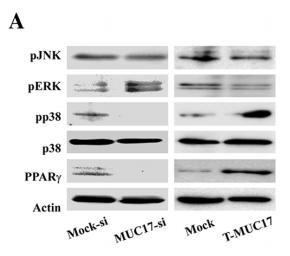
 for 24 h, and the expression levels of (A) iNOS and COX‑2 were detected using western blot analysis. (B) Effects of 11‑O-acetylcyathatriol on the phosphorylation of ERK1/2, JNK and p38 proteins. RAW 264.7 cells were treated with 1 µg/ml LPS with 11‑O‑acetylcyathatriol (12.5, 25, 50 and 100 µM) for 30 min, and the protein expression levels of p‑ERK1/2, p‑JNK and p‑p38 were detected using western blot analysis. (C) Effects of 11‑O-acetylcyathatriol on the protein degradation of IκB-α. RAW 264.7 cells were treated with 1 µg/ml of LPS with 11-O‑acetylcyathatriol (12.5, 25, 50 and 100 µM) for 10 min, and the protein expression of IκB-α was detected using western blot analysis. iNOS, inducible nitric oxide synthase; COX‑2, cyclooxygenase‑2; LPS, lipopolysaccharide; ERK, extracellular signal‑regulated kinase; JNK, c‑Jun N‑terminal kinase; IκB-α, inhibitor of nuclear factor-κB-α; p‑, phosphorylated.")
 RAW 264.7 cells were treated with LPS 1 µg/ml with or without tiliroside (12.5, 25, 50, and 100 µM) for 45 min and the expression of p‑ERK1/2, p‑JNK and p‑p38 was assessed by western blot analysis. Detection of β‑actin was conducted to confirm the equal loading of proteins. Densitometric analysis of p‑ERK1/2 (B) p‑JNK (C) and p‑p38 (D) expression represent the mean±standard deviation of three separate experiments. Data were normalized with respect to β‑actin levels. **P")
 The effect of miR-377 overexpression on inflammation-related gene expression. *P < 0.05, **P < 0.01 vs. NC 0.1% BSA; ##P < 0.01 vs. NC 10 ng/ml TNFα; ns, not signifcant (n = 3). (B) The effect of miR-377 inhibition on inflammation-related gene expression under conditions of TNFα-induced insulin-resistance. ##P < 0.01 vs. NC 10 ng/ml TNFα(n = 3). (C) The effect of miR-377 overexpression on JNK phosphorylation under conditions of TNFα-induced insulin-resistance. *P < 0.05 vs. NC without insulin; #P < 0.05 vs. NC with insulin (n = 3). (D and E) The effect of miR-377 overexpression on AKT and ERK phosphorylation. *P < 0.05 vs. NC with 0.1% BSA and insulin; #P < 0.05 vs. NC with 10 ng/ml TNFα and insulin (n = 3). (F) The effect of miR-377 inhibition on JNK phosphorylation under conditions of TNFα-induced insulin-resistance. *P < 0.05 vs. NC without insulin; #P < 0.05 vs. NC with insulin (n = 3). (G and H) The effect of miR-377 inhibition on AKT and ERK phosphorylation. *P < 0.05 vs. NC with 0.1% BSA and insulin; #P < 0.05 vs. NC with 10 ng/ml TNFα and insulin (n = 3).")

. Whole cell lysates were immunoblotted with respective target proteins. Healthy parents from the pedigree served as healthy controls")
.. d–f Western blot analysis of the protein levels of P75NTR,JNK, p-JNK, and β-actin. Quantification of P75NTR and p-JNK protein expression levels (##P < 0.01, *P < 0.05, ***P < 0.001 by one-way ANOVA followed by Bonferroni’s multiple comparison test, n = 3).")
Western blot analysis of the protein levels of ASK1-MKK3/6-p38 and ASK1-MKK4/7-JNK signaling pathways as well as corresponding phosphorylated proteins. GAPDH was used as the loading control protein.")
 Western blot analyses of ASK1, p38, JNK and the corresponding phosphorylation. b-actin was used as a loading control. (B) Quantification of the ratios of p-ASK1, p-p38, and p-JNK normalized to their corresponding unphosphorylated forms. Data were expressed as the mean ± SEM, n ¼ 3. Compared with the control þ Veh group, ##p < 0.01; compared with the TNF-a þ Veh group, *p < 0.05, **p < 0.01.")
. The red fluorescence represents Bim expression, and blue fluorescence
represents nuclei. b, c Western blot analysis of Bax, caspase-3 and cleaved caspase-3 expression in the MG, HG and HG + PGE1 groups. The
data represent the means ± SEM. *P < 0.05, **P < 0.01 versus HG group. d, e Western blot analysis of Bim, p-JNK and JNK expression in the MG,
HG and HG + PGE1 groups. f qRT-PCR was performed to determine the mRNA levels of Bim, Bax, and caspase-3 in HK-2 cells. The data
represent the means ± SEM. *P < 0.05, **P < 0.01 versus the HG group")
 The
effect of SCVP-1 on the level of phosphorylation of ERK1/2, p38 and JNK1/2 in RAW264.7 cells
by Western blotting. (B) Relative quantitative analysis of p-p38/p38, p-ERK/ERK and p-JNK/JNK
protein phosphorylation levels. (C-F) Effect of specific MAPKs inhibitors (ERK1/2 inhibitor:
CI-1040, p38 inhibitor: SB239063 and JNK1/2 inhibitor: SP600125) on SCVP-1/LPS-induced NO,
IL-1β, IL-6 and TNF-α production of RAW264.7 cells. The results were expressed as means ± SD
(n = 3). (B) *p<0.05, **p<0.01 vs. control group. (C-F) *p<0.05, **p<0.01 vs. without
anti-MAPKs antibodies")
 RAW264.7 macrophages were treated with 100 μg/ml or 400 μg/ml Riclin for
1 h, then analyzed for p38, p-p38, ERK1/2, p-ERK1/2, JNK, p-JNK and β-actin protein levels by Western blotting. (B) RAW264.7 cells were treated with PD98059 (ERK1/2
inhibitor), SB203580 (p38 inhibitor) and SP600125 (JNK inhibitor) for 1 h before Riclin treatment, Listeria infection, gentamycin sulphate treatment and a further culture for
4 h, then bacterial titers were determined for each group. (C) The mRNA expression levels of HO-1 in RAW264.7 macrophages were measured by real-time PCR after a
further culture for 4 h. (D–E) RAW264.7 cells were treated with tin protoporphyrin IX (SnPP, HO-1 inhibitor) for 1 h before Riclin treatment, Listeria infection, gentamycin
sulphate treatment and a further culture for 4 h, then mRNA expression levels of IL-1β (D) and bacterial titers (E) were determined for each group. Data are expressed as
mean ± SEM. N = 3, *p b 0.05, **p b 0.01, ***p b 0.001.")
 Cell phenotypic changes in cells treated with COMP siRNA and COMP overexpression. (B) WB analysis of
COMP and EMT-related markers in HCT116 cells under COMP knockdown or overexpression. (C) Cell wound scratch assay of HCT116, HCT-8, and SW620 cells treated with
COMP siRNA or COMP overexpression vectors. (D) Transwell assay of HCT116, HCT-8, and SW620 cells treated with COMP siRNA or COMP overexpression vectors. (E)
Immunofluorescence assay of HCT116, HCT-8, and SW620 cells treated with COMP siRNA or COMP overexpression vectors. Image J software was used to analyze the relative
intensity of E-cadherin and Vimentin. (F) HCT116 and HCT-8 cells with knocked down or overexpressed COMP were transplanted on nude mice. Tumor volumes were
measured every 4 days. (G) Tumor weight in the control, COMP knockdown, and COMP overexpression groups. (H) The in situ spleen model of the colorectal cancer cell line
SW620 showed that overexpression of COMP promoted liver metastasis of colorectal cancer, while downregulation of COMP inhibited liver metastasis of colorectal cancer. (I)
IHC staining to identify EMT biomarkers and COMP-related proteins in the control, COMP knockdown, and COMP overexpression groups. COMP knockdown displayed strong
E-cadherin staining and reduced Vimentin, Snail1, Twist1, and MMP-2 staining. The expression levels of other proteins were identical to those observed in WB experiments. (J)
Staining indices of COMP, E-cadherin, Vimentin, Snail1, Twist1, and MMP2. The error bars in all graphs represented SD, and each experiment was repeated three times. * and **
stand for P<0.05 and P<0.01, respectively.")

 Representative images of Western blot. (B–I) quantification of the Western blot data by densitometric analysis and normalization to GAPDH (n = 3 independent experiments). **P < 0.01 vs model group.")
. Significance: *P < 0.05 vs. the HC group, and #P < 0.05 vs. the KD group. f, g Effects of IL-1R8 silencing on the
activation of ERK and NFκB p65 were observed. Significance: *P < 0.05 vs. the HC group, #P < 0.05 vs. the KD group, and &P < 0.05 vs. the KD + IL-37b
group. i The nuclear translocation of NFκB p65 was observed after silencing IL-1R8 expression (n = 3). Magnification: ×200, Scale bar = 100 μm.
Experiments were done at least three times in triplicate.")
 rats. (A) Western blot was used to detect the protein levels in the brain
tissues. The full-length gels are presented in Supplementary Fig. S2. The protein expression was
evaluated by proteomic analysis. (B) Changes in the retinol-binding protein 4 (RBP4); (C) Changes in
serum albumin. *p < 0.05 and ***p < 0.001 vs. I/R.")

 on the expression of hepatic (A) XBP1, (B) phospho-IKK alpha/beta (Thr 183+Tyr 185), (C) phospho-JNK1/2/3 (Ser180/181) and (D) phospho-IRS1 (Ser 307).")


. *P <0.05 vs. control group; #P < 0.05 vs. DM group; &P < 0.05 vs. CAPE group;+P < 0.05 vs.Medium-pNO2 group. Control: the normal diet mice; DM: the HDF/STZ-induced diabeticmice; CAPE: the HDF/STZ-induced diabetic mice treated with CAPE (10 mg/kg/day);Low-pNO2: the HDF/STZ-induced diabetic mice treated with CAPE-pNO2 (5 mg/kg/day);Medium-pNO2: the HDF/STZ-induced diabetic mice treated with CAPE-pNO2 (10mg/kg/day); High-pNO2: the HDF/STZ-induced diabetic mice treated with CAPE-pNO2 (15mg/kg/day).")
 Western
blot analysis of the protein levels of ASK1-MKK3/6-p38 and ASK1-MKK4/7-JNK signaling pathways as well as corresponding phosphorylated proteins. GAPDH was used as the
loading control protein. (B) Quantitative analysis of the ratios of phosphorylated proteins normalized to their unphosphorylated form. (C) The levels of inflammatory cytokines IL-1b,
IL-6, TNF-a were detected using ELISA kits. Data are expressed as mean ± SEM (n ¼ 5e6 per group). Compared with the control group: # P < 0.05, ## P < 0.01, ### P < 0.001;
Compared with the DSS þ Veh group: * P < 0.05, ** P < 0.01, *** P < 0.001.")
 Western blot analyses of total tissue lysate for p-JNK, JNK, p-c-Jun, c-Jun,p-c-Fos, c-Fos, and β-actin (loading control).")

. B‐E, The ERK, P38, JNK, and their phosphorylation forms protein levels of
EA.hy926 cells treated with culture supernatants of HK‐2 cells were measured by western blot (n = 6 for each group). F,G, The eNOS and p‐eNOS
protein levels of EA.hy926 cells treated with culture supernatants of HK‐2 cells were measured by western blot (n = 6 for each group). Data are
presented as mean ± SEM. *P < .05 and **P < .01")
 AMPK activation, (B) ERK
activation, (C) JNK activation, and (D) P38 activation. The blots showed representative samples. All samples derived from the same experiment and that blots were
processed in parallel. AMPK and p-AMPK were normalized to the expression of GAPDH on the same membrane. Each phosphoprotein was normalized to the
expression of the corresponding total protein of the same sample in Fig. 5B–D. Results are expressed as mean ± S.E.M. (n = 6–8 for each group). *P < 0.05,
**P < 0.01, ***P < 0.001, ****P < 0.0001 according to one-way ANOVA followed by Bonferroni test.")
, p-ERK (B), p-JNK (C), p-p38 (G), p-STAT3 (D), p-p65 (E), SIRT1 (F), and PGC-1α (H) were
determined by western blotting in lung tissues. The lung tissues were dissected 24 h after LPS challenge and the levels of protein expressions were evaluated by
densitometric analysis of the blots. The blots showed representative samples. All samples derived from the same experiment and that blots were processed in parallel.
SIRT1 and PGC-1α were normalized to the expression of GAPDH on the same membrane. Each phosphoprotein was normalized to the expression of the corresponding
total protein of the same sample. Results were expressed as mean ± SD (n = 6 for each group). *P < 0.05, **P < 0.01, ***P < 0.001, ****P < 0.0001 according
to one-way ANOVA followed by Tukey’s multiple comparisons test.")
 Effects of arsenite on the relative protein expressions of IREB2,
SLC7A11, VDAC3 and GPX4. (C) Effects of arsenite on the relative mRNA expressions of Ireb2, Slc7a11, Vdac3 and Gpx4. (D–E) Effects of arsenite on the relative
protein expressions of p-ERK1/2 and p-JNK1/2. (F–G) Effects of arsenite on the relative protein expressions of CHOP and GRP78. Data were reported as mean ± S.D.
“*” denoted p < 0.05, compared with the control group.")
 Western blot analysis of p-ERK1/2, total ERK1/
2, p-P38 MAPK, total P38 MAPK, p-JNK, and total JNK. These results were used
to calculate (B) p-ERK1/2/β-actin and total ERK1/2/β-actin ratios, (C) p-P38
MAPK/β-actin and total P38 MAPK/β-actin ratios, (D) p-JNK/β-actin and total
JNK/β-actin ratios. The differences were statistically significant (p < 0.05).
Asterisk indicates significant difference in comparison to untreated cell")
 and quantification data (right) of ROS levels in hAT-MSCs after exposure to HCC-CM or normal medium; b Representative histogram (left) and quantification data (right) of ROS levels in hAT-MSCs after withdrawal of HCC-CM treatment; c HIF-1α, phosphorylated AKT, total AKT, phosphorylated JNK, total JNK, phosphorylated p38, total p38, phosphorylated ERK and total ERK protein expression of hAT-MSCs after exposure to HCC-CM or normal medium; d HIF-1α, phosphorylated AKT, total AKT, phosphorylated JNK, total JNK, phosphorylated p38, total p38, phosphorylated ERK and total ERK protein expression of hAT-MSCs after withdrawal of HCC-CM treatment; e ROS levels in hAT-MSCs after exposure to HCC-CM, normal medium or pre-treated with NAC; f HIF-1α, phosphorylated AKT, total AKT, phosphorylated JNK, total JNK, phosphorylated p38, total p38, phosphorylated ERK and total ERK protein expression of hAT-MSCs after exposure to HCC-CM, normal medium or pre-treated with NAC. *p < 0.05, **p < 0.01, vs NC, n = 2")
: Western blot analysis of phosphorylated JNK (p-JNK), JNK and β-ACTIN in hPDLSCs transfected with si-NC or si-GDF5. Histogram showed the quantification of the band intensities. (B): Western blot analyses of phosphorylated p38 (p-p38), p38 mitogen-activated protein kinase (p38), and β-ACTIN in hPDLSCs transfected with si-NC or si-GDF5. Histogram showed the quantification of the band intensities. (C): Western blot analyses of phosphorylated ERK (p-ERK), ERK and β-ACTIN in hPDLSCs transfected with si-NC or si-GDF5. Histogram showed the quantification of the band intensities. *p <0.05. hPDLSCs, human periodontal ligament stem cells; GDF5: growth differentiation factor 5; si-NC: small interfering RNA negative control; si-GDF5: the small interfering RNAs that target GDF5; NC, negative control; JNK, c-Jun N-terminal kinase; ERK, intracellular signal-regulated kinase 1/2.")
 Whole cell lysates were prepared following stimulation with LPS, NAC and TA. The protein expression levels of apoptosis‑associated proteins (p‑JNK, JNK, Bax, cleaved caspase‑9 and cleaved caspase‑3) and an anti‑apoptotic protein (Bcl‑2) were detected in H9C2 cells treated with LPS, NAC + LPS, TA + LPS and TA + NAC + LPS by western blot analysis.")
, JNK (c, d) and
p38 (e, f) proteins of the MAPKs pathway in LPS-activated RAW 264.7 cells. β-actin
was used as internal reference to confirm the equal loading of proteins. Densitometric
analysis data were standardized on the basis of levels of unphosphorylated forms
(ERK, JNK, p38). The results of phosphorylated ERK protein, phosphorylated JNK
protein, phosphorylated p38 protein are presented as mean ± S.E.M. from three
independent experiments. ##P < 0.01 vs. untreated group, **P < 0.01 vs. LPS treatment
group.")
 Whole cell lysates were prepared following stimulation with LPS, NAC and TA. The protein expression levels of apoptosis-associated proteins (p-JNK, JNK, Bax, cleaved caspase-9 and cleaved caspase-3) and an anti-apoptotic protein (Bcl-2) were detected in H9C2 cells treated with LPS, NAC + LPS, TA + LPS and TA + NAC + LPS by western blot analysis. Semi-quantification of the protein expression levels of (B) p-JNK/JNK and cleaved caspase-3, and (C) Bax/Bcl-2 and cleaved caspase-9 in H9C2 cells. The mRNA expression levels of (D) Bax, (E) caspase-3, (F) caspase-12 and (G) Cyt c in H9C2 cells normalized to Gapdh. Data are presented as the mean ± standard error of the mean (n=3). *P<0.05, **P<0.01 and ***P<0.001 vs. control; #P<0.05 and ##P<0.01 vs. LPS group; §P<0.05 and §§P<0.01 vs. TA + LPS. TA, tannic acid; LPS, lipopolysaccharide; NAC, N-acetylcysteine; p, phosphorylated; JNK, c-Jun N-terminal kinase; Cyt c, cytochrome c.")

 NX increased
the ratio of Bcl-2/BAX as assessed by western
blotting analysis. (B) NX reduced the levels of
cleaved caspase-3 by western blotting. (C) NX
increased the expressions of p-AMPK evaluated
by western blotting analysis. (D) NX reduced
the levels of p-JNK by western blotting. Data
presented are individual values with means ±
SD; n = 3. *P < 0.05 versus sham group, #P <
0.05 versus TAC group, &P < 0.05 versus
TAC+NX group.")
 The levels of p-JNK and JNK in HTR-8/SVneo cells with IR, overexpression or silence of ANGPTL8 or/and insulin stimulation.")
 Relative protein expression of JIP2 and p‐JNK was detected with Western blotting.")
 Expression of TLR4, phosphorylated p38 (P-p38), p38, P-ERK1/2, ERK1/2, P-JNK, JNK, P-p65, p65 was shown. (B) The analysis of grey scale value. Con was control group, M was model group, H was the high dose OPA group, L was low dose OPA group, P was positive group. Data were expressed as the mean ± SD, the statistical analyses were done with t-test; *** p < 0.001, ** p < 0.01, * p < 0.05 compared with the model group; ### p < 0.001, ## p < 0.01, # p < 0.05 compared with the control group.")
 CHS-IVa inhibited the activation of extracellular regulated protein
kinases (ERK), (B) c-Jun N-terminal kinase (JNK) (C) and p38 in LPS-stimulated RAW264.7 cells. Results are representative of three independent
experiments. Data are presented as means ± SD. Values with different letters are significantly different, p < 0.05.")
 knockdown inhibited the activation of the mitogen-activated protein kinase (MAPK) pathway 3 days after myocardial infarction (MI). The expression levels of p-JNK, c-Jun N-terminal kinase (JNK), p-p38, p-38, p-ERK, and ERK were determined using western blot analysis. β-actin served as the loading control.")
 Luciferase assays of CAGA-NIH3T3 cells. Cells were pretreated with Remdesivir (0–50 μM) for 30 min and then incubated with TGF-β1 (5 ng ml−1) for 24 h, then analyzed with luciferase assay. SB431542 is a TGF-β1/Smad pathway inhibitor and serves as a positive control (B) NIH-3T3 cells were co-treated with TGF-β1 (5 ng ml−1) and Remdesivir (12.5, 25, 50 μM) for 1 h. P-Smad3 and Smad3 were assessed using western blot. GAPDH was used as the internal control (C) PPF cells were co-treated with TGF-β1 (5 ng ml−1) and Remdesivir (12.5, 25, 50 μM) for 1 h. P-Smad3 and Smad3 were assessed using western blot. GAPDH was used as the internal control (D) NIH-3T3 cells were co-treated with TGF-β1 (5 ng ml−1) and Remdesivir (12.5, 25, 50 μM) for 1 h and the phosphorylation levels of P-38, JNK, ERK and Akt were analyzed by Western blot. β-tubulin was used as a loading control in grayscale analysis. Scale bar = 60 μm. Data was presented as the means ± SD, n = 3. * p < 0.05, ** p < 0.01, *** p < 0.001, **** p < 0.0001.")
 with or without physagulin A
(0.78, 1.56, 3.12, 6.25 μM), physagulin C (0.39,
0.78, 1.56, 3.12 μM), physagulin H (1.56, 3.12,
6.25, 12.5 μM) for 1 h (detection of
phosphorylated-JNK) or 15 min (detection of
phosphorylated-ERK and phosphorylated-p38),
and the expressions of p-ERK, p-JNK, p-p38 proteins were determined by Western blot analysis.
Unphosphorylated form of ERK, JNK, p38 proteins
were used as internal reference to confirm the
equal loading of proteins. Densitometric analysis
data were standardized on the basis of ERK, JNK,
p38 protein levels. The results of phosphorylated
JNK protein (a, b, c), phosphorylated ERK protein
(d, e, f), phosphorylated p38 protein (g, h, i) are
presented as mean ± S.E.M. from three independent experiments. ##P < 0.01 vs. untreated group,
**P < 0.01 vs. LPS treatment group.")
 Quantification of pro-apoptotic (p-JNK, JNK, Bax) and anti-apoptotic (p-AKT, AKT, Bcl-2) protein expressions in shRac1 and shLuci BMMSCs post-OGD 12 h. β-actin was used as the housekeeping protein. (N.S. no significance, *P < 0.05, **P < 0.01, ***P < 0.001, statistically analyzed by the Student’s t-test, n = 4). (C, D) Pro-apoptotic (p-JNK, JNK, and Bax) and anti-apoptotic (p-AKT, AKT, and Bcl-2) protein expressions in shP67 and shLuci BMMSCs post-OGD 12 h, β-actin was used as the housekeeping protein. (N.S. no significance, *P < 0.05, ***P < 0.001, statistically analyzed by the Student’s t-test, n = 4). (E–H) p53 expression and activation as reflected by total and phospho-p53 protein expressions. In shRac1- and shP67-transfected BMMSCs post-OGD treatment, p53 expression and activity were found to be decreased. (N.S. no significance, *P < 0.05, **P < 0.01, statistically analyzed by the Student’s t-test, n = 4). (I–M) Flow cytometry measured apoptosis of shLuci, shRac1, shP67 BMMSCs and wild type BMMSCs. In comparison with shLuci, shP67 and wild type BMMSCs, shRac1 BMMSCs showed better anti-apoptotic ability. (N.S. no significance, *P < 0.05, **P < 0.01, ***P < 0.001, statistically analyzed by one-way ANOVA followed by the Bonferroni correction, n = 3). Data are presented as mean ± SD.")
 The levels of p-JNK and JNK in HTR-8/SVneo cells with IR, overexpression or silence of ANGPTL8 or/and insulin stimulation. (C, D) The levels of ROCK I, ROCK II, p-MYPT1 and MYPT1 in HTR-8/SVneo cells. (E) Glucose consumption was determined in HTR-8/SVneo cells with ANGPTL8 knockdown, JNK agonist Anisomycin (1 μg/ml) or JNK antagonist SP600125 (20 μM), and insulin stimulation. (F) The levels of insulin signaling related molecules were determined in HTR-8/SVneo cells. (*p < 0.05, **p < 0.01, ns. no significance).")
 NR activated the JAK2/Stat3 pathway by increasing the levels of p-JAK2 and pStat3. Relative levels of p-JAK2 and p-Stat3 were normalized to the JAK2 and Stat3 levels, respectively. (c-d) NR inhibited the H2O2-induced phosphorylation of MAPK.
Relative levels of p-JNK and p-ERK 1/2 were normalized to the JNK and ERK 1/2 levels, respectively (**p < .01, ***p < .001, and ****p < .0001 compared to the H2O2
group).")
 The protein expression of p-p65, p65, p-ERK1/2, ERK1/2, p-p38, p38, p-JNK, and JNK were determined by Western blot and quantified. GAPDH was conducted as a loading control. (c) The nuclei translocation of p65 (red) was detected by the immunofluorescence assay. Nuclei (blue) were stained with DAPI. One-way ANOVA, ##p < 0.01, compared with control cells; $p < 0.05, $$p < 0.01, compared with IL-1β-treated cells.")
 The levels of LVEF and LVFS were detected by echocardiograph as indexes for cardiac function at the end of the 24 h reperfusion. (C) Representative images of stained myocardial tissues of mice in all groups. (D) The percentage of myocardial infarction area of all groups. (E, F) Cardiomyocyte apoptosis was determined by using TUNEL assay. (G–I) The production of ROS, MDA and cTn-I was determined. (J, K) Western blotting was performed to analyze the protein expression of CTRP3, LAMPA, JIP2, JNK and p-JNK in myocardial tissue.")

 Western blotting was performed to detect the expression of p65 and p-p65 in TNF-α-stimulated FLSs. (B) Expression levels of ERK1/2, p-ERK1/2, JNK, p-JNK, p38 and p-p38. NF-κB, nuclear factor-kappa B; MAPK, mitogen-activated protein kinase. The data represent the mean ± SD (n=3). ##P<0.01 vs. Control; **P<0.01 vs. TNF-α+NC siRNA.")
 in CD4+ T cells were quantified by western blotting and showed as the relative expression values of β-actin, which was used as a loading control to normalize protein levels. For highlighting the activation level of p-Akt and p-JNK in this signaling pathway, the ratio of p-Akt to Akt (p/t Akt) and p-JNK to JNK (p/t JNK) were used for statistics. Data are shown as Mean ± SD (n = 6). Statistically significant differences were determined by one-way analysis of variance (ANOVA) followed by Dunn’s/Tukey’s test. *P < 0.05, **P < 0.01, ***P < 0.001, ****P < 0.0001")
 and over-expression (B). Cell viability was analyzed by MTT assay in HCC1806 cells with SHMT2 knockdown treated with an ERK inhibitor FR180204 (C) or a p38 inhibitor SB202190 (D). ZR-75-1 cells with SHMT2 overexpression were treated with an ERK inhibitor FR180204 (E) or a p38 inhibitor SB202190 (F). # represents the comparison between inhibitor plus SHMT2 over-expression group and SHMT2 over-expression group. * represents the comparison between treatment group and control group. #, *P < 0.05. Student’s t-test. The data are presented as the mean ± SD. The error bars show the SD.")
 AOPPs (200 µg/mL, 24 h) significantly increased phosphorylation of p38, ERK1/2 and JNK in AF cells. (D–F) p38 MAPK inhibitor (SB202190, 10 µM), ERK1/2 inhibitor (FR180204, 10 µM) and JNK inhibitor (SP600125, 10 µM) AOPPs-induced (200 µg/mL, 24 h) senescence in AF cells. (G–I) Activation of AOPPs-induced (200 µg/mL, 24 h) MAPK phosphorylation was ameliorated by GKT (20 µM) and APO (100 µM). *P < 0.05 compared to the control. ** P < 0.01 compared to the control. #P < 0.05 compared to the AOPPs group. Error bars represent standard errors.")

 Expressions of ER stress-associated proteins in HepG2 cells after treatment with 2 μM C20/C22 for 0, 6, 12, and 24 h. All proteins were normalized with the expression of Gapdh (n = 3). (C,D) Cells were stained with PI after 24 h of C20/C22 incubation, and flow cytometry was used to determine the proportion of cells in each stage of the cell cycle (n = 3). The reported data correspond to the mean ± SD of three independent experiments. The p-value was analyzed by one-way ANOVA followed by Tukey’s test using GraphPad Prism version 8.00. * p < 0.05, ** p < 0.01, *** p < 0.001, and **** p < 0.0001 vs. 0 h/control group.")
 enhanced osteogenesis via Erk1/2 MAPK signaling pathway: (A) Schematic representation of the relative linear location in which the FGFR2 mutation is identified as illustrated by the large red arrow shown in the context of the protein structure. (B) CCK-8 assay was carried out to assess cell proliferation. Proliferation of MC3T3-E1 cells was more active in the MT group. (C) Relative expressions of osteogenic marker measured by qPCR. The expressions of Alp, ColIα2, Runx2, Opn and Ocn mRNA in differentiated MC3T3-e1 were remarkably increased in the MT group. Western blot analysis showed that the level of ALP, COLI and RUNX2 were also increased in the MT group. (D) ALP staining, alizarin red staining and quantitative tests. ALP staining showed increased crystal violet-staining cells in the MT group compared with the WT group. Quantitative experiment demonstrated that ALP activity is more active in the MT group. Alizarin red staining and quantitative test showed there were more mineralized nodules and mineral content in the MT group than in the WT group. (E) Western blot analysis demonstrated that the levels of p-FGFR2 and p-Erk1/2 were increased in the MT group. There was no significant change in the expression of key proteins in other downstream pathways. The western blot results of FGF/FGFR2-Erk1/2 was circled by the red frame. p values were significant at * p < 0.05, ** p < 0.01, *** p < 0.001 and **** p < 0.0001.")
 Western blotting detected the expression levels of proteins related to the MEK–ERK signaling pathway, including p-MEK, p-ERK, p-p38, and p-JNK. (c,d) Western blotting detected the expression level of p-ERK in HNE-3 cells overexpressing CK13 after introducing ERK activator (TBHQ 20 μM). (e) The CCK-8 test analyzed the survival of HNE-3 cells after introducing ERK activator (TBHQ 20 μM) for 72 h under 4 Gy or 0 Gy irradiation. *p")
 The mRNA expression level of IL-1β, IL-6, TNF-α and MCP-1 was examined in RAW264.7 cells treated with SP600125 (JNK inhibitor, 5 μmol/L), SCH772984 (ERK1/2 inhibitor, 300 nmol/L) and SB203580 (p38 inhibitor, 5 μmol/L), respectively. (B) The expression of phosphorylation of JNK, p38 and ERK was detected after butyrate treatment. (C) The mRNA expression of phosphatases MKP-1, PP1, PP2, PP2A, PTP1B and SHP2 in RAW264.7 after butyrate treatment. (D) The expression of MKP-1 was evaluated by using western blot. (E) The effect of MKP-1 knockdown was assessed by analyzing the phosphorylation level of JNK, p38 and ERK and expression of IL-6 and TNF-α. Values are expressed as the mean ± SEM. Significance: * P < 0.05, ** P < 0.01, *** P < 0.001, vs. the control group; # P < 0.05, ## P < 0.01, ### P < 0.01 vs. the CAWS group; ns means no significance.")
, 50 μm (white).")
 The expression of FGF2, EREG, and HSP70 was assessed by qRT−PCR. (B) Western blot analysis of the protein expression of FGF2, EREG, and HSP70 and the phosphorylation of JUN and p53. (C) AZD4547 combined with nab-PTX inhibited phosphorylation of the JNK, ERK, and p38 proteins. *: p< 0.05, **: p< 0.01, ****: p< 0.0001.")
 Whole cell lysates were prepared following stimulation with LPS, NAC and TA. The protein expression levels of apoptosis-associated proteins (p-JNK, JNK, Bax, cleaved caspase-9 and cleaved caspase-3) and an anti-apoptotic protein (Bcl-2) were detected in H9C2 cells treated with LPS, NAC + LPS, TA + LPS and TA + NAC + LPS by western blot analysis. Semi-quantification of the protein expression levels of (B) p-JNK/JNK and cleaved caspase-3, and (C) Bax/Bcl-2 and cleaved caspase-9 in H9C2 cells. The mRNA expression levels of (D) Bax, (E) caspase-3, (F) caspase-12 and (G) Cyt c in H9C2 cells normalized to Gapdh. Data are presented as the mean ± standard error of the mean (n=3). *P")
 Relative protein expression of p-ERK1/2, p-JNK, and p-p38 in vitro. (B) Relative mRNA expression levels of MAPK1, MAPK8, and MAPK14 in vitro. (C) Relative protein expression of p-c-Fos and p-p65 in vitro. (D) Relative mRNA expression levels of FOS, JUN, and NFKB1A in vitro. *P")
 The 79 intersection targets of SEC acting on BC (Venn diagram analysis). (B) The PPI network of the intersection targets. (C) The top 20 hub targets in the PPI network ranked by MCC value. (D, E) The molecular docking models of SEC with MAPK8 (JNK), MAPK14 (p38) and GSK3β. (F) The effect of SEC on JNK, p38 and Wnt/β-catenin signaling pathways of BC cells (Western blot). Data are shown as mean ± SD from three independent experiments.")
 ERK activity was calculated from the ratio of phosphorylated ERK/total ERK relative to the 21% group. (B) JNK activity was calculated from the ratio of phosphorylated JNK/total JNK relative to the 21% group. The upper panel shows Western blotting banding pictures, and the lower panel shows their relative quantification. All comparisons were made relative to the 21% group. ** p < 0.01. Each bar represents mean ± SD. tERK: total ERK; pERK: phospho-ERK; tJNK: total JNK; pJNK: phospho-JNK.")
 The serum inflammatory cytokines (i.e., TNF-α, IL-1β, IL-6, and IL-10) in serum were measured by ELISA. (E–J) Representative images of phosphorylation levels of P38, Erk1/2, JNK, NF-κB and IKK α/β were examined by western blot and the fold activation data analysis. Data are expressed as mean ± SD (at least n = 6/group), #p < 0.05 (vs. Sham group), *p < 0.05 (vs. CLP group).")


, ERK, p-JNK (Thr183/Tyr185), JNK, p-p38 (Thr180/Tyr182) determined by western-blot. b The quantitative analysis of p-ERK/total ERK expression level. c The quantitative analysis of p-JNK/total JNK expression level. d The quantitative analysis of p-p38/total p38 expression level. Values are expressed as mean ± SD. n = 6. Compared with control group: #P")
 Male or female WT and fat-1 mice were injected with 400 mg/kg APAP, and livers were collected at 6 hours post-APAP injection (n = 5). (a) Flow cytometry analysis of the intercellular ROS level by the fluorescent probe DCFH-DA in hepatocytes was carried out. (b) The mitochondrial membrane potential in hepatocytes was measured by the JC-1 dye staining for flow cytometry analysis. (c) Phosphorylation of JNK was evaluated by immunoblotting analysis at the indicated time after APAP challenge. (d) The protein levels of LC3 and p62 were examined by immunoblotting analysis. (e–g) HepaRG cells were pretreated with 50 μM DHA with or without100 nM E2 prior to stimulation with 20 mM APAP. (e) The ROS level in the cells was analyzed by flow cytometry labeling with fluorescent probe DCFH-DA at 6 hours following APAP administration. (f) Phosphorylation of JNK expression in the APAP-treated cells was evaluated by immunoblotting analysis. (g) The hepatic levels of LC3 and p62 were determined by immunoblotting analysis. ∗p < 0.05, ∗∗p < 0.01. The data represent three independent experiments with similar results.")
 GO and KEGG enrichment analysis of DEGs in DSF+Cu2+ group. (C) GSEA plots showing pathways upregulated and downregulated after DSF+Cu2+ treatment. (D) GSVA plots showing pathways upregulated and downregulated after DSF treatment. (E,F) Western blot analysis of p-p38MAPK, p38MAPK, p-JNK MAPK, JNK MAPK, p-NF-Κb, NF-Κb, and NOTCH protein levels in LenR Huh7 cells after incubation with different groups. (G1: corn oil, G2: DSF, G3: CuO NPs, G4: Len@CuO, and G5: DSF@CuO). Data are presented as mean ± SD and are representative of three independent experiments. *** p < 0.001, ns, not significant.")
 by inhibiting SMAD4 expression. (A–C) RT-qPCR analysis was performed to detect the mRNA levels of SMAD4 in LINC00909-overexpressing cells (A) and LINC00909-knockdown cells (B, C). (D–F) Western blotting analysis was conducted to examine the protein expression of SMAD4 in PDAC cells. (G, H) The efficiency of SMAD4 knockdown in PANC-1 (G) and AsPC-1 cells (H) was detected by RT-qPCR. (I, J) Representative images of sphere-formation assays in PANC-1/KD and AsPC-1/KD cells transfected with siSMAD4-2. (K, L) The protein levels of SMAD4, KLF4, c-Myc, JNK, p-JNK, JUN and p-JUN in PANC-1/LINC00909-KD (K) and AsPC-1/LINC00909-KD cells (L) transfected with siSMAD4-2 were analyzed by western blotting. (M) PANC-1 cells transfected with EV or vector encoding LINC00909 were treated with actinomycin D (1 mg/ml) at the indicated time point. (N, O) PANC-1 or AsPC-1 cells transfected with siNC, siLINC00909-3, or siLINC00909-6 were treated with actinomycin D (1 mg/ml) at the indicated time point. Total RNA was extracted and analyzed by RT-qPCR to examine the relative levels of SMAD4 mRNA (M–O). All *P")

, respectively. (A) The acetylation level of GADD45A was detected using IP assay with acetyl lysine antibody. (B) The protein expression of acetylated histone H3, H4, and GADD45A was detected using Western blot. T98G cells and LN229 cells were transfected with GADD45A-OE plasmid. (C) LN229 cells were transfected with pGL3-basic-GADD45A-promoter or pGL3-basic-vector plasmids. Subsequently, cells were treated with DMSO or PXD101 (1µM); then, the transcriptional activity of GADD45A was detected using dual-luciferase assay. (D) The GADD45A expression was detected using Western blot. (E) The protein expressions of GADD45A, AKT, p-AKT, P38, p-P38, JNK, p-JNK, STAT3, and p-STAT3 were analyzed using Western blot. (F) T98G cells and LN229 cells were treated with PXD101 (5µM, 1µM), respectively; the protein expressions of GADD4A, P38, and p-P38 were detected using Western blot. NC: negative control")


 in myomentrium of LPS induced preterm labor and PBS treated control group. (A) The representative sections of TLR4 expression in LPS induced preterm labor and PBS treated control myomentrium. Myometrium samples were obtained from LPS induced mice preterm labor and PBS treated control. (A) Recorded length of labour in mice after LPS and PBS treatments. (B) Cytokine (IL-1β、IL-6、IL-8、IL-10、TNF-α) expression in mice myometrium. (C–F) The protein level of TLR4, MAPK/NK-κB activation as well as expression of UAPs (OTR, CX43 were determined by Western blotting analysis. Representative bands were listed on top of histogram. Data were expressed as mean ± SEM. (n= 6). *P < 0.05, **P < 0.01, ***P < 0.001; P>0.05, not significant.")
. c Immunoblotting assay for pERK1/2, ERK1/2, pJNK, JNK, p-p38, and p38 of lysates from HK-2 cells treated with vehicle or MLN4924 for the indicated different doses. n = 3, mean ± SD, *P")
 The docking stick model of griffithazanone A combining with the PHE-254/PRO-241/GLU-247/GLN-252/ARG-250 domain of PIM1. Molecular docking score achieved −7.69. (b, c) Cellular thermal shift assay (CETSA): Used PBS as a negative control, quantitatively analyzed of the binding of griffithazanone A and PIM1 in A549 cells at different temperatures, and evaluated the expression using grayscale analysis. (d–g) Western blot analysis: p-ASK1, ASK1, p-JNK, JNK, p-p38, p38, p-BAD, BAD, p-foxo3a, foxo3a, Bax, Bcl-2, caspase 3 and cleaved-caspase 3 expression and its gray level in A549 cells. Take Tubulin as the internal parameter. (h) qRT-PCR showed the mRNA expression levels of Bax and Bcl-2 in A549 cells. (i) A549 cells were incubated with griffithazanone A (0.5 1 and 2 μM), and then detect the ROS level using a fluorescence enzyme-linked immunosorbent assay.")
 Western blot analysis of JUK1/2/3, ERK1/2, and their phosphorylation levels in different cell groups. (B) Histogram of quantized analysis of each strip in the MAPK–ERK signal path. (C) Western blot analysis of AKT, PI3K, and their phosphorylation levels in different cell groups. (D) Histogram of quantized analysis of each strip in the PI3K–AKT signal path. * was considered to indicate a significant difference (p < 0.05); **** was considered to indicate a highly significant difference (p < 0.01); and ns was considered to indicate no significant difference (p ≥ 0.05).")
 and the mRNA expressions (B) of β-catenin, N-cadherin and α-catenin were assayed by Western Blot and qPCR, respectively. Phosphorylation level (C) and the ubiquitination level (D) of β-catenin were detected by Western Blot. (E) Phosphorylation level of JNK was assayed by Western Blot. (F) ROS levels in KK-1 cells were examined by flow cytometry. (G) Expression of ASK1 was detected by qPCR. * P")
 Representative western blot images of TGFBR1, p-MEK1/2, p-ERK1/2, p-p38, and p-JNK1/2 in the left-ventricular samples from each group of mice; n = 4 mice per group. (B) Quantification of the protein expression of TGFBR1, p-MEK1/2, p-ERK1/2, p-p38, and p-JNK1/2 in (A). (C) Representative images of myocardial phalloidin staining. The nuclei were counterstained with DAPI (blue). Bar = 20 μm. (D) Quantification of cell surface area; n = 100 cells per group. (E) Representative western blot images of TGFBR1, p-MEK1/2, p-ERK1/2, p-p38, and p-JNK1/2 expression in the groups of H9C2 cells; n = 4 per group. (F) Quantification of the protein expression of TGFBR1, p-MEK1/2, p-ERK1/2, p-p38, and p-JNK1/2 in (E). The normality of data distribution was tested using the Shapiro–Wilk method. One-way ANOVA was applied in (B), (D), and (F). *p")
 The surface expression levels of CD44 in U251 and SHG44 cells transfected with shANXA5 or shCtrl were detected by flow cytometry. (B) Western blot for the expression of ANXA5 and CD44 in ANXA5 knock-downed U251 and SHG44 cells. (C) Representative IHC images of the intracranial glioma tissue from U251 tumor-bearing mice stained by CD44. The ANXA5 stable knockdown U251 and SHG44 cells were transfected with either ANXA5 overexpression plasmid (oe_ANXA5) or normal control plasmid (oe_ANXA5_NC). The expression of CD44 was knocked down by siRNA (si_CD44) silencing. (D) Western blot assays of ANXA5 and CD44 protein expression in transfected cells. (E-F) The proliferation and migration of transfected U251 and SHG44 cells were assessed by using colony formation and Transwell assays. Scale bar, 100 μm. (G) Western blot for the effect of ANXA5 knockdown on the expression of p-p38, p38, p-JNK, JNK, p-ERK1/2, and ERK1/2 in transfected U251 and SHG44 cells. (H) The U251 and SHG44 cells were treated with a p38 inhibitor (SB203580, 30 μM) for 24h. Western blot assays of CD44, p-p38, and p38 expression in SB203580-treated U251 and SHG44 cells. (I) The expression levels of CD44, p-p38, and p38 in ANXA5 stable knockdown U251 and SHG44 cells with transfection of ANXA5 overexpression plasmid were analyzed using Western blots. (J) The expression levels of CD44, p-p38, and p38 in ANXA5-overexpressed U251 and SHG44 cells with the inhibition of p38 MAPK. Data are shown as mean ± SD. ▲▲ P")
, which is a mitochondria-targeted superoxide dismutase mimetic, reduced the effects of TIMM8B on the ASK1/JNK signaling pathway, as determined by Western blotting assays; **P")
 Western blot analysis of JNK and pJNK in MCF-7 (MT and R1KO) cells (A) and MDA-MB-231 (shCtrl and shR1) cells (B). (C) Western blot analysis of JNK and pJNK in RHBDF1-HA-overexpressing MCF-7 cells. Representative images (left) and data summaries (right) from three experiments are shown. (D,E) Western blot analysis of FoxO3 and pFoxO3 in MCF-7 cells (D) and MDA-MB-231 cells (E) treated with JNKi (10 μM). Representative images (left) and data summaries (right) from three experiments are shown. (F) Western blot analysis of the PERK/peIF2α pathway in breast cancer cells treated with JNKi (10 μM). Representative images (up) and data summaries (down) from three experiments are shown. (G) Western blot analysis of the PERK/peIF2α and FoxO3/pFoxO3 pathways in RHBDF1-HA-overexpressing MCF-7 cells with or without JNKi treatment (10 μM, 2 h). Representative images (left) and data summaries (right) from three experiments are shown. (H) Immunofluorescence staining with anti-FoxO3 antibodies for determining FoxO3 sublocalization in JNKi-treated MCF-7 cells and RHBDF1 knockout MCF-7 cells. The control MCF-7 cells (MT) were treated with JNKi (10 μM) for 2 h. (I) Western blot analysis of FoxO3 in the cytoplasm and nucleus of JNKi (10 μM, 2 h)-treated MCF-7 cells and RHBDF1-knockout MCF-7 cells. Representative images (left) and data summaries (right) from three experiments are shown. (J) Western blot analysis of FoxO3 in the cytoplasm and nucleus of RHBDF1-overexpressing MCF-7 cells with or without JNKi treatment (10 μM, 2 h). Representative images (left) and data summaries (right) from three experiments are shown. Data are presented as the mean ± SD, n = 3.")
 and cell proliferation were analyzed using immunoblotting and immunofluorescence staining. (A) Representative immunoblots demonstrate activation of the JNK signaling pathway in SiO2-exposed BEAS-2B cells, while SP600125 markedly inhibits JNK signaling activation. This effect is evidenced by decreased expression of key pathway components, including p-JNK and p-c-Jun. Additionally, the TGF-β/Smad signaling component p-Smad3 shows reduced levels in BEAS-2B cells pretreated with SP600125 compared to those treated solely with SiO2. (B,C) Quantitative densitometric analysis of the immunoblot results in (A) indicates the relative abundance of specified proteins. (D) BEAS-2B cells pretreated with SP600125 displayed a reduction in the abundance of mesenchymal markers, such as Vimentin and SMA, alongside increased expression of the epithelial marker E-cadherin (E-Cad), as demonstrated by Western blot analysis. (E,F) Quantitative densitometric analysis of protein expression in (D) further corroborates these findings. (G,I) Immunofluorescence staining for the proliferation marker Ki67 (red) illustrates diminished proliferative capacity in SP600125-pretreated BEAS-2B cells compared to SiO2-treated cells. Quantification of Ki67-positive cells supports a significant reduction in proliferation. (H) The migratory capacity of BEAS-2B cells was assessed via a cell scratch assay. Representative images demonstrate reduced cell migration in SP600125-pretreated cells compared to SiO2 controls. (J) Quantification of the wound closure in the cell scratch assay confirms attenuated migratory behavior in the SP600125-treated group. Data are presented as the mean ± SD of three independent biological experiments, each performed in triplicate (n = 3).")
 peptidase inhibitor, clade A, member 3C (Serpina3c) inhibited endoplasmic reticulum overoxidation and its downstream in adipocytes at least partially by suppressing hypoxia-inducible factor 1α (HIF1α)/endoplasmic reticulum oxidoreductase 1α (Ero1α)-protein disulfide isomerase (PDI) signaling. (A) The effect of transcription regulatory factor HIF1α on the promoter activity of protein disulfide isomerase family A member 3 (PDIA3) and PDIA4 in 293T cells was detected by Dual-luciferase reporter assay. (B-H) Serpina3c knockdown (3cKD) 3T3-L1 adipocytes were pretreated with 100 ng/mL recombinant mouse Serpina3c protein (rSerpina3c) or 5 μM HIF1α inhibitor acriflavine (ACF) for 2 hours before being stimulated with 500 μM palmitic acid (PA) for 48 hours. (B) The mRNA levels of indicated genes in 3cKD 3T3-L1 adipocytes. (C, D) The protein levels of indicated genes in 3cKD 3T3-L1 adipocytes and quantification of the relative protein band density (n=3 for each group). (E) Hydrogen peroxide (H2O2) level in 3cKD 3T3-L1 adipocytes was determined. (F, G) The protein levels of mitogen-activated protein kinase (MAPK) signaling pathway in 3cKD 3T3-L1 adipocytes and quantification of the relative protein band density (n=3 for each group). (H) Caspase-3 activity in 3cKD 3T3-L1 adipocytes was determined. (I-L) 3cKD 3T3-L1 adipocytes were pretreated with 10 μM Ero1α inhibitor EN460 or 0.4 μM PDI inhibitor propynoic acid carbamoyl methyl amide 31 (PACMA31) alone or in combination for 2 hours before being stimulated with 500 μM PA for 48 hours. (I) The mRNA levels of indicated genes in 3T3-L1 adipocytes. (J) H2O2 level in 3cKD 3T3-L1 adipocytes was measured. (K) The protein levels of MAPK signaling pathway in 3cKD 3T3-L1 adipocytes and quantification of the relative protein band density (n=3 for each group). (L) Caspase-3 activity in 3cKD 3T3-L1 adipocytes was determined. Data were presented as mean±standard error of the mean. MCS, multiple cloning site; 6xHis, hexahistidine; IL-6, interleukin-6; CCL2 or CCL5, C-C motif chemokine ligand 2 or 5; CXLC1 or CXCL10, C-XC motif chemokine ligand 1 or 10; GRP78, glucose regulated protein 78; CHOP, C/EBP homologous protein; p-JNK, phosphorylated c-Jun N-terminal kinase; p-ERK, phosphorylated extracellular signal-regulated kinase; NS, no significance.")
 After 6 days of inducing adipogenic differentiation, porcine intramuscular preadipocytes differentiation was evaluated by the production of lipid droplets, as stained with Oil Red O, n = 10. Quantification of the lipid content of porcine intramuscular preadipocytes at 510 nm. (B) The mRNA expression of lipid synthesis genes. (C) The mRNA expression of lipolysis genes. (D) Protein expressions and abundance analysis for lipid synthesis and lipolysis and (E) MAPK signaling pathway-related proteins in porcine intramuscular preadipocytes after 6 days’ differentiation from negative control and overexpression of Myh3, n = 3. * indicates p")
 IHC staining for fibrosis markers (n = 4). Scale bars, 50 μm (40×). (C and D) Immunoblot analysis of the selected proteins under the treatment of IL-27 and IGF-1(n = 3). (E and F) Immunoblot analysis of the selected proteins under the treatment of IL-27 and TGF-β1 (n = 3). (G–I) Immunoblot analysis of the selected pathway proteins under the treatment of IL-27 and TGF-β1 (n = 3). (J and K) Wound-healing assay of TAO-OFs (n = 3). Scale bars, 500 μm. (L and M) Immunoblot analysis of the indicated proteins (n = 3). (N) GESA-GO analysis based on IL-27 expression in orbital connective tissues from GSE58331. (O) GESA-KEGG analysis based on IL-27 expression in orbital connective tissues from GSE58331. Data are presented as means ± SEM; each data point represents an individual experiment; p value by one way ANOVA with Tukey’s test (D, F, H, I, K, M); unpaired two-tailed t test (B).")
. (A, B) Protein expression levels of GPR39 and PLCβ1. (C–E) Phosphorylated protein expression levels of PI3K, AKT, and mTOR. (F–H) Phosphorylated protein expression levels of ERK, JNK, and p38 MAPK. (I) Phosphorylated protein expression level of PKC. (J) Representative immunoblots of GPR39, PLCβ1, PI3K, p-PI3K, AKT, p-AKT, mTOR, p-mTOR, ERK, p-ERK, JNK, p-JNK, p38 MAPK, p-p38 MAPK, PKC, p-PKC, and β-actin. NT-C: normal temperature-control, 40°C; HT-C: high temperature-control, 44°C; HT-ZnS: Zn sulfate under high temperature; HT-Zn-Prot M: Zn proteinate with moderate chelation strength (Qf = 51.6) under high temperature; GPR39, G protein-coupled receptor 39; PLCβ1, phospholipase C β 1; PI3K: Phosphatidylinositol 3-kinase; p-PI3K: phosphorylated phosphatidylinositol 3-kinase; AKT: serine threonine kinase; p-AKT: phosphorylated serine threonine kinase; mTOR: mammalian target of rapamycin; p-mTOR: phosphorylated mammalian target of rapamycin; ERK: extracellular regulated protein kinase; p-ERK: phosphorylated extracellular regulated protein kinase; JNK: C-jun N-terminal kinase; p-JNK: phosphorylated C-jun N-terminal kinase; p38 MAPK: mitogen activated protein kinase; p-p38 MAPK: phosphorylated mitogen activated protein kinase; PKC: protein kinase C; p-PKC: phosphorylated protein kinase C. Values of target protein expression levels were expressed by the relative quantities (RQ) of target protein band intensities to the internal reference band intensity of β-actin. Values of phosphorylated protein expression levels of target genes were calculated as the RQ of phosphorylated protein band intensities of target genes to their corresponding protein band intensities. Different letters indicate significant differences among treatments (P < 0.05). *P < 0.05. **P < 0.01. Data are means ± SE (n = 5–6).")
. (A) The expression of MAM-related proteins and Cytc in the hippocampus (n = 6); (B) the ultrastructure of MAM in the hippocampus (scale bar = 500 nm) and quantitative analysis (n = 10); (C) the apoptosis of hippocampal neurons reflected by TUNEL (n = 3). Data met normality and homogeneity of variance assumptions. One-way ANOVA with Tukey's post hoc test was used for multiple comparisons. aP")
 Viability of BV2 cells decreased with increasing glycyrrhizic acid concentration (0, 10, 20, 30, 40, 50 µM) for 24 h (n = 6). (B) Representative immunoblots of pro-inflammatory cytokines TNF-α, IL6, 1L1β and Cox2 (encoded by PTGS2) in BV2 microglial cells. (C-F) Semiquantitative analysis of the relative levels of TNF-α, IL6, 1L1β and Cox2 by densitometric analysis (n = 3). (G) Western blotting assays of the expression of total and phosphorylated JNK (p-JNK, Thr183/Tyr185). (H) Quantification of JNK/β-actin ratio (n = 3).")
 Representative immunoblots demonstrating the phosphorylation levels of MAPK pathway components (p38, JNK, ERK), PI3K, and AKT, as well as p65 phosphorylation and IκB-α degradation status. (B) Quantitative analysis of Western blot results showing statistically significant differences in phosphorylation intensity. Data are presented as mean ± standard deviation from three independent experiments. *P < 0.05, **P < 0.01, ***P < 0.001 compared to the RANKL-stimulated control group (0 μM INS). (n = 3 independent experiments).")
| Product: | Phospho-JNK1/2/3 (Thr183+Tyr185) Antibody |
| Catalog: | AF3318 |
| Description: | Rabbit polyclonal antibody to Phospho-JNK1/2/3 (Thr183+Tyr185) |
| Application: | WB IHC IF/ICC |
| Cited expt.: | WB, IF/ICC |
| Reactivity: | Human, Mouse, Rat |
| Prediction: | Pig, Bovine, Horse, Sheep, Rabbit, Dog, Chicken, Xenopus |
| Mol.Wt.: | 46,54kDa(Observed); 48kD,53kD(Calculated). |
| Uniprot: | P45983 | P45984 | P53779 |
| RRID: | AB_2834737 |
Control Products

Related Downloads
Protocols
Product Info
*The optimal dilutions should be determined by the end user. For optimal experimental results, antibody reuse is not recommended.
*Tips:
WB: For western blot detection of denatured protein samples. IHC: For immunohistochemical detection of paraffin sections (IHC-p) or frozen sections (IHC-f) of tissue samples. IF/ICC: For immunofluorescence detection of cell samples. ELISA(peptide): For ELISA detection of antigenic peptide.
Cite Format: Affinity Biosciences Cat# AF3318, RRID:AB_2834737.
Fold/Unfold
C Jun kinase 2; c Jun N terminal kinase 1; c Jun N terminal kinase 2; c Jun N terminal kinase 3; c-Jun N-terminal kinase 1; JNK 46; JNK 55; JNK; JNK-46; JNK1; JNK1A2; JNK2; JNK21B1/2; JNK2A; JNK2ALPHA; JNK2B; JNK2BETA; JNK3 alpha protein kinase; JNK3; JNK3A; Jun kinase; JUN N terminal kinase; MAP kinase 10; MAP kinase 8; MAP kinase 9; MAP kinase p49 3F12; MAPK 10; MAPK 8; MAPK 9; MAPK10; mapk8; MAPK9; Mitogen activated protein kinase 10; Mitogen activated protein kinase 8; Mitogen activated protein kinase 8 isoform JNK1 alpha1; Mitogen activated protein kinase 8 isoform JNK1 beta2; Mitogen activated protein kinase 9; Mitogen-activated protein kinase 8; MK08_HUMAN; p493F12; p54a; p54aSAPK; p54bSAPK; PRKM10; PRKM8; PRKM9; SAPK; SAPK(beta); SAPK1; SAPK1a; SAPK1b; SAPK1c; Stress activated protein kinase 1; Stress activated protein kinase 1a; Stress activated protein kinase 1b; Stress activated protein kinase 1c; Stress activated protein kinase beta; Stress activated protein kinase JNK1; Stress activated protein kinase JNK2; Stress activated protein kinase JNK3; Stress-activated protein kinase 1c; Stress-activated protein kinase JNK1; c Jun kinase 2; C Jun N terminal kinase 2; c-Jun N-terminal kinase 2; JNK 55; JNK-55; JNK2 alpha; JNK2; JNK2 beta; JNK2A; JNK2alpha; JNK2B; JNK2BETA; Jun kinase; MAP kinase 9; MAPK 9; Mapk9; Mitogen activated protein kinase 9; Mitogen-activated protein kinase 9; MK09_HUMAN; P54a; p54aSAPK; PRKM9; Protein kinase, mitogen-activated, 9; SAPK alpha; SAPK; SAPK1a; Stress activated protein kinase 1a; Stress-activated protein kinase JNK2; c Jun kinase 3; c-Jun N-terminal kinase 3; cJun N terminal kinase 3; FLJ12099; FLJ33785; JNK3 alpha protein kinase; JNK3; JNK3A; MAP kinase 10; MAP kinase; MAP kinase p49 3F12; MAPK 10; Mapk10; MGC50974; mitogen activated protein kinase 10; Mitogen-activated protein kinase 10; MK10_HUMAN; p493F12; p54bSAPK; PRKM10; protein kinase mitogen activated 10; SAPK1b; Stress activated protein kinase 1b; stress activated protein kinase beta; Stress activated protein kinase JNK3; Stress-activated protein kinase JNK3;
Immunogens
A synthesized peptide derived from human JNK1/2/3 around the phosphorylation site of Thr183+Tyr185.
Specific to a subset of neurons in the nervous system. Present in the hippocampus and areas, cerebellum, striatum, brain stem, and weakly in the spinal cord. Very weak expression in testis and kidney.
- P45983 MK08_HUMAN:
- Protein BLAST With
- NCBI/
- ExPASy/
- Uniprot
MSRSKRDNNFYSVEIGDSTFTVLKRYQNLKPIGSGAQGIVCAAYDAILERNVAIKKLSRPFQNQTHAKRAYRELVLMKCVNHKNIIGLLNVFTPQKSLEEFQDVYIVMELMDANLCQVIQMELDHERMSYLLYQMLCGIKHLHSAGIIHRDLKPSNIVVKSDCTLKILDFGLARTAGTSFMMTPYVVTRYYRAPEVILGMGYKENVDLWSVGCIMGEMVCHKILFPGRDYIDQWNKVIEQLGTPCPEFMKKLQPTVRTYVENRPKYAGYSFEKLFPDVLFPADSEHNKLKASQARDLLSKMLVIDASKRISVDEALQHPYINVWYDPSEAEAPPPKIPDKQLDEREHTIEEWKELIYKEVMDLEERTKNGVIRGQPSPLGAAVINGSQHPSSSSSVNDVSSMSTDPTLASDTDSSLEAAAGPLGCCR
- P45984 MK09_HUMAN:
- Protein BLAST With
- NCBI/
- ExPASy/
- Uniprot
MSDSKCDSQFYSVQVADSTFTVLKRYQQLKPIGSGAQGIVCAAFDTVLGINVAVKKLSRPFQNQTHAKRAYRELVLLKCVNHKNIISLLNVFTPQKTLEEFQDVYLVMELMDANLCQVIHMELDHERMSYLLYQMLCGIKHLHSAGIIHRDLKPSNIVVKSDCTLKILDFGLARTACTNFMMTPYVVTRYYRAPEVILGMGYKENVDIWSVGCIMGELVKGCVIFQGTDHIDQWNKVIEQLGTPSAEFMKKLQPTVRNYVENRPKYPGIKFEELFPDWIFPSESERDKIKTSQARDLLSKMLVIDPDKRISVDEALRHPYITVWYDPAEAEAPPPQIYDAQLEEREHAIEEWKELIYKEVMDWEERSKNGVVKDQPSDAAVSSNATPSQSSSINDISSMSTEQTLASDTDSSLDASTGPLEGCR
- P53779 MK10_HUMAN:
- Protein BLAST With
- NCBI/
- ExPASy/
- Uniprot
MSLHFLYYCSEPTLDVKIAFCQGFDKQVDVSYIAKHYNMSKSKVDNQFYSVEVGDSTFTVLKRYQNLKPIGSGAQGIVCAAYDAVLDRNVAIKKLSRPFQNQTHAKRAYRELVLMKCVNHKNIISLLNVFTPQKTLEEFQDVYLVMELMDANLCQVIQMELDHERMSYLLYQMLCGIKHLHSAGIIHRDLKPSNIVVKSDCTLKILDFGLARTAGTSFMMTPYVVTRYYRAPEVILGMGYKENVDIWSVGCIMGEMVRHKILFPGRDYIDQWNKVIEQLGTPCPEFMKKLQPTVRNYVENRPKYAGLTFPKLFPDSLFPADSEHNKLKASQARDLLSKMLVIDPAKRISVDDALQHPYINVWYDPAEVEAPPPQIYDKQLDEREHTIEEWKELIYKEVMNSEEKTKNGVVKGQPSPSGAAVNSSESLPPSSSVNDISSMSTDQTLASDTDSSLEASAGPLGCCR
Predictions
Score>80(red) has high confidence and is suggested to be used for WB detection. *The prediction model is mainly based on the alignment of immunogen sequences, the results are for reference only, not as the basis of quality assurance.
High(score>80) Medium(80>score>50) Low(score<50) No confidence
Research Backgrounds
Serine/threonine-protein kinase involved in various processes such as cell proliferation, differentiation, migration, transformation and programmed cell death. Extracellular stimuli such as proinflammatory cytokines or physical stress stimulate the stress-activated protein kinase/c-Jun N-terminal kinase (SAP/JNK) signaling pathway. In this cascade, two dual specificity kinases MAP2K4/MKK4 and MAP2K7/MKK7 phosphorylate and activate MAPK8/JNK1. In turn, MAPK8/JNK1 phosphorylates a number of transcription factors, primarily components of AP-1 such as JUN, JDP2 and ATF2 and thus regulates AP-1 transcriptional activity. Phosphorylates the replication licensing factor CDT1, inhibiting the interaction between CDT1 and the histone H4 acetylase HBO1 to replication origins. Loss of this interaction abrogates the acetylation required for replication initiation. Promotes stressed cell apoptosis by phosphorylating key regulatory factors including p53/TP53 and Yes-associates protein YAP1. In T-cells, MAPK8 and MAPK9 are required for polarized differentiation of T-helper cells into Th1 cells. Contributes to the survival of erythroid cells by phosphorylating the antagonist of cell death BAD upon EPO stimulation. Mediates starvation-induced BCL2 phosphorylation, BCL2 dissociation from BECN1, and thus activation of autophagy. Phosphorylates STMN2 and hence regulates microtubule dynamics, controlling neurite elongation in cortical neurons. In the developing brain, through its cytoplasmic activity on STMN2, negatively regulates the rate of exit from multipolar stage and of radial migration from the ventricular zone. Phosphorylates several other substrates including heat shock factor protein 4 (HSF4), the deacetylase SIRT1, ELK1, or the E3 ligase ITCH. Phosphorylates the CLOCK-ARNTL/BMAL1 heterodimer and plays a role in the regulation of the circadian clock. Phosphorylates the heat shock transcription factor HSF1, suppressing HSF1-induced transcriptional activity. Phosphorylates POU5F1, which results in the inhibition of POU5F1's transcriptional activity and enhances its proteosomal degradation (By similarity). Phosphorylates JUND and this phosphorylation is inhibited in the presence of MEN1 (By similarity). In neurons, phosphorylates SYT4 which captures neuronal dense core vesicles at synapses (By similarity).
JNK1 isoforms display different binding patterns: beta-1 preferentially binds to c-Jun, whereas alpha-1, alpha-2, and beta-2 have a similar low level of binding to both c-Jun or ATF2. However, there is no correlation between binding and phosphorylation, which is achieved at about the same efficiency by all isoforms.
Dually phosphorylated on Thr-183 and Tyr-185 by MAP2K7 and MAP2K4, which activates the enzyme. Phosphorylated by TAOK2. May be phosphorylated at Thr-183 and Tyr-185 by MAP3K1/MEKK1. Phosphorylated form is more concentrated at synapses than none-phosphorylated (By similarity).
Cytoplasm. Nucleus. Cell junction>Synapse.
Note: In the cortical neurons, predominantly cytoplasmic and associated with the Golgi apparatus and endosomal fraction. Increased neuronal activity increases phosphorylated form at synapses (By similarity). Colocalizes with POU5F1 in the nucleus.
The TXY motif contains the threonine and tyrosine residues whose phosphorylation activates the MAP kinases.
Belongs to the protein kinase superfamily. CMGC Ser/Thr protein kinase family. MAP kinase subfamily.
Serine/threonine-protein kinase involved in various processes such as cell proliferation, differentiation, migration, transformation and programmed cell death. Extracellular stimuli such as proinflammatory cytokines or physical stress stimulate the stress-activated protein kinase/c-Jun N-terminal kinase (SAP/JNK) signaling pathway. In this cascade, two dual specificity kinases MAP2K4/MKK4 and MAP2K7/MKK7 phosphorylate and activate MAPK9/JNK2. In turn, MAPK9/JNK2 phosphorylates a number of transcription factors, primarily components of AP-1 such as JUN and ATF2 and thus regulates AP-1 transcriptional activity. In response to oxidative or ribotoxic stresses, inhibits rRNA synthesis by phosphorylating and inactivating the RNA polymerase 1-specific transcription initiation factor RRN3. Promotes stressed cell apoptosis by phosphorylating key regulatory factors including TP53 and YAP1. In T-cells, MAPK8 and MAPK9 are required for polarized differentiation of T-helper cells into Th1 cells. Upon T-cell receptor (TCR) stimulation, is activated by CARMA1, BCL10, MAP2K7 and MAP3K7/TAK1 to regulate JUN protein levels. Plays an important role in the osmotic stress-induced epithelial tight-junctions disruption. When activated, promotes beta-catenin/CTNNB1 degradation and inhibits the canonical Wnt signaling pathway. Participates also in neurite growth in spiral ganglion neurons. Phosphorylates the CLOCK-ARNTL/BMAL1 heterodimer and plays a role in the regulation of the circadian clock. Phosphorylates POU5F1, which results in the inhibition of POU5F1's transcriptional activity and enhances its proteosomal degradation (By similarity).
MAPK9 isoforms display different binding patterns: alpha-1 and alpha-2 preferentially bind to JUN, whereas beta-1 and beta-2 bind to ATF2. However, there is no correlation between binding and phosphorylation, which is achieved at about the same efficiency by all isoforms. JUNB is not a substrate for JNK2 alpha-2, and JUND binds only weakly to it.
Dually phosphorylated on Thr-183 and Tyr-185 by MAP2K7 and MAP2K4, which activates the enzyme. Autophosphorylated in vitro.
Cytoplasm. Nucleus.
Note: Colocalizes with POU5F1 in the nucleus.
The TXY motif contains the threonine and tyrosine residues whose phosphorylation activates the MAP kinases.
Belongs to the protein kinase superfamily. CMGC Ser/Thr protein kinase family. MAP kinase subfamily.
Serine/threonine-protein kinase involved in various processes such as neuronal proliferation, differentiation, migration and programmed cell death. Extracellular stimuli such as proinflammatory cytokines or physical stress stimulate the stress-activated protein kinase/c-Jun N-terminal kinase (SAP/JNK) signaling pathway. In this cascade, two dual specificity kinases MAP2K4/MKK4 and MAP2K7/MKK7 phosphorylate and activate MAPK10/JNK3. In turn, MAPK10/JNK3 phosphorylates a number of transcription factors, primarily components of AP-1 such as JUN and ATF2 and thus regulates AP-1 transcriptional activity. Plays regulatory roles in the signaling pathways during neuronal apoptosis. Phosphorylates the neuronal microtubule regulator STMN2. Acts in the regulation of the amyloid-beta precursor protein/APP signaling during neuronal differentiation by phosphorylating APP. Participates also in neurite growth in spiral ganglion neurons. Phosphorylates the CLOCK-ARNTL/BMAL1 heterodimer and plays a role in the photic regulation of the circadian clock. Phosphorylates JUND and this phosphorylation is inhibited in the presence of MEN1.
Dually phosphorylated on Thr-221 and Tyr-223 by MAP2K4 and MAP2K7, which activates the enzyme. MAP2K7 shows a strong preference for Thr-221 while MAP2K4 phosphorylates Tyr-223 preferentially. Weakly autophosphorylated on threonine and tyrosine residues in vitro.
Palmitoylation regulates subcellular location and axonal development.
Cytoplasm. Membrane>Lipid-anchor. Nucleus. Mitochondrion.
Note: Palmitoylation regulates MAPK10 trafficking to cytoskeleton. Recruited to the mitochondria in the presence of SARM1 (By similarity).
Specific to a subset of neurons in the nervous system. Present in the hippocampus and areas, cerebellum, striatum, brain stem, and weakly in the spinal cord. Very weak expression in testis and kidney.
The TXY motif contains the threonine and tyrosine residues whose phosphorylation activates the MAP kinases.
Belongs to the protein kinase superfamily. CMGC Ser/Thr protein kinase family. MAP kinase subfamily.
Research Fields
· Cellular Processes > Transport and catabolism > Autophagy - animal. (View pathway)
· Cellular Processes > Cell growth and death > Apoptosis. (View pathway)
· Cellular Processes > Cell growth and death > Apoptosis - multiple species. (View pathway)
· Cellular Processes > Cell growth and death > Necroptosis. (View pathway)
· Cellular Processes > Cellular community - eukaryotes > Focal adhesion. (View pathway)
· Cellular Processes > Cellular community - eukaryotes > Tight junction. (View pathway)
· Environmental Information Processing > Signal transduction > MAPK signaling pathway. (View pathway)
· Environmental Information Processing > Signal transduction > ErbB signaling pathway. (View pathway)
· Environmental Information Processing > Signal transduction > Ras signaling pathway. (View pathway)
· Environmental Information Processing > Signal transduction > cAMP signaling pathway. (View pathway)
· Environmental Information Processing > Signal transduction > FoxO signaling pathway. (View pathway)
· Environmental Information Processing > Signal transduction > Sphingolipid signaling pathway. (View pathway)
· Environmental Information Processing > Signal transduction > Wnt signaling pathway. (View pathway)
· Environmental Information Processing > Signal transduction > TNF signaling pathway. (View pathway)
· Genetic Information Processing > Folding, sorting and degradation > Protein processing in endoplasmic reticulum. (View pathway)
· Human Diseases > Drug resistance: Antineoplastic > Endocrine resistance.
· Human Diseases > Endocrine and metabolic diseases > Type II diabetes mellitus.
· Human Diseases > Endocrine and metabolic diseases > Insulin resistance.
· Human Diseases > Endocrine and metabolic diseases > Non-alcoholic fatty liver disease (NAFLD).
· Human Diseases > Infectious diseases: Bacterial > Epithelial cell signaling in Helicobacter pylori infection.
· Human Diseases > Infectious diseases: Bacterial > Shigellosis.
· Human Diseases > Infectious diseases: Bacterial > Salmonella infection.
· Human Diseases > Infectious diseases: Bacterial > Pertussis.
· Human Diseases > Infectious diseases: Parasitic > Chagas disease (American trypanosomiasis).
· Human Diseases > Infectious diseases: Parasitic > Toxoplasmosis.
· Human Diseases > Infectious diseases: Bacterial > Tuberculosis.
· Human Diseases > Infectious diseases: Viral > Hepatitis C.
· Human Diseases > Infectious diseases: Viral > Hepatitis B.
· Human Diseases > Infectious diseases: Viral > Influenza A.
· Human Diseases > Infectious diseases: Viral > HTLV-I infection.
· Human Diseases > Infectious diseases: Viral > Herpes simplex infection.
· Human Diseases > Infectious diseases: Viral > Epstein-Barr virus infection.
· Human Diseases > Cancers: Overview > Pathways in cancer. (View pathway)
· Human Diseases > Cancers: Specific types > Colorectal cancer. (View pathway)
· Human Diseases > Cancers: Specific types > Pancreatic cancer. (View pathway)
· Human Diseases > Cancers: Overview > Choline metabolism in cancer. (View pathway)
· Organismal Systems > Development > Osteoclast differentiation. (View pathway)
· Organismal Systems > Immune system > Toll-like receptor signaling pathway. (View pathway)
· Organismal Systems > Immune system > NOD-like receptor signaling pathway. (View pathway)
· Organismal Systems > Immune system > RIG-I-like receptor signaling pathway. (View pathway)
· Organismal Systems > Immune system > IL-17 signaling pathway. (View pathway)
· Organismal Systems > Immune system > Th1 and Th2 cell differentiation. (View pathway)
· Organismal Systems > Immune system > Th17 cell differentiation. (View pathway)
· Organismal Systems > Immune system > T cell receptor signaling pathway. (View pathway)
· Organismal Systems > Immune system > Fc epsilon RI signaling pathway. (View pathway)
· Organismal Systems > Nervous system > Neurotrophin signaling pathway. (View pathway)
· Organismal Systems > Nervous system > Retrograde endocannabinoid signaling. (View pathway)
· Organismal Systems > Nervous system > Dopaminergic synapse.
· Organismal Systems > Sensory system > Inflammatory mediator regulation of TRP channels. (View pathway)
· Organismal Systems > Endocrine system > Insulin signaling pathway. (View pathway)
· Organismal Systems > Endocrine system > Progesterone-mediated oocyte maturation.
· Organismal Systems > Endocrine system > Prolactin signaling pathway. (View pathway)
· Organismal Systems > Endocrine system > Adipocytokine signaling pathway.
· Organismal Systems > Endocrine system > Relaxin signaling pathway.
References

Application: WB Species: Human Sample:

Application: IF/ICC Species: Human Sample: BEAS-2B cells
Application: WB Species: human Sample: AGS cells

Application: WB Species: Mouse Sample:
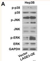
Application: WB Species: Human Sample: Hep-3B cells

Application: WB Species: Mouse Sample: RAW264.7 cell

Application: WB Species: rat Sample:
Restrictive clause
Affinity Biosciences tests all products strictly. Citations are provided as a resource for additional applications that have not been validated by Affinity Biosciences. Please choose the appropriate format for each application and consult Materials and Methods sections for additional details about the use of any product in these publications.
For Research Use Only.
Not for use in diagnostic or therapeutic procedures. Not for resale. Not for distribution without written consent. Affinity Biosciences will not be held responsible for patent infringement or other violations that may occur with the use of our products. Affinity Biosciences, Affinity Biosciences Logo and all other trademarks are the property of Affinity Biosciences LTD.