, using Fas Ligand Antibody at 1/1000 dilution.")
, using Fas Ligand Antibody at 1/1000 dilution.")
 inhibited cell proliferation and induced apoptosis in tumor tissues. (A) Ki-67 staining of tumor sections obtained from mice treated with (a) vehicle control or (b) QX. Quantification of Ki-67-positive cells in the tumor sections (c). (B)TUNEL staining of tumor sections obtained from mice treated with (a) vehicle control or (b) QX. Quantification of TUNEL-positive cells in the tumor sections (c). (C) Western blotting of proapoptotic proteins Bim, FasL, and cleaved caspase-3 expression in tumor tissues of QX-treated mice or vehicle control-treated mice. Data are presented as mean ± SD. *P < .05, **P < .01 versus vehicle control.")
Control Products

Product Info
*The optimal dilutions should be determined by the end user. For optimal experimental results, antibody reuse is not recommended.
*Tips:
WB: For western blot detection of denatured protein samples. IHC: For immunohistochemical detection of paraffin sections (IHC-p) or frozen sections (IHC-f) of tissue samples. IF/ICC: For immunofluorescence detection of cell samples. ELISA(peptide): For ELISA detection of antigenic peptide.
Cite Format: Affinity Biosciences Cat# AF0157, RRID:AB_2833338.
Fold/Unfold
ALPS1B; Apoptosis (APO 1) antigen ligand 1; Apoptosis antigen ligand 1; Apoptosis antigen ligand; APT1LG1; APTL; CD178; CD178 antigen; CD95 ligand; CD95-L; CD95L; CD95L protein; Fas antigen ligand; Fas L; Fas ligand (TNF superfamily member 6); Fas ligand; FASL; Fasl Fas ligand (TNF superfamily member 6); FASLG; Generalized lymphoproliferative disease; Gld; soluble form; TNFL6_HUMAN; TNFSF6; Tumor necrosis factor (ligand) superfamily member 6; Tumor necrosis factor ligand superfamily member 6;
Immunogens
A synthesized peptide derived from human Fas Ligand, corresponding to a region within the internal amino acids.
- P48023 TNFL6_HUMAN:
- Protein BLAST With
- NCBI/
- ExPASy/
- Uniprot
MQQPFNYPYPQIYWVDSSASSPWAPPGTVLPCPTSVPRRPGQRRPPPPPPPPPLPPPPPPPPLPPLPLPPLKKRGNHSTGLCLLVMFFMVLVALVGLGLGMFQLFHLQKELAELRESTSQMHTASSLEKQIGHPSPPPEKKELRKVAHLTGKSNSRSMPLEWEDTYGIVLLSGVKYKKGGLVINETGLYFVYSKVYFRGQSCNNLPLSHKVYMRNSKYPQDLVMMEGKMMSYCTTGQMWARSSYLGAVFNLTSADHLYVNVSELSLVNFEESQTFFGLYKL
Predictions
Score>80(red) has high confidence and is suggested to be used for WB detection. *The prediction model is mainly based on the alignment of immunogen sequences, the results are for reference only, not as the basis of quality assurance.
High(score>80) Medium(80>score>50) Low(score<50) No confidence
Research Backgrounds
Cytokine that binds to TNFRSF6/FAS, a receptor that transduces the apoptotic signal into cells. Involved in cytotoxic T-cell-mediated apoptosis, natural killer cell-mediated apoptosis and in T-cell development. Initiates fratricidal/suicidal activation-induced cell death (AICD) in antigen-activated T-cells contributing to the termination of immune responses (By similarity). TNFRSF6/FAS-mediated apoptosis has also a role in the induction of peripheral tolerance (By similarity). Binds to TNFRSF6B/DcR3, a decoy receptor that blocks apoptosis.
Induces FAS-mediated activation of NF-kappa-B, initiating non-apoptotic signaling pathways (By similarity). Can induce apoptosis but does not appear to be essential for this process.
Cytoplasmic form induces gene transcription inhibition.
The soluble form derives from the membrane form by proteolytic processing. The membrane-bound form undergoes two successive intramembrane proteolytic cleavages. The first one is processed by ADAM10 producing an N-terminal fragment, which lacks the receptor-binding extracellular domain. This ADAM10-processed FasL (FasL APL) remnant form is still membrane anchored and further processed by SPPL2A that liberates the FasL intracellular domain (FasL ICD). FasL shedding by ADAM10 is a prerequisite for subsequent intramembrane cleavage by SPPL2A in T-cells.
N-glycosylated. Glycosylation enhances apoptotic activity.
Phosphorylated by FGR on tyrosine residues; this is required for ubiquitination and subsequent internalization.
Monoubiquitinated.
Cell membrane>Single-pass type II membrane protein. Cytoplasmic vesicle lumen. Lysosome lumen.
Note: Is internalized into multivesicular bodies of secretory lysosomes after phosphorylation by FGR and monoubiquitination (PubMed:17164290). Colocalizes with the SPPL2A protease at the cell membrane (PubMed:17557115).
Secreted.
Note: May be released into the extracellular fluid by cleavage from the cell surface.
Nucleus.
Note: The FasL ICD cytoplasmic form is translocated into the nucleus.
Belongs to the tumor necrosis factor family.
Research Fields
· Cellular Processes > Cell growth and death > Apoptosis. (View pathway)
· Cellular Processes > Cell growth and death > Necroptosis. (View pathway)
· Environmental Information Processing > Signal transduction > MAPK signaling pathway. (View pathway)
· Environmental Information Processing > Signal transduction > Ras signaling pathway. (View pathway)
· Environmental Information Processing > Signaling molecules and interaction > Cytokine-cytokine receptor interaction. (View pathway)
· Environmental Information Processing > Signal transduction > FoxO signaling pathway. (View pathway)
· Environmental Information Processing > Signal transduction > PI3K-Akt signaling pathway. (View pathway)
· Human Diseases > Drug resistance: Antineoplastic > Platinum drug resistance.
· Human Diseases > Endocrine and metabolic diseases > Non-alcoholic fatty liver disease (NAFLD).
· Human Diseases > Endocrine and metabolic diseases > Type I diabetes mellitus.
· Human Diseases > Infectious diseases: Parasitic > Chagas disease (American trypanosomiasis).
· Human Diseases > Infectious diseases: Parasitic > African trypanosomiasis.
· Human Diseases > Infectious diseases: Viral > Hepatitis B.
· Human Diseases > Infectious diseases: Viral > Measles.
· Human Diseases > Infectious diseases: Viral > Influenza A.
· Human Diseases > Infectious diseases: Viral > Human papillomavirus infection.
· Human Diseases > Infectious diseases: Viral > Herpes simplex infection.
· Human Diseases > Cancers: Overview > Pathways in cancer. (View pathway)
· Human Diseases > Cancers: Overview > Proteoglycans in cancer.
· Human Diseases > Immune diseases > Autoimmune thyroid disease.
· Human Diseases > Immune diseases > Allograft rejection.
· Human Diseases > Immune diseases > Graft-versus-host disease.
· Organismal Systems > Immune system > Natural killer cell mediated cytotoxicity. (View pathway)
· Organismal Systems > Nervous system > Neurotrophin signaling pathway. (View pathway)
References

Application: WB Species: Mouse Sample: MDSCs
Application: WB Species: Rat Sample: testes tissue

Application: WB Species: human Sample:
Application: WB Species: Sample: heart

Application: WB Species: Mouse Sample: lung cancer
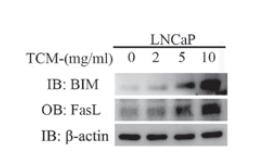
Application: WB Species: human Sample: LNCaP cells
Restrictive clause
Affinity Biosciences tests all products strictly. Citations are provided as a resource for additional applications that have not been validated by Affinity Biosciences. Please choose the appropriate format for each application and consult Materials and Methods sections for additional details about the use of any product in these publications.
For Research Use Only.
Not for use in diagnostic or therapeutic procedures. Not for resale. Not for distribution without written consent. Affinity Biosciences will not be held responsible for patent infringement or other violations that may occur with the use of our products. Affinity Biosciences, Affinity Biosciences Logo and all other trademarks are the property of Affinity Biosciences LTD.