

 G. Flow cytometry was used for cell distribution in each phase. H. The expression levels of
several cell cycle proteins were detected. (The western blot bands were cropped from Fig. S3, S4, S5 and S6.) I. Flow cytometry was performed
for detection of cell apoptosis. J. The expression levels of several apoptosis proteins were examined after MT1JP overexpression in HCCC-9810
cells. (The western blot bands were cropped from Fig. S7 and S8.) (*p < 0.05, **p < 0.01, ***p < 0.001, compared with normal, pcDNA3.1 or siRNA
NC; the original images of western blot were shown in supplementary Fig. 1–8)")
 Western blot analysis was performed to examine the cell‑cycle proteins, including p27, cyclin D1 and cyclin E1. eIF4E, eukaryotic translation initiation factor 4; shRNA, short hairpin RNA; contr, control; p27, cyclin‑dependent kinase inhibitor 1B. Statistical analyses were performed using one‑way analysis of variance followed by Bonferroni's correction post‑hoc test.")
 The immunohistochemical staining of cell cycle‑associated proteins, Ki 67 and eIF4E, in tumors. Scale bar, 50 µm. eIF4E, eukaryotic translation initiation factor 4; shRNA, short hairpin RNA; contr, control; p27, cyclin‑dependent kinase inhibitor 1B.")
; Cell viability was determined by CCK-8 assay (b); Cell cycle distribution and apoptosis were
measured by flow cytometry (c and e); Relative protein levels of cyclin D and cyclin E were calculated (d). Caspase 3 and 9 enzymatic activities were analyzed (f). *P
< 0.05, **P < 0.01, and ***P < 0.001 vs parental group. % P < 0.05 and %%% P < 0.001 vs sh-NC group. n = 3.")
 IHC staining was carried out to delineate the expression of PCNA protein in Skov3 cells. Real-time PCR was employed to detect the transcriptional levels of (B) P53 and (C) P21. (C, D, E, and F) Western blot analysis was conducted to measure key proteins of several signaling pathways. *, p<0.05 vs control group, * *, p<0.01 vs control group")
 The protein levels of c-myc, p21, cyclin D, CDK6, cyclin E, Bax, and Bcl-2 were determined by Western blot in stable Tet-on inducible RECQL4 knockdown cell lines (KYSE30 and TE-1 cells) (+Dox) and controls (–Dox). Experiments were independently repeated 3 times. All data indicate the mean ±SD. *P < 0.05; **P < 0.01; ***P < 0.001.")
 Depletion of RECQL4 by siRNA. RECQL4 protein levels were measured by Western blot. KYSE30 and TE-1 cells were transfected with siRNA duplexes (200 nM) specific to RECQL4 or negative oligo in serum-free medium for 4 h, then replaced with complete medium for 24 h. Whole cell extracts were collected for Western blot analysis using RECQL4 antibodies. (B) Cell cycle distributions in RECQL4 knockdown cell lines (KYSE30 and TE-1 cells) and controls were determined by flow cytometry. (C) Cellular senescence was examined by SA-β-gal staining. Microscopic magnification (×200), Scale bar: 50 μm. (D) The protein levels of c-myc, p21, cyclin D, CDK6, cyclin E, Bax, and Bcl-2 were determined by Western blot in stable Tet-on inducible RECQL4 knockdown cell lines (KYSE30 and TE-1 cells) (+Dox) and controls (–Dox). Experiments were independently repeated 3 times. All data indicate the mean ± SD. *P < 0.05; **P < 0.01; ***P < 0.001.")
 Cell viability was decreased by hsa_circ_0001666 siRNAs in TPC-1 and IHH-4 cells, as shown by Cell Counting Kit 8 assay. (B) 5-Ethynyl-2′-deoxyuridine staining showed that hsa_circ_0001666 knockdown decreased the proportion of cells in the S phase. (C) Knockdown of hsa_circ_0001666 arrested the cell cycle at the G1 phase, as shown by flow cytometry analysis. (D) Protein expression levels of cyclin D1 and E were decreased by hsa_circ_0001666 silencing. (E) Hsa_circ_0001666 knockdown increased cell apoptosis, as shown by flow cytometry. (F) Protein expression levels of caspase-3, and caspase-9 were increased by hsa_circ_0001666 siRNAs. *P<0.05, **P<0.01 vs. si-NC. si, small interfering; NC, negative control; Abs, absorbance; circ, circular.")
 The relationship between OTUB1 and Cyclin E1 was analyzed via gene MANIA database. (B) The protein expression of Cyclin E1 in BPH, ADPC, and CRPC was detected by IHC staining. (C) Western blotting detecting the expression of Cyclin E1 in PC3 and C4-2 cells transfected with otub1 overexpression and otub1 c91s. (D) Western blotting detecting the expression of Cyclin E1 in PC3 and C4-2 cells transfected with OTUB1 siRNA. (E) Western blotting detecting the expression of Cyclin E1 in PC3 and C4-2 cells transfected with an increasing gradient OTUB1.")
 PC3 cells transfected with OTUB1 overexpression and otub1 c91s were transplanted subcutaneously in nude mice. The effect of otub1 overexpression, otub1 c91s, and RO-3306 on the growth of PCa. (C) The tumor volume was measured with a caliper. (D) The tumor volumes were measured daily when the diameter grew into 2 mm. (E) IHC staining detecting the expression of OTUB1, ki-67, and Cyclin E1 in these tumors from nude mice. ****P < 0.0001.")
. The protein-protein interaction (PPI) network of the KRT17 from STRING online database (https://string-db.org) was constructed (b). The protein levels in AMC-HN-8 cells were evaluated using western blot (c). The protein levels in TU177 cells were estimated using western blot. ∗A significant difference compared with the sh-NC group. ∗∗P < 0.01 and ∗∗∗P < 0.001.")
 LAMC2 overexpression promotes integrin β1/FAK/Src/AKT protein expression in TU177 cells. (b) LAMC2 knockdown inhibited integrin β1/FAK/Src/AKT protein expression in AMC-HN-8 cells. Data were expressed as mean ± SD, n = 3. Compared to the vector group, #P < 0.05, ##P < 0.01. Compared to the shNC group, ∗P < 0.05, ∗∗P < 0.01.")
 or empty vector, while NCI-H292 cells were transfected with two small interfering RNAs targeting HOXD9 (siHOXD9#1, siHOXD9#2) or none-specific sequence (siNC). A After 48 h of transfection, HOXD9 mRNA expression in two NSCLC cells (NCI-H661 and NCI-H292) was evaluated by real time-PCR assay. B After 48 h of transfection, HOXD9 protein expression in two NSCLC cells was evaluated by western blot assay. C After 48 h of transfection, NSCLC cells were seeded into 96-well microplates and analyzed with CCK-8 reagents at indicated time. D After 48 h of transfection, NSCLC cells were subjected into the EdU incorporation assay. The new generation cells were stained via EdU (red). DAPI stained nuclei in blue. Scale bar = 50 μm. Quantification of EdU-positive cells was performed to assess cell proliferation. E Cell cycle progression in two NSCLC cells was evaluated by flow cytometry analysis. F Apoptosis in NCI-H292 cells was evaluated by flow cytometry analysis. The apoptotic rate was quantified by adding the percentages of early apoptotic cells (Annexin V+/PI− in Q4 quadrant) and late apoptotic cells (Annexin V+/PI+ in Q2 quadrant). G Expression of protein markers involved in apoptosis and cell cycle regulation in NCI-H292 cells was evaluated by western blot assay. * p")
 Analysis of the cell cycle distribution of AGS and MGC-803 cells overexpressing PPM1B and mock control plasmids by flow cytometry. (B) The protein levels of overexpressed PPM1B GC cell cycle-related markers, such as p-CDK2, CDK2, CDK4, Cyclin D1, Cyclin E1, were detected by WB. (C, E) Flow cytometry analysis of the cell cycle distribution of AGS and MGC-803 cells with knockdown, knockout PPM1B and mock control plasmids. (D, F) The protein levels of cell cycle-related markers of knockdown and knockout PPM1B GC cells were detected by WB.")
 Effects of M. oleifera leaves proteins on the expression of apoptosis, cyclin and autophagy proteins in Jurkat cells. (b) Bax/Bcl-2, (c) cleaved CasPase-3, (d) CyclinE1, (e) CyclinD1 and (f) ATG5 statistical analysis of protein expression. Compared with the blank control group (0 μg/mL), the differences between the groups are represented by *P")
| Product: | Cyclin E1 Antibody |
| Catalog: | AF0144 |
| Description: | Rabbit polyclonal antibody to Cyclin E1 |
| Application: | WB IHC IF/ICC |
| Cited expt.: | WB, IHC |
| Reactivity: | Human, Mouse, Rat |
| Mol.Wt.: | 49kDa(Observed); 47kD(Calculated). |
| Uniprot: | P24864 |
| RRID: | AB_2833326 |
Control Products

Related Downloads
Protocols
Product Info
*The optimal dilutions should be determined by the end user. For optimal experimental results, antibody reuse is not recommended.
*Tips:
WB: For western blot detection of denatured protein samples. IHC: For immunohistochemical detection of paraffin sections (IHC-p) or frozen sections (IHC-f) of tissue samples. IF/ICC: For immunofluorescence detection of cell samples. ELISA(peptide): For ELISA detection of antigenic peptide.
Cite Format: Affinity Biosciences Cat# AF0144, RRID:AB_2833326.
Fold/Unfold
CCNE; Ccne1; CCNE1_HUMAN; cyclin E variant ex5del; cyclin E variant ex7del; Cyclin E1; Cyclin Es; Cyclin Et; CyclinE; G1/S specific cyclin E; G1/S-specific cyclin-E1;
Immunogens
A synthesized peptide derived from human Cyclin E1, corresponding to a region within C-terminal amino acids.
Highly expressed in testis and placenta. Low levels in bronchial epithelial cells.
- P24864 CCNE1_HUMAN:
- Protein BLAST With
- NCBI/
- ExPASy/
- Uniprot
MPRERRERDAKERDTMKEDGGAEFSARSRKRKANVTVFLQDPDEEMAKIDRTARDQCGSQPWDNNAVCADPCSLIPTPDKEDDDRVYPNSTCKPRIIAPSRGSPLPVLSWANREEVWKIMLNKEKTYLRDQHFLEQHPLLQPKMRAILLDWLMEVCEVYKLHRETFYLAQDFFDRYMATQENVVKTLLQLIGISSLFIAAKLEEIYPPKLHQFAYVTDGACSGDEILTMELMIMKALKWRLSPLTIVSWLNVYMQVAYLNDLHEVLLPQYPQQIFIQIAELLDLCVLDVDCLEFPYGILAASALYHFSSSELMQKVSGYQWCDIENCVKWMVPFAMVIRETGSSKLKHFRGVADEDAHNIQTHRDSLDLLDKARAKKAMLSEQNRASPLPSGLLTPPQSGKKQSSGPEMA
Research Backgrounds
Essential for the control of the cell cycle at the G1/S (start) transition.
Phosphorylation of both Thr-395 by GSK3 and Ser-399 by CDK2 creates a high affinity degron recognized by FBXW7, and accelerates degradation via the ubiquitin proteasome pathway. Phosphorylation at Thr-77 creates a low affinity degron also recognized by FBXW7.
Ubiquitinated by UHRF2; appears to occur independently of phosphorylation.
Nucleus.
Highly expressed in testis and placenta. Low levels in bronchial epithelial cells.
Belongs to the cyclin family. Cyclin E subfamily.
Research Fields
· Cellular Processes > Cell growth and death > Cell cycle. (View pathway)
· Cellular Processes > Cell growth and death > Oocyte meiosis. (View pathway)
· Cellular Processes > Cell growth and death > p53 signaling pathway. (View pathway)
· Cellular Processes > Cell growth and death > Cellular senescence. (View pathway)
· Environmental Information Processing > Signal transduction > PI3K-Akt signaling pathway. (View pathway)
· Human Diseases > Infectious diseases: Viral > Hepatitis B.
· Human Diseases > Infectious diseases: Viral > Measles.
· Human Diseases > Infectious diseases: Viral > Human papillomavirus infection.
· Human Diseases > Cancers: Overview > Pathways in cancer. (View pathway)
· Human Diseases > Cancers: Overview > Viral carcinogenesis.
· Human Diseases > Cancers: Overview > MicroRNAs in cancer.
· Human Diseases > Cancers: Specific types > Prostate cancer. (View pathway)
· Human Diseases > Cancers: Specific types > Small cell lung cancer. (View pathway)
· Human Diseases > Cancers: Specific types > Gastric cancer. (View pathway)
References
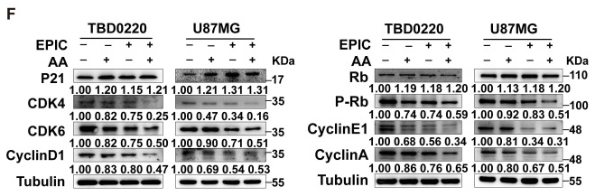
Application: WB Species: Human Sample: TBD0220 and U87MG cells

Application: WB Species: Human Sample: HCT116 and SW480 cells
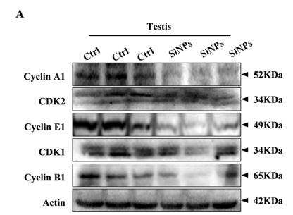
Application: WB Species: mouse Sample: testis
Restrictive clause
Affinity Biosciences tests all products strictly. Citations are provided as a resource for additional applications that have not been validated by Affinity Biosciences. Please choose the appropriate format for each application and consult Materials and Methods sections for additional details about the use of any product in these publications.
For Research Use Only.
Not for use in diagnostic or therapeutic procedures. Not for resale. Not for distribution without written consent. Affinity Biosciences will not be held responsible for patent infringement or other violations that may occur with the use of our products. Affinity Biosciences, Affinity Biosciences Logo and all other trademarks are the property of Affinity Biosciences LTD.