
, using MIF Antibody. The lane on the left was treated with blocking peptide.")
, using MIF Antibody at 1/1000 dilution.
5ug/NC membrane strip.
Exposure for 5s with Affinity™ ECL Kit(#KF8001).
Bands result from membrane strip incubation.")

| Product: | MIF Antibody |
| Catalog: | DF6404 |
| Description: | Rabbit polyclonal antibody to MIF |
| Application: | WB IHC |
| Cited expt.: | WB, IHC |
| Reactivity: | Human, Mouse, Rat |
| Prediction: | Pig, Zebrafish, Bovine, Horse, Sheep, Rabbit, Chicken, Xenopus |
| Mol.Wt.: | 12kDa(Observed); 12kD(Calculated). |
| Uniprot: | P14174 |
| RRID: | AB_2838367 |
Control Products

Related Downloads
Protocols
Product Info
*The optimal dilutions should be determined by the end user. For optimal experimental results, antibody reuse is not recommended.
*Tips:
WB: For western blot detection of denatured protein samples. IHC: For immunohistochemical detection of paraffin sections (IHC-p) or frozen sections (IHC-f) of tissue samples. IF/ICC: For immunofluorescence detection of cell samples. ELISA(peptide): For ELISA detection of antigenic peptide.
Cite Format: Affinity Biosciences Cat# DF6404, RRID:AB_2838367.
Fold/Unfold
GIF; GLIF; Glycosylation inhibiting factor; Glycosylation-inhibiting factor; L-dopachrome isomerase; L-dopachrome tautomerase; Macrophage migration inhibitory factor (glycosylation-inhibiting factor); Macrophage migration inhibitory factor; MIF; MIF protein; MIF_HUMAN; MMIF; Phenylpyruvate tautomerase;
Immunogens
A synthesized peptide derived from human MIF, corresponding to a region within C-terminal amino acids.
- P14174 MIF_HUMAN:
- Protein BLAST With
- NCBI/
- ExPASy/
- Uniprot
MPMFIVNTNVPRASVPDGFLSELTQQLAQATGKPPQYIAVHVVPDQLMAFGGSSEPCALCSLHSIGKIGGAQNRSYSKLLCGLLAERLRISPDRVYINYYDMNAANVGWNNSTFA
Predictions
Score>80(red) has high confidence and is suggested to be used for WB detection. *The prediction model is mainly based on the alignment of immunogen sequences, the results are for reference only, not as the basis of quality assurance.
High(score>80) Medium(80>score>50) Low(score<50) No confidence
Research Backgrounds
Pro-inflammatory cytokine. Involved in the innate immune response to bacterial pathogens. The expression of MIF at sites of inflammation suggests a role as mediator in regulating the function of macrophages in host defense. Counteracts the anti-inflammatory activity of glucocorticoids. Has phenylpyruvate tautomerase and dopachrome tautomerase activity (in vitro), but the physiological substrate is not known. It is not clear whether the tautomerase activity has any physiological relevance, and whether it is important for cytokine activity.
Secreted. Cytoplasm.
Note: Does not have a cleavable signal sequence and is secreted via a specialized, non-classical pathway. Secreted by macrophages upon stimulation by bacterial lipopolysaccharide (LPS), or by M.tuberculosis antigens.
Belongs to the MIF family.
Research Fields
· Metabolism > Amino acid metabolism > Tyrosine metabolism.
· Metabolism > Amino acid metabolism > Phenylalanine metabolism.
References

Application: WB Species: Mouse Sample:
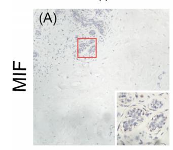
Application: IHC Species: human Sample: breast

Application: IHC Species: Human Sample: breast cancer

Application: IF/ICC Species: human Sample:
Restrictive clause
Affinity Biosciences tests all products strictly. Citations are provided as a resource for additional applications that have not been validated by Affinity Biosciences. Please choose the appropriate format for each application and consult Materials and Methods sections for additional details about the use of any product in these publications.
For Research Use Only.
Not for use in diagnostic or therapeutic procedures. Not for resale. Not for distribution without written consent. Affinity Biosciences will not be held responsible for patent infringement or other violations that may occur with the use of our products. Affinity Biosciences, Affinity Biosciences Logo and all other trademarks are the property of Affinity Biosciences LTD.