
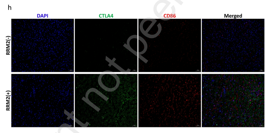
, using CD86 Antibody.")

 by IF/ICC. The samples were fixed with PFA and permeabilized in 0.1% Triton X-100,then blocked in 10% serum for 45 minutes at 25°C. Samples were then incubated with primary Ab(DF6332) and mouse anti-beta tubulin Ab(T0023) for 1 hour at 37°C. An AlexaFluor594 conjugated goat anti-rabbit IgG(H+L) Ab(Red) and an AlexaFluor488 conjugated goat anti-mouse IgG(H+L) Ab(Green) were used as the secondary antibody.
The nuclear counter stain is DAPI (blue).")
. b–e The inflammatory marker proteins immunofluorescence staining and histograms at 14 days, including CD86, GFAP, TNF-α, iNOS(magnification × 400). All data presented as mean ± SEM in each group. *p < 0.05 vs. SCI + saline group, **p < 0.01 vs. SCI + saline group. #p < 0.05 comparison between SCI + EPO group and SCI + EPO + PD98059, ##p < 0.01 comparison between SCI + EPO group and SCI + EPO + PD98059")
 (a) and M2(CD206+/iba1+) (b). The representative double immunofluorescence labeling image of CD86 (red)/Iba1 (green) in brain tissue, and the nuclei were stained with DAPI (blue). The representative double immunofluorescence labeling image of CD206 (red)/Iba1 (green) in brain tissue and the nuclei were stained with DAPI (blue). The quantity of the percentage of CD86+/Iba1+ cell (c) and CD206+/Iba1+ cell. (d) The effects of emodin on M1 microglia cell subsets mRNA relative expression. Mice were sacrificed on 21 dpi. The CNS tissues are collected. The IL-6 (e), TGF-β (f), IL-17A (g), and RORγt mRNA relative expression were analyzed by qRT-PCR, respectively. All data were expressed as mean ± S.D (n = 5). Comparisons among each group were analyzed by one-way ANOVA in C, D, F, and G. Comparisons among each group were analyzed by Kruskal-Wallis test using Bonferroni comparisons post hoc tests in E and H. *P < 0.05, **P < 0.01 versus NC group, #P < 0.05, ##P < 0.01 versus EAE group.")
 The contents of CD86 in Y79 tumors after different treatments. (C) TNF-α levels in Y79 tumors after different treatments (**p<.01).")
 Observation of cell morphology, (b) Screening the effective concentration (p")
 GL261 glioma-bearing C57BL/6 to undergo xenograft tumor and brain treatment and MRI tests on days 0 and 24. (B,D) At the end of the experiment (30 days), the mouse brains were made into sections for immunofluorescence staining (CD86 and CD206); (C,E) analysis of the immunofluorescence intensity showed that the SG group induced more CD86 expression in the gliomas than CD206 expression.")
 Immunohistochemistry of CD138, CD86, and PD-L1. Case 49: − (CD138), − (CD86), and 0 (PD-L1); Case 52: + (CD138), + (CD86), and 2 (PD-L1); Case 14: − (CD138), − (CD86), and 1 (PD-L1); Case 13: − (CD138), + (CD86), and 2 (PD-L1). (B) Schematic diagram of the balance of immune cells and PD-L1. The red solid line represented that the PD-L1 expression was positively correlated with M1 macrophages, plasma cells, and CD8+ T cells, while the blue dotted line represented that the PD-L1 expression was negatively correlated with resting mast cells and regulatory T cells.")
. All images were taken at 400× magnification. B-D Correlation of MICA protein expression with CD68, CD86, CD206 level in HCC tumor and background liver tissues. E-H Correlation of EHHADH protein expression with CD68, CD86, CD206, MICA level in HCC tumor and background liver tissues.")
 and wild type. B EHHADH and PPAR-α mRNA expression are showed in MICA+Huh-7 cells compared to NC+Huh-7 cells. C Representative immunofluorescence staining for EHHADH in MICA+Huh-7 cells compared to NC+Huh-7 cells and statistically quantificational results. D-G The CD86, CD206, TNF-α and IL-10 mRNA expression are showed in macrophages co-cultured with MICA+Huh-7 cells or NC cells for 24 h or 72 h. H-I Representative immunofluorescence staining for CD86, CD206, TNF-α, and IL-10 in macrophages co-cultured with MICA+ Huh-7 cells or NC cells for 24 h or 72 h and statistically quantificational results. All images were taken at 400× magnification.")
 and F4/80 (green) immunofluorescence staining (scale bar, 50μm). B: CD206 (red) and F4/80 (green) immunofluorescence staining (scale bar, 50μm). C: Statistics of the percentage of CD86 positive macrophages in the lung tissues of mice in each group (n=3). D: Statistics of the percentage of CD206 positive macrophages in the lung tissues of mice in each group (n=3). E: mRNA levels of M1 macrophage-related markers (n=8). F: mRNA levels of M2 macrophage-related markers (n=8). G, H: Expression levels of IL-12 in lung cancer tissues (n=6). G, I: Expression levels of IL-10 in lung cancer tissues (n=6). J, K, L: Plasma levels of IL-12(J), IL-10(K) and IFN-γ(L) (n=8). Note: &, && respectively represent a significant difference compared with the Saline group (P")
 Chemical structure of AST. (b) Cell viability assays of HUVECs and RAW264.7 macrophages treated with various concentrations of AST, showing no significant cytotoxicity. (c) Immunofluorescence staining of RAW264.7 macrophages for CD86 (M1 marker) and CD206 (M2 marker) under control, LPS, and LPS + AST conditions, demonstrating AST-induced M2 polarization. 3D intensity plots confirm increased CD206 expression with AST treatment. (d) Flow cytometry analysis of CD86 and CD206 expression in macrophages, showing a significant increase in M2 (CD206+) and decrease in M1 (CD86+) populations following AST treatment. (e) Scratch assay of HUVECs under high-glucose (HG) conditions with and without AST treatment, indicating enhanced wound closure with AST. (f) Transwell migration assay demonstrating increased HUVEC migration upon AST treatment. (g) Tube formation assay showing improved angiogenic capacity of HUVECs after AST treatment. (h) Western blot analysis of angiogenesis markers (CD31 and VEGF) in HUVECs, showing upregulated expression following AST treatment. The data are represented as mean ± SD (n = 3). ns: not significant, ∗P < 0.05, ∗∗P < 0.01, ∗∗∗P < 0.001, ∗∗∗∗P < 0.0001.")
 and IFN-γ (3 ng/mL) and treated with Nintedanib (1 μM) or Ruxolitinib (5 μM and 10 μM) for 12 h. (A) RT-qPCR analysis of mRNA levels of CD80, Tnf-α, and CD86 in different treatment groups (n = 3 per group). (B) Immunofluorescence staining of CD80 in RAW264.7 cells. (C) Flow cytometry analysis of the percentage of CD86-positive pro-inflammatory macrophages. (D) Western blot analysis of CD80, CD86, iNOS, p-JAK1, and p-STAT1 protein expression levels (n = 3 per group). Data are presented as mean ± SEM (n = 3). *P < 0.05, **P < 0.01, ***P < 0.001.")
 Representative immunofluorescence images (400 × ) of CD86 (magenta) and Iba1 (green) in ischemic penumbra cortex after 3 and 7 days of intervention, scale bar = 25 μm. (B, D) Representative immunofluorescence images (400 × ) of CD206 (magenta) and Iba-1 (green) in ischemic penumbra cortex after 3 and 7 days of intervention, scale bar = 25 μm. (E, F) Proportion of M1 and M2 microglia of rats after 3 and 7 days of intervention in each group (3 d: M1, n = 3, p = 0.000, F = 44.762, df = 2; M2, n = 3, p = 0.000, F = 47.263, df = 2. 7 d: M1, n = 3, p = 0.000, F = 47.579, df = 2; M2, n = 3, p = 0.000, F = 29.017, df = 2.). Data were obtained from three independent replicate experiments and expressed as median with range, the “box” depicts the value and median for 3 independent samples with a total of 6 slices, while the “whiskers” show the minimum and maximum values. ***p < 0.001 versus MCAO group at the same time; ###p < 0.001 versus EA group at the same time.")
 The mRNA expression levels of IL-1α (A), IL-6 (B), COX2 (C), and TGF-α (D) in each group were determined by qPCR. (E,F) Immunofluorescence staining of ICAM1 (E) and TGF-α (F) in lung tissues from each group. Scale bar: 20 μm. (G) TUNEL staining showing the degree of apoptosis. Scale bar: 20 μm. (H) IHC staining of CD86, CD163, and Gr-1 in lung tissues from each group. Scale bar: 20 μm. (I) Protein expression levels of IL-1β, NOS2, TLR2, CD86, CD115, CD206, ARG1, and CD163 in each group, as detected by western blot analysis. Sham: control group; I/RI: mice whose superior mesenteric artery was completely clamped; I/RI + Ger: I/RI group administered with germacrone. Data are shown as the mean ± SD. *P")
 Immunofluorescent staining of CD206 in the mPFC brain region.(n = 3, scale bar = 20 μm) blue for DAPI, red for CD206, green for iba1. (B) CD206 in immunofluorescence semi-quantitative analysis of the mPFC. (C-E) Western blot in the mPFC. (F-G) Immunofluorescence analysis shows changes in the expression of iba1 (green) and CD86 (red) or CD206 (red) in HMC-3 cells following a 12 h LPS treatment (1 μg/mL) combined with different doses of p-SYN (5, 10, 20 μM) (n = 3, scale bar = 50 μm). Magnification: 400 × . (H) Semi-quantitative analysis of CD86 and CD206 immunofluorescence intensity. (I-J) Western blot in the HMC-3.(n = 3, P < 0.05).")
, n = 6 for (A–E).")
 and anti-Mφ (CD163, CD206) marker protein level in the testis of WT and KO mice. B The mRNA of Ym1 and IL10 in the testis of WT and KO mice. C Multiple immunofluorescences staining of CD74, CD86, and CD163 in the testis of WT and KO groups, scale bar is 200 µm and 20 µm, respectively. D Changes of mRNA level of spermatogenic cells in different stages of mice testis in WT and KO groups, including Plzf, Grfα1, Id4, c-kit, and Scp3. E The western blot and analysis of Plzf, c-kit, and Scp3 in WT and KO groups. F Multiple immunofluorescences of Plzf, c-kit, and Scp3 in testicles of WT and KO, scale bar is 200 µm and 50 µm, respectively. The western blot (G) and immunofluorescence (H) were used to detect the expression of γ-H2AX in the testis of WT and KO, scale bar 20 µm. All values were presented as the mean ± SD. Student’s t-test; *P")
 Flow cytometric analysis quantified CD86/CD11c expression in LPS and LPS + NorCA groups. (b,c) Flow cytometry-derived CD86/CD11c expression in LPS vs LPS + NorCA groups (bar graphs). (d,e) qRT-PCR analysis quantified CD86/CD11c expression in LPS vs LPS + NorCA groups. (f–h) The expression levels of CD86 and CD11c in the LPS group and LPS + NorCA group were detected by western blotting. *p < 0.05, **p < 0.01, ***p < 0.001. LPS, lipopolysaccharide; qRT-PCR, quantitative reverse transcription PCR; FITC-A, fluorescein isothiocyanate - area; FSC-A, forward scatter - area.")
 and protein (B) expression in sh-NC/sh-ETS2-transfected RAW264.7 cells with LPS/IFN-γ induction. **P")
 Schematic illustration of the preparation processes for CM1, CM2, and CM3. (B) Immunofluorescence images showing CD86 or CD206 expression in TAMs treated with CM1, CM2, and CM3. (C) Relative fluorescence intensity of immunofluorescence signals. (D) Protein expression levels of CD86 and CD206 in TAMs treated with CM1, CM2, and CM3. (E) Protein expression levels of IL-6, TNF-α, and TGF-β1. *p < 0.05, **p < 0.01, ***p < 0.001, ****p < 0.0001; “ns” indicates no significant difference.")
. Rats were treated as described in Figure 1 legend. (A). Graphs of Western Blot experiment results; (B–E). analysis of iNOS, CD86,Arg-1, and IL-10 protein expression results; (F–I). analysis of IL-6, IL-1β, TNF-α, and IL-10 gene expression data; (J–L). results and data analysis of immunodouble fluorescence staining of macrophage M1-type polarization markers. “*” indicates significant dqinghuidaifference compared with CON group, p < 0.05. “**” indicates highly significant difference compared with CON group, p < 0.01. “#” means significant difference compared with CRS group, p < 0.05. “##” means extremely significant difference compared with CRS group, p < 0.01.")
 Images of immunofluorescence staining for CD163 (green), CD86 (green), and F4/80 (red) in the aorta. The scale bar in the figure is 200 μm. (B) Percentage of CD86+ and CD163+ macrophages. n = 3. (C) Expression of CD86 and CD163 in the aorta was revealed by western blot. (D) Qualification of CD86+ and CD163+. n = 3. (E) Images of immunofluorescence staining for α-SMA (green) and F4/80 (red) in the aorta. The scale bar in the figure is 300 μm, and the image in the right frame is a ×400 magnification of the point pointed. n = 3. (F) Percentage of F4/80+α-SMA+ cells. n = 3. (G) Expression of NF-κB p65, NF-κB pp65, NLRP3, and Caspase-1 p20 in the aorta by western blot. (H) Qualification of NF-κB p65, NF-κB pp65, NLRP3, and Caspase-1 p20. n = 3. Multi-group comparison was measured by one-way ANOVA. The data are presented as the mean ± SD. *p")
 Images of immunofluorescence staining for CD163 (green), CD86 (green), and F4/80 (red) in the aorta. The scale bar in the figure is 200 μm. (B) Percentage of CD86+ and CD163+ macrophages. n = 3. (C) Expression of CD86 and CD163 in the aorta was revealed by western blot. (D) Qualification of CD86+ and CD163+. n = 3. (E) Images of immunofluorescence staining for α-SMA (green) and F4/80 (red) in the aorta. The scale bar in the figure is 300 μm, and the image in the right frame is a ×400 magnification of the point pointed. n = 3. (F) Percentage of F4/80+α-SMA+ cells. n = 3. (G) Expression of NF-κB p65, NF-κB pp65, NLRP3, and Caspase-1 p20 in the aorta by western blot. (H) Qualification of NF-κB p65, NF-κB pp65, NLRP3, and Caspase-1 p20. n = 3. Multi-group comparison was measured by one-way ANOVA. The data are presented as the mean ± SD. *p")
Control Products

Related Downloads
Protocols
Product Info
*The optimal dilutions should be determined by the end user. For optimal experimental results, antibody reuse is not recommended.
*Tips:
WB: For western blot detection of denatured protein samples. IHC: For immunohistochemical detection of paraffin sections (IHC-p) or frozen sections (IHC-f) of tissue samples. IF/ICC: For immunofluorescence detection of cell samples. ELISA(peptide): For ELISA detection of antigenic peptide.
Cite Format: Affinity Biosciences Cat# DF6332, RRID:AB_2838296.
Fold/Unfold
Activation B7-2 antigen 3; Activation B7-2 antigen; B-lymphocyte activation antigen B7-2 2; B-lymphocyte activation antigen B7-2; B7 2; B7; B7-2; B7.2; B70; B72 antigen; BU63; CD28 antigen ligand 2 2; CD28 antigen ligand 2; Cd28l2; CD28LG2; CD86; CD86 antigen (CD28 antigen ligand 2, B7-2 antigen) 1, 2; CD86 antigen (CD28 antigen ligand 2, B7-2 antigen); CD86 antigen; CD86 molecule; CD86_HUMAN; CLS1; CTLA-4 counter-receptor B7.2 2, 3; CTLA-4 counter-receptor B7.2; Early T-cell costimulatory molecule 1; ETC-1; FUN 1; FUN-1; LAB72; Ly-58; Ly58; MB7; MB7-2; Membrane glycoprotein; MGC34413; T-lymphocyte activation antigen CD86; TS/A-2;
Immunogens
A synthesized peptide derived from human CD86, corresponding to a region within the internal amino acids.
- P42081 CD86_HUMAN:
- Protein BLAST With
- NCBI/
- ExPASy/
- Uniprot
MDPQCTMGLSNILFVMAFLLSGAAPLKIQAYFNETADLPCQFANSQNQSLSELVVFWQDQENLVLNEVYLGKEKFDSVHSKYMGRTSFDSDSWTLRLHNLQIKDKGLYQCIIHHKKPTGMIRIHQMNSELSVLANFSQPEIVPISNITENVYINLTCSSIHGYPEPKKMSVLLRTKNSTIEYDGVMQKSQDNVTELYDVSISLSVSFPDVTSNMTIFCILETDKTRLLSSPFSIELEDPQPPPDHIPWITAVLPTVIICVMVFCLILWKWKKKKRPRNSYKCGTNTMEREESEQTKKREKIHIPERSDEAQRVFKSSKTSSCDKSDTCF
Research Backgrounds
Receptor involved in the costimulatory signal essential for T-lymphocyte proliferation and interleukin-2 production, by binding CD28 or CTLA-4. May play a critical role in the early events of T-cell activation and costimulation of naive T-cells, such as deciding between immunity and anergy that is made by T-cells within 24 hours after activation. Isoform 2 interferes with the formation of CD86 clusters, and thus acts as a negative regulator of T-cell activation.
(Microbial infection) Acts as a receptor for adenovirus subgroup B.
Polyubiquitinated; which is promoted by MARCH8 and results in endocytosis and lysosomal degradation.
Cell membrane>Single-pass type I membrane protein.
Expressed by activated B-lymphocytes and monocytes.
Research Fields
· Environmental Information Processing > Signaling molecules and interaction > Cell adhesion molecules (CAMs). (View pathway)
· Human Diseases > Endocrine and metabolic diseases > Type I diabetes mellitus.
· Human Diseases > Cancers: Overview > Transcriptional misregulation in cancer.
· Human Diseases > Immune diseases > Autoimmune thyroid disease.
· Human Diseases > Immune diseases > Systemic lupus erythematosus.
· Human Diseases > Immune diseases > Rheumatoid arthritis.
· Human Diseases > Immune diseases > Allograft rejection.
· Human Diseases > Immune diseases > Graft-versus-host disease.
· Human Diseases > Cardiovascular diseases > Viral myocarditis.
· Organismal Systems > Immune system > Toll-like receptor signaling pathway. (View pathway)
· Organismal Systems > Immune system > Intestinal immune network for IgA production. (View pathway)
References

Application: WB Species: Mouse Sample:
Restrictive clause
Affinity Biosciences tests all products strictly. Citations are provided as a resource for additional applications that have not been validated by Affinity Biosciences. Please choose the appropriate format for each application and consult Materials and Methods sections for additional details about the use of any product in these publications.
For Research Use Only.
Not for use in diagnostic or therapeutic procedures. Not for resale. Not for distribution without written consent. Affinity Biosciences will not be held responsible for patent infringement or other violations that may occur with the use of our products. Affinity Biosciences, Affinity Biosciences Logo and all other trademarks are the property of Affinity Biosciences LTD.