Hexokinase II Antibody - #DF6176
.
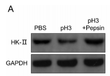
Bands result from membrane strip incubation.")
.
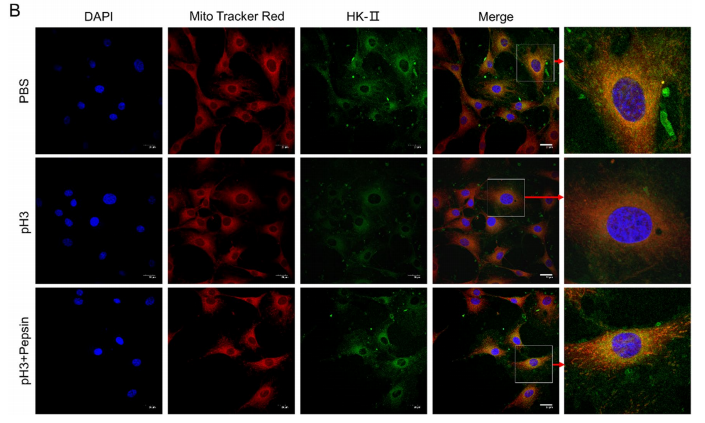
Bands result from membrane strip incubation.")
.
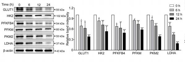
Bands result from membrane strip incubation.")

, diluted 1/600 was used as secondary antibody.")
 of triplicate determination from three independent experiments.")
 The expressions of LDHA, PFKM, HK2 and MACC1 in HT-29 cells were detected by western blots. (B) The gray value of the strip was measured with ImageJ. (C) The expressions of LDHA, PFKM, HK2 and MACC1in HCT-116 cells were detected by western blots. (D) The gray value of the strip was measured with ImageJ. Data were expressed as mean ± standard deviation (SD), n = 3. *P")
 Western blot experiment measuring the relative protein expression levels of HK2 and LDHA. (* indicates statistical significance compared between the two groups.")
| Product: | Hexokinase II Antibody |
| Catalog: | DF6176 |
| Description: | Rabbit polyclonal antibody to Hexokinase II |
| Application: | WB IHC IF/ICC |
| Cited expt.: | WB, IF/ICC |
| Reactivity: | Human, Mouse, Rat |
| Prediction: | Pig, Bovine, Horse, Sheep, Dog, Chicken, Xenopus |
| Mol.Wt.: | 102kDa(Observed); 102kD(Calculated). |
| Uniprot: | P52789 |
| RRID: | AB_2838143 |
Related Downloads
Protocols
Product Info
*The optimal dilutions should be determined by the end user. For optimal experimental results, antibody reuse is not recommended.
*Tips:
WB: For western blot detection of denatured protein samples. IHC: For immunohistochemical detection of paraffin sections (IHC-p) or frozen sections (IHC-f) of tissue samples. IF/ICC: For immunofluorescence detection of cell samples. ELISA(peptide): For ELISA detection of antigenic peptide.
Cite Format: Affinity Biosciences Cat# DF6176, RRID:AB_2838143.
Fold/Unfold
DKFZp686M1669; Hexokinase 2; Hexokinase 2 muscle; Hexokinase type II; Hexokinase-2; HK 2; HK II; HK2; HKII; HxK 2; HxK2; HXK2_HUMAN; Muscle form hexokinase;
Immunogens
A synthesized peptide derived from human Hexokinase II, corresponding to a region within C-terminal amino acids.
Predominant hexokinase isozyme expressed in insulin-responsive tissues such as skeletal muscle.
- P52789 HXK2_HUMAN:
- Protein BLAST With
- NCBI/
- ExPASy/
- Uniprot
MIASHLLAYFFTELNHDQVQKVDQYLYHMRLSDETLLEISKRFRKEMEKGLGATTHPTAAVKMLPTFVRSTPDGTEHGEFLALDLGGTNFRVLWVKVTDNGLQKVEMENQIYAIPEDIMRGSGTQLFDHIAECLANFMDKLQIKDKKLPLGFTFSFPCHQTKLDESFLVSWTKGFKSSGVEGRDVVALIRKAIQRRGDFDIDIVAVVNDTVGTMMTCGYDDHNCEIGLIVGTGSNACYMEEMRHIDMVEGDEGRMCINMEWGAFGDDGSLNDIRTEFDQEIDMGSLNPGKQLFEKMISGMYMGELVRLILVKMAKEELLFGGKLSPELLNTGRFETKDISDIEGEKDGIRKAREVLMRLGLDPTQEDCVATHRICQIVSTRSASLCAATLAAVLQRIKENKGEERLRSTIGVDGSVYKKHPHFAKRLHKTVRRLVPGCDVRFLRSEDGSGKGAAMVTAVAYRLADQHRARQKTLEHLQLSHDQLLEVKRRMKVEMERGLSKETHASAPVKMLPTYVCATPDGTEKGDFLALDLGGTNFRVLLVRVRNGKWGGVEMHNKIYAIPQEVMHGTGDELFDHIVQCIADFLEYMGMKGVSLPLGFTFSFPCQQNSLDESILLKWTKGFKASGCEGEDVVTLLKEAIHRREEFDLDVVAVVNDTVGTMMTCGFEDPHCEVGLIVGTGSNACYMEEMRNVELVEGEEGRMCVNMEWGAFGDNGCLDDFRTEFDVAVDELSLNPGKQRFEKMISGMYLGEIVRNILIDFTKRGLLFRGRISERLKTRGIFETKFLSQIESDCLALLQVRAILQHLGLESTCDDSIIVKEVCTVVARRAAQLCGAGMAAVVDRIRENRGLDALKVTVGVDGTLYKLHPHFAKVMHETVKDLAPKCDVSFLQSEDGSGKGAALITAVACRIREAGQR
Predictions
Score>80(red) has high confidence and is suggested to be used for WB detection. *The prediction model is mainly based on the alignment of immunogen sequences, the results are for reference only, not as the basis of quality assurance.
High(score>80) Medium(80>score>50) Low(score<50) No confidence
Research Backgrounds
Catalyzes the phosphorylation of hexose, such as D-glucose and D-fructose, to hexose 6-phosphate (D-glucose 6-phosphate and D-fructose 6-phosphate, respectively). Mediates the initial step of glycolysis by catalyzing phosphorylation of D-glucose to D-glucose 6-phosphate. Plays a key role in maintaining the integrity of the outer mitochondrial membrane by preventing the release of apoptogenic molecules from the intermembrane space and subsequent apoptosis.
Mitochondrion outer membrane>Peripheral membrane protein. Cytoplasm>Cytosol.
Note: The mitochondrial-binding peptide (MBP) region promotes association with the mitochondrial outer membrane (PubMed:29298880). The interaction with the mitochondrial outer membrane via the mitochondrial-binding peptide (MBP) region promotes higher stability of the protein (PubMed:29298880). Release from the mitochondrial outer membrane into the cytosol induces permeability transition pore (PTP) opening and apoptosis (PubMed:18350175).
Predominant hexokinase isozyme expressed in insulin-responsive tissues such as skeletal muscle.
The N- and C-terminal halves of the protein contain a hexokinase domain (PubMed:29298880). In contrast to hexokinase-1 and -3 (HK1 and HK3, respectively), both hexokinase domains display catalytic activity (PubMed:29298880). The region connecting the two hexokinase domains is required for the catalytic activity of the N-terminal hexokinase domain (PubMed:29298880). The N-terminal half regulates stability of the whole enzyme (PubMed:29298880).
Belongs to the hexokinase family.
Research Fields
· Environmental Information Processing > Signal transduction > HIF-1 signaling pathway. (View pathway)
· Human Diseases > Endocrine and metabolic diseases > Type II diabetes mellitus.
· Human Diseases > Cancers: Overview > Central carbon metabolism in cancer. (View pathway)
· Metabolism > Carbohydrate metabolism > Glycolysis / Gluconeogenesis.
· Metabolism > Carbohydrate metabolism > Fructose and mannose metabolism.
· Metabolism > Carbohydrate metabolism > Galactose metabolism.
· Metabolism > Carbohydrate metabolism > Starch and sucrose metabolism.
· Metabolism > Carbohydrate metabolism > Amino sugar and nucleotide sugar metabolism.
· Metabolism > Biosynthesis of other secondary metabolites > Neomycin, kanamycin and gentamicin biosynthesis.
· Metabolism > Global and overview maps > Metabolic pathways.
· Metabolism > Global and overview maps > Carbon metabolism.
· Organismal Systems > Endocrine system > Insulin signaling pathway. (View pathway)
· Organismal Systems > Digestive system > Carbohydrate digestion and absorption.
References
Application: WB Species: human Sample: MIA PaCa-2 cells

Application: WB Species: Human Sample: SHSY-5Y cells

Application: WB Species: Mouse Sample:
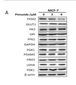
Application: WB Species: Human Sample: MCF-7 cells
Restrictive clause
Affinity Biosciences tests all products strictly. Citations are provided as a resource for additional applications that have not been validated by Affinity Biosciences. Please choose the appropriate format for each application and consult Materials and Methods sections for additional details about the use of any product in these publications.
For Research Use Only.
Not for use in diagnostic or therapeutic procedures. Not for resale. Not for distribution without written consent. Affinity Biosciences will not be held responsible for patent infringement or other violations that may occur with the use of our products. Affinity Biosciences, Affinity Biosciences Logo and all other trademarks are the property of Affinity Biosciences LTD.