Product: IGF1 Antibody
Catalog: DF6096
Description: Rabbit polyclonal antibody to IGF1
Application: WB IHC
Cited expt.: WB, IHC
Reactivity: Human, Mouse, Rat
Prediction: Pig, Zebrafish, Bovine, Sheep, Rabbit, Dog, Chicken, Xenopus
Mol.Wt.: 17kDa; 22kD(Calculated).
Uniprot: P05019
RRID: AB_2838064

View similar products>>

   Size Price Inventory
 100ul $280 In stock
 200ul $350 In stock

Lead Time: Same day delivery

For pricing and ordering contact:
Local distributors

Product Info

Source:
Rabbit
Application:
WB 1:500-1:2000, IHC 1:50-1:200
*The optimal dilutions should be determined by the end user.
*Tips:

WB: For western blot detection of denatured protein samples. IHC: For immunohistochemical detection of paraffin sections (IHC-p) or frozen sections (IHC-f) of tissue samples. IF/ICC: For immunofluorescence detection of cell samples. ELISA(peptide): For ELISA detection of antigenic peptide.

Reactivity:
Human,Mouse,Rat
Prediction:
Pig(100%), Zebrafish(86%), Bovine(100%), Sheep(100%), Rabbit(100%), Dog(100%), Chicken(100%), Xenopus(100%)
Clonality:
Polyclonal
Specificity:
IGF1 Antibody detects endogenous levels of total IGF1.
RRID:
AB_2838064
Cite Format: Affinity Biosciences Cat# DF6096, RRID:AB_2838064.
Conjugate:
Unconjugated.
Purification:
The antiserum was purified by peptide affinity chromatography using SulfoLink™ Coupling Resin (Thermo Fisher Scientific).
Storage:
Rabbit IgG in phosphate buffered saline , pH 7.4, 150mM NaCl, 0.02% sodium azide and 50% glycerol. Store at -20 °C. Stable for 12 months from date of receipt.
Alias:

Fold/Unfold

IBP1; IGF I; IGF IA; IGF IB; IGF-I; Igf1; IGF1_HUMAN; IGF1A; IGFI; IGFIA; Insulin like growth factor 1 (somatomedin C); Insulin like growth factor 1; Insulin like growth factor IA; Insulin like growth factor IB; Insulin-like growth factor I; Mechano growth factor; MGF; OTTHUMP00000195080; OTTHUMP00000195081; OTTHUMP00000195082; OTTHUMP00000195083; OTTHUMP00000195084; Somatomedia C; Somatomedin C; Somatomedin-C;

Immunogens

Immunogen:

A synthesized peptide derived from human IGF1, corresponding to a region within the internal amino acids.

Uniprot:
Gene(ID):
Description:
IGF1, also named as IBP1, MGF, IGF-IA and Somatomedin-C, belongs to the insulin family. IGF1 is structurally and functionally related to insulin but have a much higher growth-promoting activity. Altered expression or mutation of IGF-1 is associated with several human disorders, including type I diabetes and various forms of cancer. Defects in IGF1 are the cause of insulin-like growth factor I deficiency (IGF1 deficiency) which is an autosomal recessive disorder characterized by growth retardation, sensorineural deafness and mental retardation. The antibody is specific to isoform IGF-1A.
Sequence:
MGKISSLPTQLFKCCFCDFLKVKMHTMSSSHLFYLALCLLTFTSSATAGPETLCGAELVDALQFVCGDRGFYFNKPTGYGSSSRRAPQTGIVDECCFRSCDLRRLEMYCAPLKPAKSARSVRAQRHTDMPKTQKYQPPSTNKNTKSQRRKGWPKTHPGGEQKEGTEASLQIRGKKKEQRREIGSRNAECRGKKGK

Predictions

Predictions:

Score>80(red) has high confidence and is suggested to be used for WB detection. *The prediction model is mainly based on the alignment of immunogen sequences, the results are for reference only, not as the basis of quality assurance.

Species
Results
Score
Pig
100
Bovine
100
Sheep
100
Dog
100
Xenopus
100
Chicken
100
Rabbit
100
Zebrafish
86
Horse
0
Model Confidence:
High(score>80) Medium(80>score>50) Low(score<50) No confidence

Research Backgrounds

Function:

The insulin-like growth factors, isolated from plasma, are structurally and functionally related to insulin but have a much higher growth-promoting activity. May be a physiological regulator of [1-14C]-2-deoxy-D-glucose (2DG) transport and glycogen synthesis in osteoblasts. Stimulates glucose transport in bone-derived osteoblastic (PyMS) cells and is effective at much lower concentrations than insulin, not only regarding glycogen and DNA synthesis but also with regard to enhancing glucose uptake. May play a role in synapse maturation. Ca(2+)-dependent exocytosis of IGF1 is required for sensory perception of smell in the olfactory bulb (By similarity). Acts as a ligand for IGF1R. Binds to the alpha subunit of IGF1R, leading to the activation of the intrinsic tyrosine kinase activity which autophosphorylates tyrosine residues in the beta subunit thus initiatiating a cascade of down-stream signaling events leading to activation of the PI3K-AKT/PKB and the Ras-MAPK pathways. Binds to integrins ITGAV:ITGB3 and ITGA6:ITGB4. Its binding to integrins and subsequent ternary complex formation with integrins and IGFR1 are essential for IGF1 signaling. Induces the phosphorylation and activation of IGFR1, MAPK3/ERK1, MAPK1/ERK2 and AKT1.

Subcellular Location:

Secreted.

Extracellular region or secreted Cytosol Plasma membrane Cytoskeleton Lysosome Endosome Peroxisome ER Golgi apparatus Nucleus Mitochondrion Manual annotation Automatic computational assertionSubcellular location
Family&Domains:

Belongs to the insulin family.

Research Fields

· Cellular Processes > Cell growth and death > Oocyte meiosis.   (View pathway)

· Cellular Processes > Cell growth and death > p53 signaling pathway.   (View pathway)

· Cellular Processes > Cellular community - eukaryotes > Focal adhesion.   (View pathway)

· Cellular Processes > Cellular community - eukaryotes > Signaling pathways regulating pluripotency of stem cells.   (View pathway)

· Environmental Information Processing > Signal transduction > MAPK signaling pathway.   (View pathway)

· Environmental Information Processing > Signal transduction > Ras signaling pathway.   (View pathway)

· Environmental Information Processing > Signal transduction > Rap1 signaling pathway.   (View pathway)

· Environmental Information Processing > Signal transduction > HIF-1 signaling pathway.   (View pathway)

· Environmental Information Processing > Signal transduction > FoxO signaling pathway.   (View pathway)

· Environmental Information Processing > Signal transduction > mTOR signaling pathway.   (View pathway)

· Environmental Information Processing > Signal transduction > PI3K-Akt signaling pathway.   (View pathway)

· Environmental Information Processing > Signal transduction > AMPK signaling pathway.   (View pathway)

· Human Diseases > Drug resistance: Antineoplastic > EGFR tyrosine kinase inhibitor resistance.

· Human Diseases > Drug resistance: Antineoplastic > Endocrine resistance.

· Human Diseases > Cancers: Overview > Pathways in cancer.   (View pathway)

· Human Diseases > Cancers: Overview > Transcriptional misregulation in cancer.

· Human Diseases > Cancers: Overview > Proteoglycans in cancer.

· Human Diseases > Cancers: Specific types > Glioma.   (View pathway)

· Human Diseases > Cancers: Specific types > Prostate cancer.   (View pathway)

· Human Diseases > Cancers: Specific types > Melanoma.   (View pathway)

· Human Diseases > Cancers: Specific types > Breast cancer.   (View pathway)

· Human Diseases > Cardiovascular diseases > Hypertrophic cardiomyopathy (HCM).

· Human Diseases > Cardiovascular diseases > Dilated cardiomyopathy (DCM).

· Organismal Systems > Aging > Longevity regulating pathway.   (View pathway)

· Organismal Systems > Aging > Longevity regulating pathway - multiple species.   (View pathway)

· Organismal Systems > Nervous system > Long-term depression.

· Organismal Systems > Sensory system > Inflammatory mediator regulation of TRP channels.   (View pathway)

· Organismal Systems > Endocrine system > Ovarian steroidogenesis.

· Organismal Systems > Endocrine system > Progesterone-mediated oocyte maturation.

· Organismal Systems > Excretory system > Aldosterone-regulated sodium reabsorption.

References

1). Tumor-associated macrophages/C-X-C motif chemokine ligand 1 promotes breast cancer autophagy-mediated chemoresistance via IGF1R/STAT3/HMGB1 signaling. Cell death & disease, 2024 (PubMed: 39394189) [IF=8.1]

Application: WB    Species: Mouse    Sample: MDA-MB-231 cells

Fig. 5: Identification of IGF1/IGF1R as a downstream signaling for CXCL1-induced autophagy.A The heatmap and Venn diagram analyses were conducted using data from a high-throughput quantitative PCR chip to evaluate differences among the indicated four groups, with a focus on IGF1 as the primary target. B Western blotting analysis was performed to evaluate the impact of varying concentrations of CXCL1 (0–50 ng/mL), CXCL1 overexpression, or CXCL1 knockdown on the expression levels of IGF1 and its receptor IGF1R in MDA-MB-231 cells. C Immunofluorescence was utilized to examine the expression and distribution of IGF1R with 20 ng/mL CXCL1 alone or its combination with 40 ng/mL IGF1 (scale bars indicate 20 μm). D, E mRFP-GFP-LC3 reporter assay was conducted to investigate the effect of 40 ng/mL IGF1 or IGF1R silence on CXCL1-mediated autophagy in MDA-MB-231 cells (n = 5, scale bars indicate 20 μm). F CoIP analysis was conducted to assess the effect of CXCL1 treatment (20 ng/mL) on protein interaction between IGF1 and IGF1R in MDA-MB-231 cells. G, H Western blotting assay was utilized to examine whether the change of IGF1R expression was affected by proteasome degradation. CXCL1 dose was 20 ng/mL, IGF1 dose was 40 ng/mL, CHX dose was 10 μM, and MG132 dose was 10 μM. Values are presented as Mean ± SD, n = 3 unless otherwise indicated, *P 

2). GPNMB and ATP6V1A interact to mediate microglia phagocytosis of multiple types of pathological particles. Cell reports, 2025 (PubMed: 39992792) [IF=7.5]

3). Cathepsin S activity controls chronic stress-induced muscle atrophy and dysfunction in mice. Cellular and Molecular Life Sciences, 2023 (PubMed: 37589754) [IF=6.2]

Application: WB    Species: Mouse    Sample:

Fig. 4 CTSS deficiency ameliorated stress-related anabolic and catabolic molecular alterations. a–e: Representative immunoblotting images and quantitative data for CTSS, IGF-1, IRS-2, p-PI3K, p-Akt, p-mTOR, p-FoxO1α, MuRF-1, MAFbx1, PGC-1α, PPAR-γ, C-caspase-3, and Bcl-2 in GAS muscles at Day 14 after stress (n = 3). Data are mean ± SEM, and p-values were determined by a one-way ANOVA followed by Bonferroni post hoc tests (b–e). CW: CTSS+/+ control mice, CK: CTSS−/− control mice, SW: 14-day-stressed CTSS+/+ mice, SK: 14-day-stressed CTSS−/− mice. *p 

4). An IGF1-expressing endometrial stromal cell population is associated with human decidualization. BMC Biology, 2022 (PubMed: 36482461) [IF=5.4]

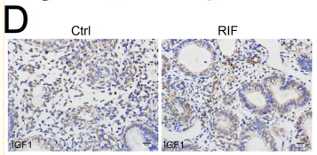
Application: IHC    Species: Human    Sample:

Fig. 7 The aberrant ratio of IGF1+SC to IGF1R+SC is observed in repeated implantation failure patients. A Cell phone heatmap showed the interaction of cells. The number indicates the number of interactions. B Heat map showing relative expression (z-score) of IGF1 and IGF1R in the total Rem_SC (Rem_SC and dRem_SC) from Endometrium and Decidua. C Heat map showing relative expression (z-score) of IGF1 and IGF1R in the stroma cells from Ctrl and RIF. D, E The expression of IGF1 and IGF1R in the endometrial stroma from controls and RIF patients at the WOI time were analyzed by immunohistochemistry (n = 3 for each group). Scale bar, 20 μm. F General diagram of decidualization process. During the time of WOI, the heterogeneity of stromal cells determines different division of labor. More specifically, IGF1+ stromal cells (Rem) start endometrial decidualization (mainly IGF1R+ stromal cells, i.e., dRem and dSec) by crosstalking with EVT and immune cells, including AREG+ NK cells; IL1B+ stromal cells (dSec) participate in the establishment and maintenance of decidua hemostasis.

5). Cardiomyocyte protective effects of thyroid hormone during hypoxia/reoxygenation injury through activating of IGF‐1‐mediated PI3K/Akt signalling. JOURNAL OF CELLULAR AND MOLECULAR MEDICINE, 2021 (PubMed: 33724692) [IF=5.3]

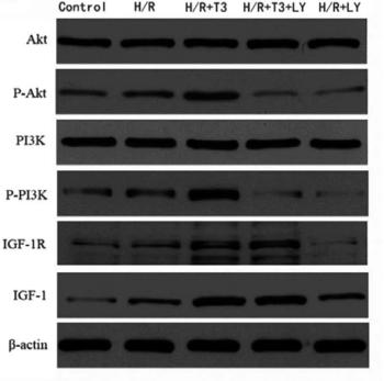
Application: WB    Species: mouse    Sample: cardiomyocytes

FIGURE 6| T3 protected cardiomyocytes from H/R-induced apoptosis through PI3K/Akt signalling pathway (A and B). A, The expression of Akt, P-Akt, PI3K, P-Akt, IGF-1, IGF-1R; β-actin protein expression was used for normalization and (B) representative bands quantified in the corresponding bar graph.

6). WISP2/IGF-1 promotes the survival of DSCs and impair the cytotoxicity of decidual NK cells. REPRODUCTION, 2021 (PubMed: 33561006) [IF=3.7]

7). Capsaicin reduces blood glucose and prevents prostate growth by regulating androgen, RAGE/IGF-1/Akt, TGF-β/Smad signalling pathway and reversing epithelial-mesenchymal transition in streptozotocin-induced diabetic mice. Naunyn-Schmiedeberg's archives of pharmacology, 2024 (PubMed: 38700794) [IF=3.1]

8). Novel_circ_003686 regulates the osteogenic differentiation of MSCs in patients with myeloma bone disease through miR-142-5p/IGF1 axis. Journal of bone oncology, 2023 (PubMed: 38021072) [IF=3.1]

Application: WB    Species: Human    Sample:

Fig. 5 Regulatory relationship between miR-142-3p and IGF1. (A) Online TargetScan (https://www.targetscan.org/vert_72/) suggests that miR-142-3p may bind to 3′ untranslated region (3′UTR) of IGF1 mRNA. (B) The binding of miR-142-3p to IGF1 was verified by dual-luciferase reporter assay. (C) IGF1 mRNA expression after miR-142-3p-mimic treatment was examined by RT-PCR analysis. (D-E) The IGF1 was silencing by si-IGF1-1, si-IGF1-2 or si-IGF1-3 in MBD-BMSC, and the protein expression was observed by WB. (F) IGF1 mRNA expression after IGF1 was silenced was examined by RT-PCR. * P < 0.05, ** P < 0.01 and *** P < 0.001.

9). OSI-906 restores the sensitivity of ovarian clear cell carcinoma to cisplatin by targeting the IGF1R/AKT pathway. MEDICAL ONCOLOGY, 2022 (PubMed: 34982265) [IF=2.8]

10). Downregulated lncRNA HOTAIR ameliorates polycystic ovaries syndrome via IGF-1 mediated PI3K/Akt pathway. Gynecological endocrinology : the official journal of the International Society of Gynecological Endocrinology, 2023 (PubMed: 37356454) [IF=2.0]

Load more

Restrictive clause


Affinity Biosciences tests all products strictly. Citations are provided as a resource for additional applications that have not been validated by Affinity Biosciences. Please choose the appropriate format for each application and consult Materials and Methods sections for additional details about the use of any product in these publications.

For Research Use Only.
Not for use in diagnostic or therapeutic procedures. Not for resale. Not for distribution without written consent. Affinity Biosciences will not be held responsible for patent infringement or other violations that may occur with the use of our products. Affinity Biosciences, Affinity Biosciences Logo and all other trademarks are the property of Affinity Biosciences LTD.