Product: Transferrin Receptor Antibody
Catalog: AF5343
Description: Rabbit polyclonal antibody to Transferrin Receptor
Application: WB IHC IF/ICC
Reactivity: Human, Mouse, Monkey
Prediction: Rat, Pig, Bovine, Horse, Sheep, Rabbit, Dog, Chicken
Mol.Wt.: 50kD(reduced),80~130kDa; 85kD(Calculated).
Uniprot: P02786
RRID: AB_2837828

View similar products>>

   Size Price Inventory
 100ul $280 In stock
 200ul $350 In stock

Lead Time: Same day delivery

For pricing and ordering contact:
Local distributors

Product Info

Source:
Rabbit
Application:
WB 1:500-1:2000, IHC 1:50-1:200, IF/ICC 1:100-1:500
*The optimal dilutions should be determined by the end user.
*Tips:

WB: For western blot detection of denatured protein samples. IHC: For immunohistochemical detection of paraffin sections (IHC-p) or frozen sections (IHC-f) of tissue samples. IF/ICC: For immunofluorescence detection of cell samples. ELISA(peptide): For ELISA detection of antigenic peptide.

Reactivity:
Human,Mouse,Monkey
Prediction:
Rat(%), Pig(100%), Bovine(100%), Horse(100%), Sheep(100%), Rabbit(100%), Dog(100%), Chicken(89%)
Clonality:
Polyclonal
Specificity:
Transferrin Receptor Antibody detects endogenous levels of total Transferrin Receptor.
RRID:
AB_2837828
Cite Format: Affinity Biosciences Cat# AF5343, RRID:AB_2837828.
Conjugate:
Unconjugated.
Purification:
The antiserum was purified by peptide affinity chromatography using SulfoLink™ Coupling Resin (Thermo Fisher Scientific).
Storage:
Rabbit IgG in phosphate buffered saline , pH 7.4, 150mM NaCl, 0.02% sodium azide and 50% glycerol. Store at -20 °C. Stable for 12 months from date of receipt.
Alias:

Fold/Unfold

CD 71; CD71; CD71 antigen; IMD46; OTTHUMP00000208523; OTTHUMP00000208524; OTTHUMP00000208525; p90; sTfR; T9; TFR 1; TfR; TfR1; TFR1_HUMAN; TFRC; TR; Transferrin receptor (p90 CD71); Transferrin receptor protein 1, serum form; Trfr;

Immunogens

Immunogen:
Uniprot:
Gene(ID):
Sequence:
MMDQARSAFSNLFGGEPLSYTRFSLARQVDGDNSHVEMKLAVDEEENADNNTKANVTKPKRCSGSICYGTIAVIVFFLIGFMIGYLGYCKGVEPKTECERLAGTESPVREEPGEDFPAARRLYWDDLKRKLSEKLDSTDFTGTIKLLNENSYVPREAGSQKDENLALYVENQFREFKLSKVWRDQHFVKIQVKDSAQNSVIIVDKNGRLVYLVENPGGYVAYSKAATVTGKLVHANFGTKKDFEDLYTPVNGSIVIVRAGKITFAEKVANAESLNAIGVLIYMDQTKFPIVNAELSFFGHAHLGTGDPYTPGFPSFNHTQFPPSRSSGLPNIPVQTISRAAAEKLFGNMEGDCPSDWKTDSTCRMVTSESKNVKLTVSNVLKEIKILNIFGVIKGFVEPDHYVVVGAQRDAWGPGAAKSGVGTALLLKLAQMFSDMVLKDGFQPSRSIIFASWSAGDFGSVGATEWLEGYLSSLHLKAFTYINLDKAVLGTSNFKVSASPLLYTLIEKTMQNVKHPVTGQFLYQDSNWASKVEKLTLDNAAFPFLAYSGIPAVSFCFCEDTDYPYLGTTMDTYKELIERIPELNKVARAAAEVAGQFVIKLTHDVELNLDYERYNSQLLSFVRDLNQYRADIKEMGLSLQWLYSARGDFFRATSRLTTDFGNAEKTDRFVMKKLNDRVMRVEYHFLSPYVSPKESPFRHVFWGSGSHTLPALLENLKLRKQNNGAFNETLFRNQLALATWTIQGAANALSGDVWDIDNEF

Predictions

Predictions:

Score>80(red) has high confidence and is suggested to be used for WB detection. *The prediction model is mainly based on the alignment of immunogen sequences, the results are for reference only, not as the basis of quality assurance.

Species
Results
Score
Pig
100
Horse
100
Bovine
100
Sheep
100
Dog
100
Rabbit
100
Chicken
89
Xenopus
0
Zebrafish
0
Model Confidence:
High(score>80) Medium(80>score>50) Low(score<50) No confidence

PTMs - P02786 As Substrate

Site PTM Type Enzyme
S7 Phosphorylation
S10 Phosphorylation
S19 Phosphorylation
Y20 Phosphorylation P12931 (SRC)
T21 Phosphorylation
S24 Phosphorylation P17252 (PRKCA)
S34 Phosphorylation
K39 Ubiquitination
T52 Phosphorylation
K53 Acetylation
K53 Ubiquitination
K58 Ubiquitination
K60 Ubiquitination
T104 O-Glycosylation
S106 Phosphorylation
Y123 Phosphorylation
K128 Acetylation
K128 Ubiquitination
K134 Ubiquitination
K145 Ubiquitination
K161 Ubiquitination
Y168 Phosphorylation
K177 Ubiquitination
K189 Ubiquitination
K193 Ubiquitination
S195 Phosphorylation
S199 Phosphorylation
K205 Ubiquitination
Y219 Phosphorylation
K224 Ubiquitination
K231 Ubiquitination
K240 Ubiquitination
K241 Ubiquitination
N251 N-Glycosylation
K261 Ubiquitination
Y282 Phosphorylation
T286 Phosphorylation
N317 N-Glycosylation
S326 Phosphorylation
S327 Phosphorylation
S338 Phosphorylation
K344 Ubiquitination
K358 Ubiquitination
K371 Ubiquitination
K374 Ubiquitination
K382 Ubiquitination
K385 Ubiquitination
K394 Acetylation
Y402 Phosphorylation
K418 Acetylation
K418 Ubiquitination
S419 Phosphorylation
Y481 Phosphorylation
K486 Ubiquitination
S499 Phosphorylation
Y503 Phosphorylation
K508 Ubiquitination
K514 Ubiquitination
Y523 Phosphorylation
K531 Ubiquitination
T572 Phosphorylation
Y573 Phosphorylation
K585 Ubiquitination
Y611 Phosphorylation
S616 Phosphorylation
S620 Phosphorylation
T658 Phosphorylation
K665 Ubiquitination
Y683 Phosphorylation
S687 Phosphorylation
S691 Phosphorylation
K693 Ubiquitination
K717 Ubiquitination
K720 Ubiquitination

Research Backgrounds

Function:

Cellular uptake of iron occurs via receptor-mediated endocytosis of ligand-occupied transferrin receptor into specialized endosomes. Endosomal acidification leads to iron release. The apotransferrin-receptor complex is then recycled to the cell surface with a return to neutral pH and the concomitant loss of affinity of apotransferrin for its receptor. Transferrin receptor is necessary for development of erythrocytes and the nervous system (By similarity). A second ligand, the heditary hemochromatosis protein HFE, competes for binding with transferrin for an overlapping C-terminal binding site. Positively regulates T and B cell proliferation through iron uptake.

(Microbial infection) Acts as a receptor for new-world arenaviruses: Guanarito, Junin and Machupo virus.

PTMs:

N- and O-glycosylated, phosphorylated and palmitoylated. The serum form is only glycosylated.

Proteolytically cleaved on Arg-100 to produce the soluble serum form (sTfR).

Palmitoylated on both Cys-62 and Cys-67. Cys-62 seems to be the major site of palmitoylation.

Subcellular Location:

Cell membrane>Single-pass type II membrane protein. Melanosome.
Note: Identified by mass spectrometry in melanosome fractions from stage I to stage IV.

Secreted.

Extracellular region or secreted Cytosol Plasma membrane Cytoskeleton Lysosome Endosome Peroxisome ER Golgi apparatus Nucleus Mitochondrion Manual annotation Automatic computational assertionSubcellular location
Subunit Structure:

Homodimer; disulfide-linked. Binds one transferrin or HFE molecule per subunit. Binds the HLA class II histocompatibility antigen, DR1. Interacts with SH3BP3. Interacts with STEAP3; facilitates TFRC endocytosis in erythroid precursor cells.

(Microbial infection) Interacts with Guanarito, Junin and Machupo arenavirus glycoprotein complex.

Family&Domains:

Belongs to the peptidase M28 family. M28B subfamily.

Research Fields

· Cellular Processes > Transport and catabolism > Endocytosis.   (View pathway)

· Cellular Processes > Transport and catabolism > Phagosome.   (View pathway)

· Cellular Processes > Cell growth and death > Ferroptosis.   (View pathway)

· Environmental Information Processing > Signal transduction > HIF-1 signaling pathway.   (View pathway)

· Organismal Systems > Immune system > Hematopoietic cell lineage.   (View pathway)

References

1). A cancer-specific activatable theranostic nanodrug for enhanced therapeutic efficacy via amplification of oxidative stress. Theranostics, 2020 (PubMed: 31903126) [IF=12.4]

2). Defective ferritinophagy and imbalanced iron metabolism in PBDE-47-triggered neuronal ferroptosis and salvage by Canolol. The Science of the total environment, 2024 (PubMed: 38750757) [IF=9.8]

3). Endoplasmic reticulum stress-triggered ferroptosis via the XBP1-Hrd1-Nrf2 pathway induces EMT progression in diabetic nephropathy.. Biomedicine & Pharmacotherapy, 2023 (PubMed: 37224754) [IF=7.5]

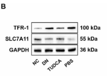
Application: WB    Species: Mouse    Sample: renal tissues

Fig. 4. The characteristic changes of ferroptosis were rescued by TUDCA treatment in DN mice. The qRT-PCR results of SLC7A11 and TFR-1 in each group (n = 3) (A). The WB results of SLC7A11 and TFR-1 among groups (n = 3) (B - C). Immunohistochemistry and quantitative analysis of SLC7A11 and TFR-1 in NC, DN, and TUDCA group (n = 3) (D - E). The content of iron (F), MDA (G), and GSH (H) in renal tissues (n = 6). Data are expressed as the mean ± SEM.

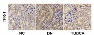
Application: IHC    Species: Mouse    Sample: renal tissues

Fig. 4. The characteristic changes of ferroptosis were rescued by TUDCA treatment in DN mice. The qRT-PCR results of SLC7A11 and TFR-1 in each group (n = 3) (A). The WB results of SLC7A11 and TFR-1 among groups (n = 3) (B - C). Immunohistochemistry and quantitative analysis of SLC7A11 and TFR-1 in NC, DN, and TUDCA group (n = 3) (D - E). The content of iron (F), MDA (G), and GSH (H) in renal tissues (n = 6). Data are expressed as the mean ± SEM.

4). Biochanin A protects against iron overload associated knee osteoarthritis via regulating iron levels and NRF2/System xc-/GPX4 axis. Biomedicine & Pharmacotherapy, 2023 (PubMed: 36379122) [IF=7.5]

5). Allogeneic platelet-rich plasma inhibits ferroptosis in promoting wound repair of type 2 diabetic ulcers. Free radical biology & medicine, 2024 (PubMed: 38408545) [IF=7.4]

6). Activation of AMPKα2 attenuated doxorubicin-induced cardiotoxicity via inhibiting lipid peroxidation associated ferroptosis. Free Radical Biology and Medicine, 2023 (PubMed: 37331642) [IF=7.4]

7). GSK-3β-dependent Nrf2 antioxidant response modulates ferroptosis of lens epithelial cells in age-related cataract. Free Radical Biology and Medicine, 2023 (PubMed: 37156294) [IF=7.4]

8). Alleviated NCOA4-mediated ferritinophagy protected RA FLSs from ferroptosis in lipopolysaccharide-induced inflammation under hypoxia. Inflammation research : official journal of the European Histamine Research Society ... [et al.], 2024 (PubMed: 38189810) [IF=6.7]

9). Fine particulate matter potentiates Th17-cell pathogenicity in experimental autoimmune uveitis via ferroptosis. ECOTOXICOLOGY AND ENVIRONMENTAL SAFETY, 2024 [IF=6.2]

Application: WB    Species: Mouse    Sample:

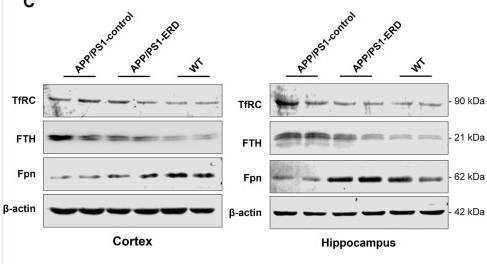
Fig. 5. PM2.5 induced iron overload and lipid peroxidation in EAU. (A) Representative fluorescence images (left) and fluorescence intensity quantification (right) of FerroOrange indicating the intracellular Fe2+ content of CD4+ T cells in EUA mice with or without PM2.5 exposure (n=6/ group; unpaired student’s t-test; scale bar: 25μm). (B) Representative flow cytometry images (left) and quantification (right) of ROS level in CD4+ T cells of EUA mice with or without PM2.5 exposure (n=5/ group; unpaired student’s t-test). (C) Representative flow cytometry images (left) and quantification (right) of oxidized and non-oxidized lipid production of CD4+ T cells in EUA mice with or without PM2.5 exposure (n=5/ group; unpaired student’s t-test). (D) The concentration of MDA in serum of EAU mice with or without PM2.5 exposure (n=3/ group; unpaired student’s t-test). (E) The concentration of MDA in CD4+ T cells of EAU mice with or without PM2.5 exposure (n=5/ group; unpaired student’s t-test). (F) GSH/GSSH ratio in CD4+ T cells of EUA mice with or without PM2.5 exposure (n=5/ group; unpaired student’s t-test). (G) Representative Western blotting images of TFRC, FTH1, FTL1, ACSL1, HMOX1, GPX4 and β-actin in vivo (left) and in vitro (right). (H) Quantification of relative expression of TFRC, FTH1, FTL1, ACSL1, HMOX1, GPX4 in vivo and in vitro (n=3/ group; one-way ANOVA). β-actin served as an internal control. Data represent mean ± SD; *, P < 0.05; **, P < 0.01; ***, P < 0.001.

10). Erythropoietin inhibits ferroptosis and ameliorates neurological function after spinal cord injury. Neural Regeneration Research, 2023 (PubMed: 36204858) [IF=6.1]

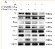
Application: WB    Species: Rat    Sample: spinal cord

Figure 3 EPO regulates the expression of ferroptotic biomarkers after SCI. (A) Western blot of the indicated proteins in injured tissues from the treatment groups. (B–H) Quantitative analysis of Tfr, Fpn, Fth, Acsl4 and 4-Hne protein expression at 14 dpi. Gapdh was used as the reference protein. (I, J) Quantitative reverse transcription polymerase chain reaction results of xCT and Gpx4 mRNA extracted from injured tissue at 7 dpi. β-Actin mRNA was used as the reference gene. (K) Quantitative analysis of reduced GSH from injured tissue at 7 dpi. All data are expressed as the mean ± SD (n = 5 in each group). *P < 0.05, **P < 0.01, ***P < 0.001 (one-way analysis of variance followed by Dunnett’s multiple comparisons test). 4-Hne: 4-Hydroxynonenal; Acsl4: acyl-coenzyme A synthetase long chain family member 4; dpi: day(s) post injury; EPO: erythropoietin; Fpn: ferroportin or solute carrier family 40 member 1; Fth: ferritin heavy chain; Gapdh: glyceraldehyde-3-phosphate dehydrogenase; Gpx4: glutathione peroxidase 4; GSH: glutathione; ns: not significant; SCI: spinal cord injury; Tfr: transferrin receptor; xCT: the solute carrier family 7 member 11.

Load more

Restrictive clause


Affinity Biosciences tests all products strictly. Citations are provided as a resource for additional applications that have not been validated by Affinity Biosciences. Please choose the appropriate format for each application and consult Materials and Methods sections for additional details about the use of any product in these publications.

For Research Use Only.
Not for use in diagnostic or therapeutic procedures. Not for resale. Not for distribution without written consent. Affinity Biosciences will not be held responsible for patent infringement or other violations that may occur with the use of our products. Affinity Biosciences, Affinity Biosciences Logo and all other trademarks are the property of Affinity Biosciences LTD.