Product: FAS Antibody
Catalog: AF5342
Description: Rabbit polyclonal antibody to FAS
Application: WB IHC IF/ICC
Cited expt.: WB, IHC, IF/ICC
Reactivity: Human, Mouse, Rat
Mol.Wt.: 37kD,50~70kD(Glycosylated); 38kD(Calculated).
Uniprot: P25445
RRID: AB_2837827

View similar products>>

   Size Price Inventory
 100ul $280 In stock
 200ul $350 In stock

Lead Time: Same day delivery

For pricing and ordering contact:
Local distributors

Product Info

Source:
Rabbit
Application:
WB 1:500-1:2000, IHC 1:50-1:200, IF/ICC 1:100-1:500
*The optimal dilutions should be determined by the end user. For optimal experimental results, antibody reuse is not recommended.
*Tips:

WB: For western blot detection of denatured protein samples. IHC: For immunohistochemical detection of paraffin sections (IHC-p) or frozen sections (IHC-f) of tissue samples. IF/ICC: For immunofluorescence detection of cell samples. ELISA(peptide): For ELISA detection of antigenic peptide.

Reactivity:
Human,Mouse,Rat
Clonality:
Polyclonal
Specificity:
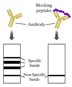
FAS Antibody detects endogenous levels of total FAS.
RRID:
AB_2837827
Cite Format: Affinity Biosciences Cat# AF5342, RRID:AB_2837827.
Conjugate:
Unconjugated.
Purification:
The antiserum was purified by peptide affinity chromatography using SulfoLink™ Coupling Resin (Thermo Fisher Scientific).
Storage:
Rabbit IgG in phosphate buffered saline , pH 7.4, 150mM NaCl, 0.02% sodium azide and 50% glycerol. Store at -20 °C. Stable for 12 months from date of receipt.
Alias:

Fold/Unfold

ALPS 1A; ALPS1A; APO 1; Apo 1 antigen; APO 1 cell surface antigen; Apo-1 antigen; APO1; Apo1 antigen; APO1 cell surface antigen; Apoptosis antigen 1; Apoptosis mediating surface antigen FAS; Apoptosis-mediating surface antigen FAS; APT 1; APT1; CD 95; CD 95 antigen; CD95; CD95 antigen; Delta Fas; Delta Fas/APO 1/CD95; Delta Fas/APO1/CD95; Fas (TNF receptor superfamily, member 6); FAS 1; FAS 827dupA; Fas AMA; Fas; FAS Antigen; Fas cell surface death receptor; FAS1; FASLG receptor; FASTM; sFAS; Surface antigen APO1; TNF receptor superfamily, member 6; TNFRSF 6; TNFRSF6; TNR6_HUMAN; Tumor necrosis factor receptor superfamily member 6;

Immunogens

Immunogen:

A synthesized peptide derived from human FAS, corresponding to a region within C-terminal amino acids.

Uniprot:
Gene(ID):
Expression:
P25445 TNR6_HUMAN:

Isoform 1 and isoform 6 are expressed at equal levels in resting peripheral blood mononuclear cells. After activation there is an increase in isoform 1 and decrease in the levels of isoform 6.

Description:
Receptor for TNFSF6/FASLG. The adapter molecule FADD recruits caspase-8 to the activated receptor. The resulting death-inducing signaling complex (DISC) performs caspase-8 proteolytic activation which initiates the subsequent cascade of caspases (aspartate-specific cysteine proteases) mediating apoptosis.
Sequence:
MLGIWTLLPLVLTSVARLSSKSVNAQVTDINSKGLELRKTVTTVETQNLEGLHHDGQFCHKPCPPGERKARDCTVNGDEPDCVPCQEGKEYTDKAHFSSKCRRCRLCDEGHGLEVEINCTRTQNTKCRCKPNFFCNSTVCEHCDPCTKCEHGIIKECTLTSNTKCKEEGSRSNLGWLCLLLLPIPLIVWVKRKEVQKTCRKHRKENQGSHESPTLNPETVAINLSDVDLSKYITTIAGVMTLSQVKGFVRKNGVNEAKIDEIKNDNVQDTAEQKVQLLRNWHQLHGKKEAYDTLIKDLKKANLCTLAEKIQTIILKDITSDSENSNFRNEIQSLV

Research Backgrounds

Function:

Receptor for TNFSF6/FASLG. The adapter molecule FADD recruits caspase-8 to the activated receptor. The resulting death-inducing signaling complex (DISC) performs caspase-8 proteolytic activation which initiates the subsequent cascade of caspases (aspartate-specific cysteine proteases) mediating apoptosis. FAS-mediated apoptosis may have a role in the induction of peripheral tolerance, in the antigen-stimulated suicide of mature T-cells, or both. The secreted isoforms 2 to 6 block apoptosis (in vitro).

PTMs:

N- and O-glycosylated. O-glycosylated with core 1 or possibly core 8 glycans.

Subcellular Location:

Cell membrane>Single-pass type I membrane protein.

Secreted.

Secreted.

Secreted.

Secreted.

Secreted.

Extracellular region or secreted Cytosol Plasma membrane Cytoskeleton Lysosome Endosome Peroxisome ER Golgi apparatus Nucleus Mitochondrion Manual annotation Automatic computational assertionSubcellular location
Tissue Specificity:

Isoform 1 and isoform 6 are expressed at equal levels in resting peripheral blood mononuclear cells. After activation there is an increase in isoform 1 and decrease in the levels of isoform 6.

Family&Domains:

Contains a death domain involved in the binding of FADD, and maybe to other cytosolic adapter proteins.

Research Fields

· Cellular Processes > Cell growth and death > p53 signaling pathway.   (View pathway)

· Cellular Processes > Cell growth and death > Apoptosis.   (View pathway)

· Cellular Processes > Cell growth and death > Necroptosis.   (View pathway)

· Environmental Information Processing > Signal transduction > MAPK signaling pathway.   (View pathway)

· Environmental Information Processing > Signaling molecules and interaction > Cytokine-cytokine receptor interaction.   (View pathway)

· Environmental Information Processing > Signal transduction > TNF signaling pathway.   (View pathway)

· Human Diseases > Drug resistance: Antineoplastic > Platinum drug resistance.

· Human Diseases > Endocrine and metabolic diseases > Non-alcoholic fatty liver disease (NAFLD).

· Human Diseases > Endocrine and metabolic diseases > Type I diabetes mellitus.

· Human Diseases > Neurodegenerative diseases > Alzheimer's disease.

· Human Diseases > Infectious diseases: Parasitic > Chagas disease (American trypanosomiasis).

· Human Diseases > Infectious diseases: Parasitic > African trypanosomiasis.

· Human Diseases > Infectious diseases: Viral > Hepatitis B.

· Human Diseases > Infectious diseases: Viral > Measles.

· Human Diseases > Infectious diseases: Viral > Influenza A.

· Human Diseases > Infectious diseases: Viral > Human papillomavirus infection.

· Human Diseases > Infectious diseases: Viral > Herpes simplex infection.

· Human Diseases > Cancers: Overview > Pathways in cancer.   (View pathway)

· Human Diseases > Cancers: Overview > Proteoglycans in cancer.

· Human Diseases > Immune diseases > Autoimmune thyroid disease.

· Human Diseases > Immune diseases > Allograft rejection.

· Human Diseases > Immune diseases > Graft-versus-host disease.

· Organismal Systems > Immune system > Natural killer cell mediated cytotoxicity.   (View pathway)

References

1). Truncated thrombospondin-1 polypeptide alleviates non-alcoholic fatty liver disease induced by palmitic acid and high-fat diets. International journal of biological macromolecules, 2025 (PubMed: 41109376) [IF=7.7]

2). Sepia pharaonis ink derived melanin ameliorated high-fat diet induced lipid dysmetabolism: Insights from microbiome and hepatic transcription. Food research international (Ottawa, Ont.), 2026 (PubMed: 41539751) [IF=7.0]

3). LINC317.5 as a novel biomarker for hypertriglyceridemia in normal glucose metabolism. Cell death discovery, 2024 (PubMed: 38670967) [IF=7.0]

Application: WB    Species: human    Sample: HepG2-IR cell

Fig. 3: Knockdown of LINC317.5 regulates the lipid metabolism-related gene, transcription factor, and protein expressions in the HepG2-IR model. A The TG level of si-lncRNA and si-NC in the HepG2-IR model. B The effect of knockdown of LINC317.5 on the activity of HepG2-IR models with CCK-8. C The effect of knockdown of LINC317.5 on apoptosis of HepG2-IR model with flow cytometry (a) si-NC; (b) si-lncRNA; (c) Relative apoptosis rate. D The TKFC relative expression after knockdown of LINC317.5 in the HepG2-IR model; (a) for gene expression by qRT-PCR and (b) for protein expression by western blot. E The effect of knockdown of LINC317.5 on transcription factors of HepG2-IR model with qRT-PCR. The housekeeping gene which was used to establish the relative expression of the analyzed genes was β-actin. F The lipid metabolism-related protein expression of knockdown of LINC317.5 in the HepG2-IR (ACADM, CPT1A, FAS, ACC1). Western blot experiments have been repeated three times (The relative expression was calculated based on the target gene expression levels in the si-NC group. The relative proliferation was calculated based on the proliferation level in the si-NC group. The relative apoptotic rate was calculated based on the apoptotic rate in the si-NC group. *P 

4). Nontargeted metabolomics combining with intestinal microbiota revealed the potential mechanisms of DHA/EPA - acetylated astaxanthin esters in regulating the abnormal lipid metabolism. Food research international (Ottawa, Ont.), 2025 (PubMed: 41606935) [IF=7.0]

Application: WB    Species: Mouse    Sample:
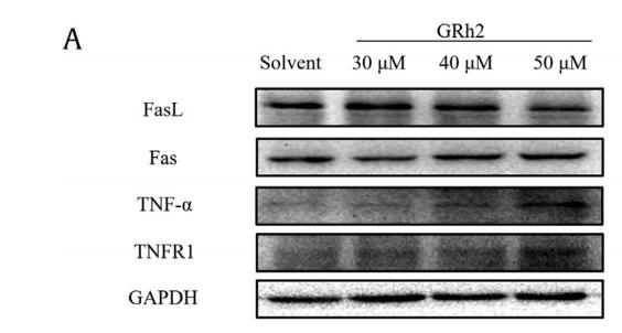
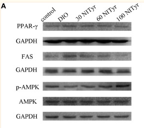
Fig. 3. DA and EA supplementation ameliorated β-oxidation and fatty acid production in HFD mice. (A) Key protein expressions of β-oxidation in the liver. (Bsingle bondC) Quantitative analysis of the CPT1A and PPAR-α. (D) Key protein expressions of fatty acid production in the liver. (E-F) Quantitative analysis of the SREBP-1 and FAS.

5). 20 (S)-Ginsenoside Rh2 induces caspase-dependent PML-RARA degradation in NB4 cells via Akt/Bax/caspase9 and TNF-α/caspase8 signaling cascades. Journal of Ginseng Research, 2021 (PubMed: 33841010) [IF=6.8]

Application: WB    Species: Human    Sample: NB4 cells
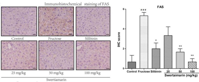
Fig. 7. GRh2 activated the TNF-a/caspase8 cascade. (A) NB4 cells were incubated with 30 mM, 40 mM, or 50 mM GRh2 for 12 h. Protein expression levels of FasL, Fas, TNF-a, and TNFR1 in NB4 cells were detected via Western blot. A representative picture of three replicates is shown. (B) Quantitative statistical graph of the relative protein expression levels. The results are shown as the mean  SD (n ¼ 3) *p < 0.05, **p < 0.01. (C) RT-PCR was used to detect the mRNA expression level of TNF-a in NB4 cells after GRh2 administration. The results are shown as the mean  SD (n ¼ 3) **p < 0.01, ***p < 0.001 versus solvent. After preincubation with 1.5 mM TNF-a inhibitor, C 87, for 2 h, 30 mM, 40 mM, or 50 mM GRh2 was applied for another 12 h. (D) CCK-8 assay measured the NB4 cell viability. The results are shown as the mean  SD (n ¼ 6) ***p < 0.001, ###p < 0.001. (E) Hoechst 33258 staining was used to observe changes in nuclear morphology of NB4 cells after GRh2 and C 87 administration (800  ). (F) Quantitative statistical graph of caspase8 cleavage activation levels in NB4 cells. The Ac-IETD-pNA method was used. The results are shown as the mean  SD (n ¼ 3) ***p < 0.001, #p < 0.05. GRh2, 20(S)-ginsenoside Rh2.

6). Amelioration of nonalcoholic fatty liver disease by swertiamarin in fructose-fed mice. Phytomedicine, 2019 (PubMed: 31005808) [IF=6.7]

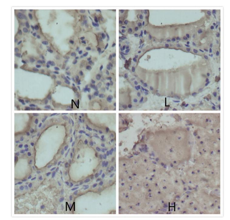
Application: IHC    Species: mouse    Sample: liver
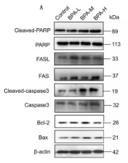
Figure.7. |Effect of swertiamarin (25, 50 and 100 mg/kg) and silibinin on (A) ACC1 expression by western blotting analysis. Immunohistochemical stainings of (B) SREBP-1 and (C) FAS in liver tissue of mice. Scale bar represents 30 μm. Representative analysis from six animals in each group.Significance is represented as ##P ≤ 0.01 and ###P ≤ 0.001 compared to the normal control group,*P ≤ 0.05 and **P ≤ 0.01 compared to the fructose group.

7). Effects of Bisphenol A on reproductive toxicity and gut microbiota dysbiosis in male rats. Ecotoxicology and Environmental Safety, 2022 (PubMed: 35567931) [IF=6.2]

Application: WB    Species: Rat    Sample: testes tissue

Fig. 4. Effects of BPA exposure on cell apoptosis in testes. (A) Western blotting photographs and relative quantitative analysis of (B) Cleaved-PARP and PARP protein, (C) FASL, (D) FAS, (E) Cleaved-caspase3, (F) Caspase3, (G) Bcl-2, (H) Bax, and (I) Bcl-2/Bax. Data are expressed as mean ± SD (n = 4). Different letters indicate significant differences between groups (P 

8). Propionate Ameliorates Alcohol-Induced Liver Injury in Mice via the Gut–Liver Axis: Focus on the Improvement of Intestinal Permeability. JOURNAL OF AGRICULTURAL AND FOOD CHEMISTRY, 2022 (PubMed: 35549256) [IF=5.7]

9). Patchouli alcohol ameliorates acute liver injury via inhibiting oxidative stress and gut-origin LPS leakage in rats. International Immunopharmacology, 2021 (PubMed: 34182243) [IF=4.8]

10). Buyang huanwu decoction alleviates stroke-induced immunosuppression in MCAO mice by reducing splenic T cell apoptosis triggered by AIM2 inflammasome. Journal of ethnopharmacology, 2024 (PubMed: 38906338) [IF=4.8]

Load more

Restrictive clause


Affinity Biosciences tests all products strictly. Citations are provided as a resource for additional applications that have not been validated by Affinity Biosciences. Please choose the appropriate format for each application and consult Materials and Methods sections for additional details about the use of any product in these publications.

For Research Use Only.
Not for use in diagnostic or therapeutic procedures. Not for resale. Not for distribution without written consent. Affinity Biosciences will not be held responsible for patent infringement or other violations that may occur with the use of our products. Affinity Biosciences, Affinity Biosciences Logo and all other trademarks are the property of Affinity Biosciences LTD.