Product: MyD88 Antibody
Catalog: AF5195
Source: Rabbit
Application: WB, IHC, IF/ICC
Reactivity: Human, Mouse, Rat, Monkey
Prediction: Pig, Zebrafish, Bovine, Horse, Sheep, Rabbit, Dog, Chicken, Xenopus
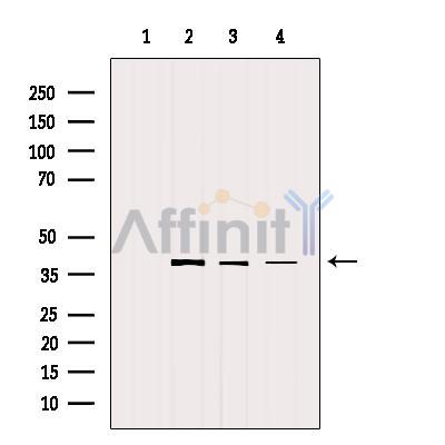
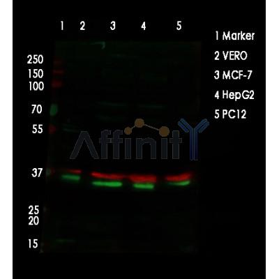
Mol.Wt.: 33 kDa; 33kD(Calculated).
Uniprot: Q99836
RRID: AB_2837681

View similar products>>

   Size Price Inventory
 100ul $280 In stock
 200ul $350 In stock

Lead Time: Same day delivery

For pricing and ordering contact:
Local distributors

Product Info

Source:
Rabbit
Application:
WB 1:500-1:2000, IHC 1:50-1:200, IF/ICC 1:100-1:500
*The optimal dilutions should be determined by the end user.
*Tips:

WB: For western blot detection of denatured protein samples. IHC: For immunohistochemical detection of paraffin sections (IHC-p) or frozen sections (IHC-f) of tissue samples. IF/ICC: For immunofluorescence detection of cell samples. ELISA(peptide): For ELISA detection of antigenic peptide.

Reactivity:
Human,Mouse,Rat,Monkey
Prediction:
Pig(100%), Zebrafish(90%), Bovine(100%), Horse(100%), Sheep(100%), Rabbit(90%), Dog(100%), Chicken(80%), Xenopus(80%)
Clonality:
Polyclonal
Specificity:
MyD88 Antibody detects endogenous levels of total MyD88.
RRID:
AB_2837681
Cite Format: Affinity Biosciences Cat# AF5195, RRID:AB_2837681.
Conjugate:
Unconjugated.
Purification:
The antiserum was purified by peptide affinity chromatography using SulfoLink™ Coupling Resin (Thermo Fisher Scientific).
Storage:
Rabbit IgG in phosphate buffered saline , pH 7.4, 150mM NaCl, 0.02% sodium azide and 50% glycerol. Store at -20 °C. Stable for 12 months from date of receipt.
Alias:

Fold/Unfold

Mutant myeloid differentiation primary response 88; MYD 88; Myd88; MYD88_HUMAN; MYD88D; Myeloid differentiation marker 88; Myeloid differentiation primary response 88; Myeloid differentiation primary response gene (88); Myeloid differentiation primary response gene 88; Myeloid differentiation primary response gene; Myeloid differentiation primary response protein MyD88; OTTHUMP00000161718; OTTHUMP00000208595; OTTHUMP00000209058; OTTHUMP00000209059; OTTHUMP00000209060;

Immunogens

Immunogen:
Uniprot:
Gene(ID):
Expression:
Q99836 MYD88_HUMAN:

Ubiquitous.

Description:
Adapter protein involved in the Toll-like receptor and IL-1 receptor signaling pathway in the innate immune response. Acts via IRAK1, IRAK2, IRF7 and TRAF6, leading to NF-kappa-B activation, cytokine secretion and the inflammatory response. Increases IL-8 transcription. Involved in IL-18-mediated signaling pathway.
Sequence:
MAAGGPGAGSAAPVSSTSSLPLAALNMRVRRRLSLFLNVRTQVAADWTALAEEMDFEYLEIRQLETQADPTGRLLDAWQGRPGASVGRLLELLTKLGRDDVLLELGPSIEEDCQKYILKQQQEEAEKPLQVAAVDSSVPRTAELAGITTLDDPLGHMPERFDAFICYCPSDIQFVQEMIRQLEQTNYRLKLCVSDRDVLPGTCVWSIASELIEKRCRRMVVVVSDDYLQSKECDFQTKFALSLSPGAHQKRLIPIKYKAMKKEFPSILRFITVCDYTNPCTKSWFWTRLAKALSLP

Predictions

Predictions:

Score>80(red) has high confidence and is suggested to be used for WB detection. *The prediction model is mainly based on the alignment of immunogen sequences, the results are for reference only, not as the basis of quality assurance.

Species
Results
Score
Pig
100
Horse
100
Bovine
100
Sheep
100
Dog
100
Zebrafish
90
Rabbit
90
Xenopus
80
Chicken
80
Model Confidence:
High(score>80) Medium(80>score>50) Low(score<50) No confidence

PTMs - Q99836 As Substrate

Site PTM Type Enzyme
K95 Ubiquitination
C113 S-Nitrosylation
K115 Ubiquitination
K119 Ubiquitination
K127 Ubiquitination
K190 Ubiquitination
K214 Ubiquitination
C216 S-Nitrosylation
K231 Ubiquitination
K238 Ubiquitination
S244 Phosphorylation
K250 Ubiquitination
K256 Ubiquitination
Y257 Phosphorylation
K262 Ubiquitination
Y276 Phosphorylation
K282 Ubiquitination
K291 Ubiquitination

Research Backgrounds

Function:

Adapter protein involved in the Toll-like receptor and IL-1 receptor signaling pathway in the innate immune response. Acts via IRAK1, IRAK2, IRF7 and TRAF6, leading to NF-kappa-B activation, cytokine secretion and the inflammatory response. Increases IL-8 transcription. Involved in IL-18-mediated signaling pathway. Activates IRF1 resulting in its rapid migration into the nucleus to mediate an efficient induction of IFN-beta, NOS2/INOS, and IL12A genes. MyD88-mediated signaling in intestinal epithelial cells is crucial for maintenance of gut homeostasis and controls the expression of the antimicrobial lectin REG3G in the small intestine (By similarity).

PTMs:

Ubiquitinated; undergoes 'Lys-63'-linked polyubiquitination. OTUD4 specifically hydrolyzes 'Lys-63'-linked polyubiquitinated MYD88.

Subcellular Location:

Cytoplasm. Nucleus.

Extracellular region or secreted Cytosol Plasma membrane Cytoskeleton Lysosome Endosome Peroxisome ER Golgi apparatus Nucleus Mitochondrion Manual annotation Automatic computational assertionSubcellular location
Tissue Specificity:

Ubiquitous.

Subunit Structure:

Homodimer. Also forms heterodimers with TIRAP. Binds to TLR2, TLR4, TLR5, IRAK1, IRAK2 and IRAK4 via their respective TIR domains. Interacts with IL18R1. Interacts with BMX, IL1RL1, IKBKE and IRF7. Interacts with LRRFIP1 and LRRFIP2; this interaction positively regulates Toll-like receptor (TLR) signaling in response to agonist. Interacts with FLII. LRRFIP1 and LRRFIP2 compete with FLII for MYD88-binding. Interacts with IRF1. Upon IL1B treatment, forms a complex with PELI1, IRAK1, IRAK4 and TRAF6; this complex recruits MAP3K7/TAK1, TAB1 and TAB2 to mediate NF-kappa-B activation. Direct binding of SMAD6 to PELI1 prevents the complex formation and hence negatively regulates IL1R-TLR signaling and eventually NF-kappa-B-mediated gene expression. May interact with PIK3AP1. Interacts (via TIR domain) with DHX9 (via H2A and OB-fold regions); this interaction is direct. Interacts with OTUD4 deubiquitinase; the interaction is direct.

(Microbial infection) In case of infection, interacts with uropathogenic E.coli protein TcpC; suppressing Toll-like receptor (TLR)-mediated cytokine production.

(Microbial infection) In case of infection, interacts with uropathogenic E.faecalis protein TcpF; suppressing Toll-like receptor (TLR)-mediated cytokine production.

Family&Domains:

The intermediate domain (ID) is required for the phosphorylation and activation of IRAK.

Research Fields

· Environmental Information Processing > Signal transduction > MAPK signaling pathway.   (View pathway)

· Environmental Information Processing > Signal transduction > NF-kappa B signaling pathway.   (View pathway)

· Human Diseases > Infectious diseases: Bacterial > Salmonella infection.

· Human Diseases > Infectious diseases: Bacterial > Pertussis.

· Human Diseases > Infectious diseases: Bacterial > Legionellosis.

· Human Diseases > Infectious diseases: Parasitic > Leishmaniasis.

· Human Diseases > Infectious diseases: Parasitic > Chagas disease (American trypanosomiasis).

· Human Diseases > Infectious diseases: Parasitic > African trypanosomiasis.

· Human Diseases > Infectious diseases: Parasitic > Malaria.

· Human Diseases > Infectious diseases: Parasitic > Toxoplasmosis.

· Human Diseases > Infectious diseases: Bacterial > Tuberculosis.

· Human Diseases > Infectious diseases: Viral > Hepatitis B.

· Human Diseases > Infectious diseases: Viral > Measles.

· Human Diseases > Infectious diseases: Viral > Influenza A.

· Human Diseases > Infectious diseases: Viral > Herpes simplex infection.

· Organismal Systems > Immune system > Toll-like receptor signaling pathway.   (View pathway)

· Organismal Systems > Immune system > NOD-like receptor signaling pathway.   (View pathway)

References

1). Li C et al. Oxyberberine, a novel gut microbiota-mediated metabolite of berberine, possesses superior anti-colitis effect: Impact on intestinal epithelial barrier, gut microbiota profile and TLR4-MyD88-NF-κB pathway. Pharmacol Res 2020 Feb;152:104603 (PubMed: 31863867) [IF=10.334]

Application: WB    Species: Mice    Sample: colonic tissues

Fig. 6. Effect of OBB on the activation of TLR4-MyD88-NF-κB signaling pathway in DSS-induced colonic tissues. (A) Representative Western blotting images of TLR4, MyD88, cytoplasmic p65, nuclear p65, p-IκBα and IκBα. Changes in the relative protein expression levels of TLR4 (B), MyD88 (C), nuclear p65 (D), cytoplasmic p65 (E), and p-IκBα/IκBα ratio (F) were measured. Data are shown as the mean ± SEM (n = 3). # P < 0.05, ## P < 0.01 vs. Control group, * P < 0.05, ** P < 0.01 vs. DSS group.

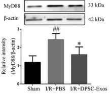
2). Li S et al. Dental pulp stem cell‐derived exosomes alleviate cerebral ischaemia‐reperfusion injury through suppressing inflammatory response. Cell Prolif 2021 Aug;54(8):e13093. (PubMed: 34231932) [IF=8.755]

Application: WB    Species: Mice    Sample:

FIGURE 3 Effect of DPSC‐Exos on the expression of TLR4, MyD88, NF‐κB p65 and HMGB1 on day 7 after cerebral I/R damage. (A) The relative expression level of TLR4. (B) The relative expression level of MyD88. (C) The relative expression level of NF‐κB p65. (D) The relative expression level of nuclear HMGB1. (E) The relative expression level of cytoplasmic HMGB1. Protein samples were acquired from the ischaemic cortex and assayed by western blot. Nuclear proteins were normalized to the intensity of Histone H3, and cytoplasmic and total proteins were normalized to the intensity of GAPDH or β‐actin. Data were expressed as means ± SD (n = 3). ## P < .01 versus the sham group; *P < .05 and **P < .01 versus the I/R + PBS group. DPSC‐Exos, dental pulp stem cell‐derived exosomes; HMGB1, high‐mobility group box 1 protein; I/R, ischaemia/reperfusion; MyD88, myeloid differentiation protein 88; NF‐κB, nuclear factor‐kappa B; PBS, phosphate‐buffered saline; TLR4, toll‐like receptor‐4

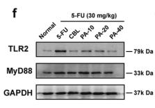
3). Wu J et al. Patchouli alcohol attenuates 5-fluorouracil-induced intestinal mucositis via TLR2/MyD88/NF-kB pathway and regulation of microbiota. Biomed Pharmacother 2020 Jan 28;124:109883 (PubMed: 32004938) [IF=7.419]

Application: WB    Species: rat    Sample:

Fig. 3.| Effect of PA on inflammtory cytokines (n = 8) and TLR2/MyD88/NF-κB pathway proteins (n = 3). (a–e) Levels of TNF-α, IL-1β, IL-6, IL-10, and MPO; (f–h)Expressions of TLR2 and MyD88 proteins

4). Zhan X et al. Polysaccharides from Garlic Protect against Liver Injury in DSS-Induced Inflammatory Bowel Disease of Mice via Suppressing Pyroptosis and Oxidative Damage. Oxid Med Cell Longev 2022 Aug 16;2022:2042163. (PubMed: 36017235) [IF=7.310]

Application: WB    Species: Mice    Sample: liver tissues

Figure 5 PSG suppressed SLI via inhibiting the LPS/TLR4/MyD88/NF-κB pathway in DSS-induced IBD mice. (a) Histological analysis for liver tissues by H&E staining. Protein expressions of the TLR4/MyD88/NF-κB pathway in the liver tissues were determined by Western blot assay (b) and quantitative analysis for Western blot results (n = 3) (c). Effects of PSG on LPS (d) and IL-10 (e) in the liver tissues of IBD mice. Results were expressed as mean ± SEM. ∗p < 0.05 and ∗∗p < 0.01 vs. the control group; #p < 0.05 and ##p < 0.01 vs. the model group (DSS only).

5). Bai C et al. Chronic intermittent hypoxia induces the pyroptosis of renal tubular epithelial cells by activating the NLRP3 inflammasome. Bioengineered 2022 Mar;13(3):7528-7540. (PubMed: 35263214) [IF=6.832]

Application: WB    Species: Rat    Sample: renal tissues

Figure 6. The expression level of Cleaved Caspase-1, Caspase-1, GSDMD, TLR4, MyD88, p-NF-κB, NF-κB, and NLRP3 in renal tissues of rats was measured by Western blotting assay following different strategies of CIH (n = 3, *p < 0.05 vs. control, #p < 0.05 vs. Mild-CIH).

6). Yang T et al. Amelioration of nonalcoholic fatty liver disease by sodium butyrate is linked to the modulation of intestinal tight junctions in db/db mice. Food Funct 2020 Dec 1;11(12):10675-10689. (PubMed: 33216087) [IF=6.317]

Application: WB    Species: Mice    Sample: liver tissue

Fig. 5 Effects of sodium butyrate on liver histology, liver injury and inflammation in db/db mice. (A) Weight changes in mice over 16 weeks. (B) Weights of the mice at week 16. (C) Liver morphology, (D) liver weights in mice. (E) The levels of serum ALT, AST and ALP activity in mice. (F) Liver Hematoxylin–Eosin (H&E) staining. (G) The expression of F4/80 in the liver through immunohistochemistry. (H) Levels of serum LPS in mice. (I) Levels of serum IL-1β, TNF-α and IL-6 in mice. (J) Levels of liver IL-1β, TNF-α and IL-6 in mice. (K) The relative protein levels of TLR4, MyD88 and NFκB in mice. (L) The expression of TLR4, MyD88 and NF-κB in the liver through immunohistochemistry. db/db + mic: db/db mice administrated with 5 × 10⁷ CFU kg−1 C. butyricum per day for 6 weeks; db/db + NaB: db/db mice administrated with 500 mg kg−1 NaB per day for 6 weeks. Each bar represents the mean ± SEM for groups of six. *P < 0.05, **P < 0.01, compared to db/m mice as indicated; #P < 0.05, ##P < 0.01, compared to db/db mice as indicated.

Application: IHC    Species: Mice    Sample: liver tissue

Fig. 5 Effects of sodium butyrate on liver histology, liver injury and inflammation in db/db mice. (A) Weight changes in mice over 16 weeks. (B) Weights of the mice at week 16. (C) Liver morphology, (D) liver weights in mice. (E) The levels of serum ALT, AST and ALP activity in mice. (F) Liver Hematoxylin–Eosin (H&E) staining. (G) The expression of F4/80 in the liver through immunohistochemistry. (H) Levels of serum LPS in mice. (I) Levels of serum IL-1β, TNF-α and IL-6 in mice. (J) Levels of liver IL-1β, TNF-α and IL-6 in mice. (K) The relative protein levels of TLR4, MyD88 and NFκB in mice. (L) The expression of TLR4, MyD88 and NF-κB in the liver through immunohistochemistry. db/db + mic: db/db mice administrated with 5 × 10⁷ CFU kg−1 C. butyricum per day for 6 weeks; db/db + NaB: db/db mice administrated with 500 mg kg−1 NaB per day for 6 weeks. Each bar represents the mean ± SEM for groups of six. *P < 0.05, **P < 0.01, compared to db/m mice as indicated; #P < 0.05, ##P < 0.01, compared to db/db mice as indicated.

7). Morsy MA et al. Paeonol Attenuates Hepatic Ischemia/Reperfusion Injury by Modulating the Nrf2/HO-1 and TLR4/MYD88/NF-κB Signaling Pathways. Antioxidants (Basel) 2022 Aug 29;11(9):1687. (PubMed: 36139761) [IF=6.312]

Application: IHC    Species: Rat    Sample: hepatic tissues

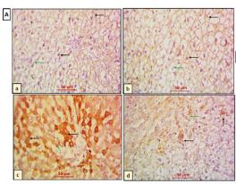
Figure 5. Effect of paeonol (PAE) on toll-like receptor 4 (TLR4) in rats’ hepatic ischemia/reperfusion (HIR) injury. (A) Photomicrographs of rat liver sections stained for detection of TLR4 (×400). (Aa,Ab) Sham and PAE + sham groups show negative expression of TLR4 in both hepatocytes (black arrows) and Kupffer cells (green arrows). (Ac) The HIR group shows hepatocytes (black arrows) and Kupffer cells (green arrow) with extensive TLR4 cytoplasmic expression. (Ad) The PAE + HIR group shows hepatocytes (black arrows) and Kupffer cells (green arrow) with minimal TLR4 cytoplasmic expression. (B) Semi-quantification of the hepatic TLR4 expression. Each value represents the mean ± SEM (n = 7). a Significant difference from the sham-operated control group and b significant difference from the HIR group, respectively, at p < 0.05.

8). Hassan HM et al. Adamantane-linked isothiourea derivatives suppress the growth of experimental hepatocellular carcinoma via inhibition of TLR4-MyD88-NF-κB signaling. Am J Cancer Res 2021 Feb 1;11(2):350-369. (PubMed: 33575076) [IF=5.942]

Application: IHC    Species: rat    Sample: hepatic tissues

Figure 9. |Effect of compounds 5 and 6 on hepatic MyD88 protein expression in TAA-administered rats. Representative microimages of immunostaining for MyD88 protein (IHC counterstained with Mayer’s hematoxylin) in hepatic tissues and statistical analysis of positive area of immunolabelling (%) are shown. Black arrows denote positive immunoexpression. Images were captured at magnification of 100× (scale bar, 100 µm) or 400× (scale bar, 50 µm). *P < 0.05 vs. control group, #P < 0.05 vs. HCC group. DOXO: doxorubicin, HCC: hepatocellular carcinoma, IHC: immunohistochemistry, MyD88: myeloid differentiation primary response-88, TAA: thioacetamide.

9). Yu P et al. Panax quinquefolius L. Saponins Protect Myocardial Ischemia Reperfusion No-Reflow Through Inhibiting the Activation of NLRP3 Inflammasome via TLR4/MyD88/NF-κB Signaling Pathway. Front Pharmacol 2021 Jan 15;11:607813. (PubMed: 33628178) [IF=5.810]

Application: WB    Species: Human    Sample:

FIGURE 6 Effects of PQS on NLRP3, ASC, procaspase-1, and caspase-1 expressions. (A) Representative examples of Western blot analysis demonstrating the expression levels of NLRP3, ASC, procaspase-1, and caspase-1; quantification of the expression levels of (B) NLRP3, (C) ASC, and (D) procaspase-1 and (E) caspase-1; (F) representative examples of Western blot analysis demonstrating the expression levels of TLR4, MyD88, p-NF-κB, and NF-κB p65; quantification of the expression levels of (G) TLR4, (H) MyD88, (I) p-NF-κB, and NF-κB p65. Data presented are the means ± SD. ##p < 0.01 compared with Sham; *p < 0.05, **p < 0.01 compared with MI/R.

10). Zhang C et al. Drug D, a Diosgenin Derive, Inhibits L-Arginine-Induced Acute Pancreatitis through Meditating GSDMD in the Endoplasmic Reticulum via the TXNIP/HIF-1α Pathway. Nutrients 2022 Jun 22;14(13):2591. (PubMed: 35807771) [IF=5.717]

Load more

Restrictive clause


Affinity Biosciences tests all products strictly. Citations are provided as a resource for additional applications that have not been validated by Affinity Biosciences. Please choose the appropriate format for each application and consult Materials and Methods sections for additional details about the use of any product in these publications.

For Research Use Only.
Not for use in diagnostic or therapeutic procedures. Not for resale. Not for distribution without written consent. Affinity Biosciences will not be held responsible for patent infringement or other violations that may occur with the use of our products. Affinity Biosciences, Affinity Biosciences Logo and all other trademarks are the property of Affinity Biosciences LTD.