PDHK1 Antibody - #DF4365
| Product: | PDHK1 Antibody |
| Catalog: | DF4365 |
| Description: | Rabbit polyclonal antibody to PDHK1 |
| Application: | WB IHC IF/ICC |
| Cited expt.: | WB |
| Reactivity: | Human, Mouse, Rat |
| Prediction: | Pig, Bovine, Horse, Sheep, Dog, Chicken |
| Mol.Wt.: | 49 KD; 49kD(Calculated). |
| Uniprot: | Q15118 |
| RRID: | AB_2836733 |
Related Downloads
Protocols
Product Info
*The optimal dilutions should be determined by the end user. For optimal experimental results, antibody reuse is not recommended.
*Tips:
WB: For western blot detection of denatured protein samples. IHC: For immunohistochemical detection of paraffin sections (IHC-p) or frozen sections (IHC-f) of tissue samples. IF/ICC: For immunofluorescence detection of cell samples. ELISA(peptide): For ELISA detection of antigenic peptide.
Cite Format: Affinity Biosciences Cat# DF4365, RRID:AB_2836733.
Fold/Unfold
[Pyruvate dehydrogenase [lipoamide]] kinase isozyme 1, mitochondrial; HGNC:8809; Mitochondrial pyruvate dehydrogenase kinase isoenzyme 1; PDH kinase 1; Pdk1; PDK1_HUMAN; Pyruvate dehydrogenase kinase isoform 1; Pyruvate dehydrogenase kinase, isoenzyme 1;
Immunogens
A synthesized peptide derived from human PDHK1, corresponding to a region within the internal amino acids.
Expressed predominantly in the heart. Detected at lower levels in liver, skeletal muscle and pancreas.
- Q15118 PDK1_HUMAN:
- Protein BLAST With
- NCBI/
- ExPASy/
- Uniprot
MRLARLLRGAALAGPGPGLRAAGFSRSFSSDSGSSPASERGVPGQVDFYARFSPSPLSMKQFLDFGSVNACEKTSFMFLRQELPVRLANIMKEISLLPDNLLRTPSVQLVQSWYIQSLQELLDFKDKSAEDAKAIYDFTDTVIRIRNRHNDVIPTMAQGVIEYKESFGVDPVTSQNVQYFLDRFYMSRISIRMLLNQHSLLFGGKGKGSPSHRKHIGSINPNCNVLEVIKDGYENARRLCDLYYINSPELELEELNAKSPGQPIQVVYVPSHLYHMVFELFKNAMRATMEHHANRGVYPPIQVHVTLGNEDLTVKMSDRGGGVPLRKIDRLFNYMYSTAPRPRVETSRAVPLAGFGYGLPISRLYAQYFQGDLKLYSLEGYGTDAVIYIKALSTDSIERLPVYNKAAWKHYNTNHEADDWCVPSREPKDMTTFRSA
Predictions
Score>80(red) has high confidence and is suggested to be used for WB detection. *The prediction model is mainly based on the alignment of immunogen sequences, the results are for reference only, not as the basis of quality assurance.
High(score>80) Medium(80>score>50) Low(score<50) No confidence
Research Backgrounds
Kinase that plays a key role in regulation of glucose and fatty acid metabolism and homeostasis via phosphorylation of the pyruvate dehydrogenase subunits PDHA1 and PDHA2. This inhibits pyruvate dehydrogenase activity, and thereby regulates metabolite flux through the tricarboxylic acid cycle, down-regulates aerobic respiration and inhibits the formation of acetyl-coenzyme A from pyruvate. Plays an important role in cellular responses to hypoxia and is important for cell proliferation under hypoxia. Protects cells against apoptosis in response to hypoxia and oxidative stress.
Phosphorylated by constitutively activated ABL1, FGFR1, FLT3 and JAK2 (in vitro), and this may also occur in cancer cells that express constitutively activated ABL1, FGFR1, FLT3 and JAK2. Phosphorylation at Tyr-243 and Tyr-244 strongly increases kinase activity, while phosphorylation at Tyr-136 has a lesser effect.
Mitochondrion matrix.
Expressed predominantly in the heart. Detected at lower levels in liver, skeletal muscle and pancreas.
Belongs to the PDK/BCKDK protein kinase family.
Research Fields
· Environmental Information Processing > Signal transduction > HIF-1 signaling pathway. (View pathway)
· Human Diseases > Cancers: Overview > Central carbon metabolism in cancer. (View pathway)
· Organismal Systems > Development > Axon guidance. (View pathway)
References
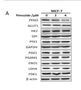
Application: WB Species: Human Sample: MCF-7 cells
Application: WB Species: Rat Sample: H9c2 cardiomyocytes
Restrictive clause
Affinity Biosciences tests all products strictly. Citations are provided as a resource for additional applications that have not been validated by Affinity Biosciences. Please choose the appropriate format for each application and consult Materials and Methods sections for additional details about the use of any product in these publications.
For Research Use Only.
Not for use in diagnostic or therapeutic procedures. Not for resale. Not for distribution without written consent. Affinity Biosciences will not be held responsible for patent infringement or other violations that may occur with the use of our products. Affinity Biosciences, Affinity Biosciences Logo and all other trademarks are the property of Affinity Biosciences LTD.


