

, using Caspase 3 Antibody at 1/1000 dilution.")


 and mouse anti-beta tubulin Ab(T0023 1:200) for 1 hour at 37°C. An AlexaFluor594 conjugated goat anti-rabbit IgG(H+L) Ab(Red) and an AlexaFluor488 conjugated goat anti-mouse IgG(H+L) Ab(Green) were used as the secondary antibody.
The nuclear counter stain is DAPI(blue).")
 Blots showing proteins in cells treated with AGO14699 and (B) quantification. (C) Blots showing proteins in cells treated with (C) BSI‑201 and (D) quantification. * P<0.05, compared with the control group; ∆P<0.05, compared with the low dose group; ∆∆P<0.05, compared with the middle dose group. CTRL, control; Bcl‑2, B‑cell lymphoma 2; BAX, Bcl‑2‑associated X protein.")


 BCL2L10 induced apoptosis in HepG2 and Huh7 cells, as determined by flow cytometry analysis following Annexin V and PI staining. The upper panel represents FACS images of HCC cells transfected with empty vector or BCL2L10, while the lower panel shows the quantitative analyses of early apoptotic and late apoptotic cells. The experiment was repeated three times in triplicate. Data are mean ± SD. (B) Protein expression of the active forms of apoptosis related genes caspase 3, caspase 8, and caspase 9 was evaluated by Western blot.")
, caspase 9, cleaved caspase 9 (phosphorylated form), Bcl-2, Bax, and β-actin was determined using Western blotting.")
 C. sakazakii-induced programmed cell death in HT-29 was ameliorated by pretreatment with ZY-312. Flow cytometry (A) and fluorescence microscope (B) were used to examine the results of staining of FITC and PI. (C) Western blot analysis shows that ZY-312 suppressed C. sakazakii-induced NEC by modulating apoptosis through caspase-3, Bax, and Bcl-2. -Actin was used as an indicator of protein loading.")
. b CircGCNL1 with/
without miR-330-3p mimics was transfected into co-cultured synoviocytes. After 2 days of co-culture, proteins were extracted from chondrocytes in
the lower chamber, and WB was performed to detect the levels of MMP13, MMP3, COL2A1, ADAMTS4, TNF-α, p65, Bcl-2, Bax, caspase-3, and cleaved
caspase-3. N = 4 (four independent experiments). Data are presented as mean ± S.D. One-way ANOVA with Bonferroni test was performed. WB
western blotting, NC normal control, MMP matrix metalloproteinase, TNF tumor necrosis factor, Bcl-2 B-cell lymphoma-2, Bax BCL2-associated X,
COL2A1 collagen type II alpha 1.")
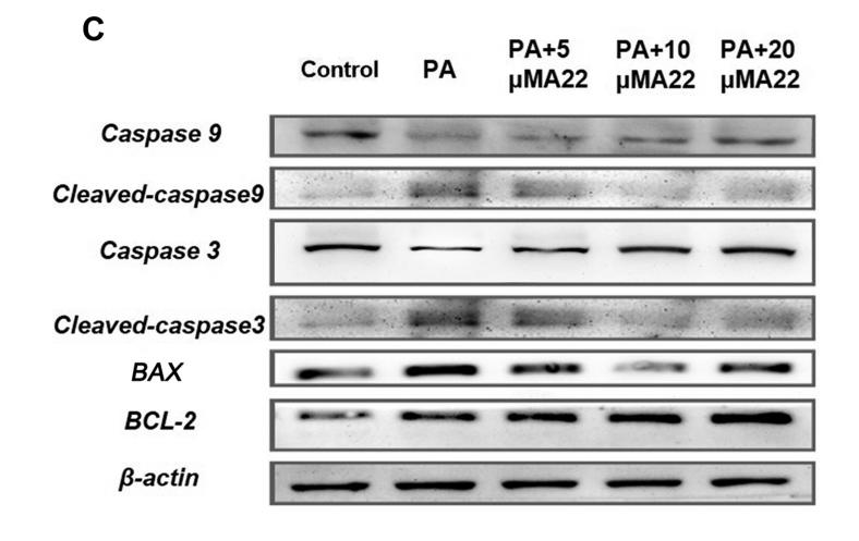
 M22 upregulated the expression of proapoptotic genes APAF1, CASP9, CASP3, BAX, and depress antiapoptotic genes BCL2 for 48 h. RNA was isolated and analysis was performed to detect by qRT-PCR. *Means P < 0.01, ***means P < 0.0001. (B) Western blot analysis showed cleavage of caspases-9 and −3 in M19 treated A549 cells afer 48 h of exposure. Equal loading was confrmed by reprobing the membrane with GAPDH. Te bands shown here are from a representative experiment repeated three times with similar results.")
 rats. (A) Western blot was used to detect the protein levels in the brain
tissues. The full-length gels are presented in Supplementary Fig. S2. The protein expression was
evaluated by proteomic analysis. (B) Changes in the retinol-binding protein 4 (RBP4); (C) Changes in
serum albumin. *p < 0.05 and ***p < 0.001 vs. I/R.")
 and (B): The relative luciferase activities were analyzed in 293T cells upon co-transfection of miR-153-5p mimic and luciferase reporter psiCHECK2/circPAN3-mut153, or co-transfection of miR-183-5p mimic and psiCHECK2/circPAN3-mut183.")
 Flow cytometry determined the rate of apoptosis in rat NP cells transfected with agomir-145-CTR or agomir-145 in the presence of H2O2 for 48 h, with untransfected cells serving as the controls. (C, D) Western blotting (C) and subsequent densitometric analysis (D) showed the protein expression of Bcl-2, Bax,caspase-3 and activated caspase-3 (A-caspase 3); GAPDH was used as a loading control. The data are presented as the mean ± SD. *P < 0.05 vs. the control group.")
 Representative photomicrographs of TUNEL staining and quantitative analyses in the indicated groups. n = 4 per group. Scale bar = 100 μm. (C)
Typical photomicrographs showing double immunofluorescence staining of PGC1α (green) and NeuN (red) in diverse experimental groups. n = 4 per group. Scale
bar = 50 μm. (D) Western blot images and quantitative analyses of p-AMPK/AMPK, PGC1α, Drp1, Nrf1, TFAM, UCP2, Cleaved caspase-3/Caspase-3, Bcl2, Bax,
Cytosolic and mitochondrial cytochrome c. n = 6 per group. Bars represent mean ± SD. **P < 0.01, *P < 0.05 vs. Sham group. ##P < 0.01, #P < 0.05 vs. SAH +
Vehicle group. &&P < 0.01, &P < 0.05 vs. SAH + hsp22+scramble siRNA. (For interpretation of the references to colour in this figure legend, the reader is referred to
the Web version of this article.)")
 inhibited cell proliferation and induced apoptosis in tumor tissues. (A) Ki-67 staining of tumor sections obtained from mice treated with (a) vehicle control or (b) QX. Quantification of Ki-67-positive cells in the tumor sections (c). (B)TUNEL staining of tumor sections obtained from mice treated with (a) vehicle control or (b) QX. Quantification of TUNEL-positive cells in the tumor sections (c). (C) Western blotting of proapoptotic proteins Bim, FasL, and cleaved caspase-3 expression in tumor tissues of QX-treated mice or vehicle control-treated mice. Data are presented as mean ± SD. *P < .05, **P < .01 versus vehicle control.")
 Nude mice tumorigenesis assay. Representative images of xenograft tumors were presented. (B) The
growth curves of tumor volumes were plotted, and the tumor weight of each mouse was measured. (C) Representative images of HE staining from control and DHA
treatment group (200×). (D) Representative IHC images of Ki-67, CASP3, BCL2L1, P53, p-KDR and p-STAT1 in control and DHA treatment groups. *P < 0.05, **P <
0.01, ***P < 0.001.")
 qRT-PCR analysis of the effects of DHA on the expression
levels of BCL2, BAX, CKD4, p53/BCL2L1/Caspase-3/-7 and STAT1/KDR/MMP9 in GC cells. (B) WB analysis of the effects of DHA on the expression of apoptosis and
cycle-related proteins including cleaved-caspase 3, caspase 3, caspase 7, BCL2L1, p27 and p53. (C) WB analysis of the effects of DHA on the expression of p-STAT1,
STAT1, p-KDR, KDR and MMP9.")
 THP-1/ADM and K562/ADM cells were pre-incubated with 5 mM 3-MA for 24 h before exposure to 2 μg/mL of ADM for additional 24 h. Apoptosis-related proteins were detected and quantified by Western blot. All data are presented as means ± SD of three independent experiments. *P < 0.05, **P < 0.01, and ***P < 0.001, compared to parental cell line (THP-1 or K562), negative control, or newly diagnosed patients. ADM: doxorubicin; GFP, green fluorescent protein; mRFP, monomeric red fluorescent protein; OD: optical density.")
.")
 Representative Western blot results for Caspase3 and Cleaved-Caspase3 in the testes of rats from all four groups.")
. Data are presented as mean ± S.D. One-way ANOVA with Bonferroni test was performed. WB western blotting, NC normal control, MMP matrix metalloproteinase, TNF tumor necrosis factor, Bcl-2 B-cell lymphoma-2, Bax BCL2-associated X,COL2A1 collagen type II alpha 1.")
 The expression of death receptor 4 (DR4), death receptor 5 (DR5), cysteinyl aspartate specific proteinase 8(caspase 8) and cysteinyl aspartate specific proteinase 3 (caspase 3) induced by Amuc_1434* in LS174T cells was dependent on tumor necrosis factor-related apoptosis-inducing ligand (TRAIL). (a) LS174T cells were treated with 8 and 64 µg/mL Amuc_1434* for 24 h, respectively. The cell lysates were analyzed by Western blot.")

 reduced the expressions of Cleaved caspase 3 and BAX/BCL‐2, and increased the expressions of Nrf2 and its downstream antioxidative genes, compared with H2O2 treatment. The relative gene expressions were normalized to GAPDH. Data are presented as mean ± SEM of three independent experiments. Different letters indicate significant differences (P < .05)")

 Western blot analysis of samples from the experimental groups were tested with the indicated antibodies.")
, Bax (red) in all of the groups' liver tissues. DAPI (blue) was used to stain the nucleus.B, Western blot analysis for the expression of Bax and Bcl‐2. C, Immunohistochemistry staining of caspase‐3 in liver tissues.")
 could overcome the resistance of CAL-62 cells.E Western blotting of active caspase-3 and caspase-3 after treatment with a combination of 80 nM PP and 150 μM ART for 48 h. Gels/blots are cropped for a clear presentation of results. Protein samples derived from the same experiment and gels/blots were processed in parallel. Black line delineates the boundary between gels. Ratio, the ratio of integrated density(treatment and untreated cells); N, groups that were not treated with ART; ART, groups treated with 150 μM ART; PP + ART, groups treated with a combination of 80 nM PP and 150 μM ART. Blue,apoptosis; red, G1/G0; yellow, G2/M; slash, S")
 Mitochondrial membrane potential (Δψm) were detected by TMRM and
JC-1 staining. Bar, 20 μm. (B and C) The red fluorescence intensity of TMRM and JC-1 was quantified, n = 6. (D) Immunoblot analysis of cytochrome C (Cyto C),
BAX, Bcl-2, cleavage of caspase 3 (CI-Casp3), and cleavage of caspase 9 (CI-Casp9) from lysates of PASMCs treated with various concentrations of apigenin, n = 4. (E)
Quantitation data of (D), n = 4. All data are mean ± SEM. The P values were evaluated by ANOVA followed by a post hoc Tukey test. (*P < 0.05, **P < 0.01).
(For interpretation of the references to colour in this figure legend, the reader is referred to the Web version of this article.)")
 The western blotting revealed that compared with the control group, caspase-3 was markedly upregulated in the
TGR5 knockdown group, and was suppressed in the TGR5 overexpression group. Glyceraldehyde phosphate dehydrogenase (GAPDH) was used as a normalizing
control. *P < 0.05 and **P < 0.01, when compared with the control group. (B) The annexin V/PI double staining revealed a higher level of apoptosis when TGR5
was knocked down by siRNA. *P < 0.05 and **P < 0.01, when compared with the control group. Glyceraldehyde phosphate dehydrogenase (GAPDH) was used as
a normalizing control.")
 The expressions of NLRP3, ASC, caspase-1, IL-18, and IL-1β. Carnosine-L: carnosine (100 mg kg−1 d−1
); carnosine-H: carnosine (200 mg kg−1 d−1
). Data represent mean ± SEM (n = 10 per group). #p < 0.05, ##p < 0.01, ###p <
0.001 vs. SAMR1db m−1
; *p < 0.05, **p < 0.01, ***p < 0.001 vs. SAMP8.")

 DRI of Cal-62, 8505C, and 8305C cell lines treated with GSK-J4 and doxorubicin (B) The effect of GSK-J4 combined with doxorubicin on the sphere-forming ability (C) of the Cal-62 cell line. Scale bar, 100 μM. YO-PRO-1/7-AAD staining show the effect of GSK-J4 combined with doxorubicin on cell apoptosis of the Cal-62 cell line (D), Western blot showed the caspase 3 and Pro-caspase 3 with the treatment of effect of GSK-J4 combined with doxorubicin (E) (DOX: GSK-J4 = 0.156:0.078 μM). DRI, Dose reduction index. n.s., no statistical difference. *, p < 0.05, **, p < 0.01, ***, p < 0.001.")
 IF and ISH combination assay revealing the co-localization of APAF1 protein with included ncRNAs in HCT116 cells. Scale bars, 5 μm. (B and C) The levels of APAF1 mRNA (B) and protein (C) were determined after interfering LNC473 expression in HCT116 and SW480 cells by qPCR and IF assays. Scale bars, 20 μm. (D) The expression of apoptosis-related proteins including APAF1 was detected after interfering LNC473 expression in HCT116 and SW480 cells by western blot assay. (E and F) Rescue experiments showing the APAF1 levels in HCT116 and SW480 cells with exposure to the co-transfection of pcDH-LNC473 vector and miR574-5p or miR15b-5p mimic by qPCR (E) and western blot (F) assays. (G) Pattern diagram of APAF1-CDS and APAF1-IRES-CDS vectors. (H and I) APAF1 protein expression was determined in HCT116 and SW480 cells treated with APAF1-IRES-CDS vector, or pcDH-LNC473 and APAF1-IRES-CD co-transfection by western blot (H) and IF assays (I). Scale bars, 5 μm. (J) The percentage (%) of cell apoptosis in cells upon co-overexpressing APAF1-CDS or APAF1-IRE-CDS and LNC473 as assayed by flow cytometry. All tests were performed at least three times. Data were expressed as mean ± SD. ns (nonsignificant), p > 0.05; ∗p < 0.05, ∗∗p < 0.01, ∗∗∗p < 0.001, ∗∗∗∗p < 0.0001.")
, negative control (NC), overexpression (OE), and knockdown (KD) groups. B-C: The colony-forming assay showed the colony-forming ability of HTR8 cells in the four groups. D-E: The scratch wound healing assay showed that the closed wound area of HTR8 cells in the four groups. F and I: Transwell assay that the migration and invasion number of HTR8 cells in the four groups. J: Western blot assay determined the expression of epithelial-mesenchymal transition (EMT) markers in the four groups. K-L: Flow cytometry showed that the percentage of apoptosis cells in the four groups. M: Western blot assay determined the expression of apoptosis-related proteins in the four groups. All experiments were conducted with three biological replicates. Data were expressed as mean ± SD. * P < 0.05 compared to NC group.")
 The interference efficiency of DUOX1 was detected by Q-PCR (A) and western blot (B). (C, D) MDA (C) and SOD (D) levels were detected by biochemical detection. (E, F) ROS level (E) and apoptosis (F) were detected by flow cytometry. (G) Protein expression of NF-κB and cleaved caspase-3 was determined by western blot. * P<0.05, ** P<0.01, *** P<0.001 versus siNC or control; # P<0.05, ## P<0.01, ### P<0.001 versus 5% O2+siNC. siNC: Negative control of DUOX1 interference; siDUOX1: DUOX1 interference; Control: rat neuron cells cultured under normoxic conditions. DUOX1 – dual oxidase 1; Q-PCR – quantitative polymerase chain reaction; MDA – malondialdehyde; SOD – superoxide dismutase; ROS – reactive oxygen species.")
. The
expression of apoptotic proteins in the liver was confirmed by immunofluorescence (IF)
staining. (A) Representative images indicating the expression of caspase3 (green) are
shown. (B) Representative images indicating the expression of cleaved caspase3 (red)
are shown. Magnification, 400×; scale bar represents 10 µm;4’,6-diamidino-2-phenylindole (DAPI): blue.")
 The target genes of miR-147a were predicted in databases (TargetScan, miRWalk, miRDB, StarBase) and there were 141 genes which overlapped in the Venn diagram. (B) PPI network analyses was performed by use of the String database, and results with a combined score of > 0.4 were selected. Among these, CDK6, AKT3, AKT2, PDPK1 were found to have had a high degree of connectivity and were selected as hub genes for miR-147a. (C) CDK6, AKT3, AKT2 expression was significantly reduced after overexpression of miR-147a, especially for CDK6. (D) There were multiple binding sites between miR-147a and CDK6. (E) The binding relationship between miR-147a and CDK6 was confirmed by dual-luciferase reporter assays. (F) miR-147a mimics markedly reduced expression of CDK6, whereas miR-147a inhibitors significantly enhanced CDK6 levels in HO8910 and A2780 cells. (G) Expression of CDK6 mRNA decreased after knockdown of circ_0072995, but was rescued by miR-147a inhibitors as determined by qRT-PCR. (H) The expression of CDK6 proteins decreased after knockdown of circ_0072995, but was rescued by miR-147a inhibitors as determined by WB.")
 and Hoechst 33,342 staining (b).
The mRNA (c) and protein (d) levels of Bax, Bcl-2, and caspase 3. Data are expressed as mean ± S.E.M. and are representative
of three independent experiments. *P\0.05 vs. control.
**P \0.01 vs. control. #
P \0.05 vs. H2O2. ##P \0.01 vs.
H2O2")
 Western blot analysis showing the protein levels of Bcl‑2, Bax, cleaved caspase‑3, caspase‑3, p‑c‑Met and c‑Met in different groups.")
 RT-qPCR quantification of COL2, COL10,
SOX9, CASP3 and Bcl-2 in the calluses of shamoperated groups and osteotomized condylar
necks of control (CON) and conditional knockout mice (CKO) on POD14. Data reflect
mean ± SEM. (b) Hematoxylin and eosin staining of control and cko groups on POD14. Lower
magnification, scale bars =200 μm; higher
magnification, scale bars =100 μm. (c, d) The
area of higher magnification in (b) shows
TUNEL and immunofluorescence staining of
COL2, COL10, Ki67, and caspase-3. Scale bars
=100 μm. ns ≥ 0.05 (no significance),
**p < 0.01, ***p < 0.001, ****p < 0.0001.")
 Western blot analysis showing caspase-3/8 expression in AT2 cells. The overexpression of SOX11 inhibited, whilst the knockdown of SOX11 increased caspase-3/8 expression. Furthermore, FAK antagonism blocked the effect of SOX11 overexpression on caspase-3/8 expression. Data are presented as the means ± standard error of the mean. *P<0.05, **P<0.01. AT2, alveolar type II; SOX, Sex-determining gene on the Y chromosome related high mobility group box; FAK, focal adhesion kinase.")
 between samples of different treatments are marked with different letters on each column.")
 Histological results of the testis in rats treated
with Cdcl2, melotonin (Mel, SIRT3 activator), 3-MA (autophagy inhibitor targeting PI3K), 3-TYP (SIRT3 inhibitor). Scale bar, 100 mm in the upper line, and 25 mm in the lower line. (B)
Activities of testicular marker enzymes LDH, ACP, AKP, g-GT. (C) A representative immunoblot of SIRT3, autophagy markers (LC3B, BECN1), apoptosis markers (Bcl-2, Caspase3,
Cleaved-caspase3). (D) Quantification analysis of SIRT3. (E) Quantification analysis of LC3B-I, LC3B-II, LC3B-II/LC3B-I, BECN1. (F) Quantification analysis of Bcl-2, Caspase3, Cleavedcaspase3, Cleaved-Caspase3/Caspase3). *p < 0.05; ns shows p > 0.05, indicating no significant difference.")

 Representative blots of pro‑caspase‑9 and pro‑caspase‑3. Semiquantitative analysis of (B) pro‑caspase‑9 and (C) pro‑caspase‑3. Data are presented as the mean ± standard deviation. n=3. ##P<0.01 vs. Sham. **P<0.01 vs. MI/R. MI/R, myocardial ischemia/reperfusion; IOE, Inonotus obliquus extract.")
 A549 cells were treated with gefitinib 40 μmol/L and cinobufotalin 0.5 mg/mL for 48 h either alone or in combination. Apoptotic rate of A549 cells was evaluated by annexin-V/PI staining and flow cytometry. (b) The percentage of apoptotic cells in the treatment groups was quantified. (c) TUNEL staining of A549 cells. A549 cells were treated with gefitinib 40 μmol/L and cinobufotalin 0.5 mg/mL either alone or in combination and subsequently stained with TUNEL (magnification ×200). Green, apoptotic cells; blue, nucleus. (d) Western blot analysis of cell apoptosis-related proteins in A549 cells treated with gefitinib, cinobufotalin, or cinobufotalin + gefitinib. ∗P < 0.05, ∗∗P < 0.01, ∗∗∗P < 0.001, and ∗∗∗∗P < 0.0001 vs. the control group; ####P < 0.0001 vs. the gefitinib group.")
 Representative western blot bands. (B–G) The relative protein expressions of ALDH-2 (F3,16 = 9.839, P = 0.001), 4HNE (F3,16 = 6.776, P = 0.004), Caspase-3 (F3,16 = 0.199, P = 0.895), cleaved caspase-3 (F3,16 = 5.436, P = 0.009), Bax (F3,16 = 6.609, P = 0.004), and Bcl-2 (F3,16 = 6.705, P = 0.004) are expressed as the optical density ratio of a target protein to β-actin. *P < 0.05, **P < 0.01.")
 Cell apoptosis in different transfection groups was assessed using a TUNEL assay. Magnification, x100. (B) Western blot analysis showing the protein levels of Bcl-2, Bax, cleaved caspase-3, caspase-3, p-c-Met and c-Met in different groups. ***P<0.001 vs. shRNA-NC; #P<0.05 and ###P<0.001 vs. shRNA-B3GALT5-AS1-1 + ov-NC. B3GALT5-AS1, β-1,3-galactosyltransferase 5-AS1; CSNK2A1, casein kinase 2 a1; p-, phosphorylated; Ov, expression; shRNA, short hairpin RNA; NC, negative control.")

 Protein expression of caspase-3, Bax and Bcl-2 in MLE-12 cells.")
 SLC25A21 promoted the efflux of α-KG from the mitochondria to the cytosol. (B) The levels of α-KG in the mitochondria affected succinate production. (C) The upregulation of SLC25A21 promoted ROS accumulation in BCa cells. (D) The upregulation of SLC25A21 decreased Δψm in both BCa cell lines. (E) Western blot assays showed that SLC25A21 induced cyto C transfer from the mitochondria to the cytosol and increased the activation of caspase-9 and caspase-3 in BCa cells. (F) Immunohistochemistry showed that SLC25A21 increased the activation of caspase-9 and caspase-3 in BCa xenograft tissues. (G) Schematic diagram showing the mechanism of action of SLC25A21 on cell apoptosis in BCa. The results were reproducible in three independent experiments. *P < 0.05, **P < 0.01, ***P < 0.001 and ****P < 0.0001.")
 Representative blots of pro-caspase-9 and pro-caspase-3. Semiquantitative analysis of (B) pro-caspase-9 and (C) pro-caspase-3. Data are presented as the mean ± standard deviation. n=3. ##P<0.01 vs. Sham. **P<0.01 vs. MI/R. MI/R, myocardial ischemia/reperfusion; IOE, Inonotus obliquus extract.")
. (A) The expression of caspase-3, caspase-9, and Bcl-2 in the hippocampus of each group; (B) The relative expression of caspase-9 in the hippocampus of each group; (C) The relative expression of caspase-3 in the hippocampus of each group; (D) The relative expression of Bcl-2 in the hippocampus of each group. **p<0.01 vs sham, #p<0.05 vs VaD, ##p<0.05 vs VaD, Δp<0.05 vs CH-I-H, ^p<0.05 vs CBL.")
 Detection of miR-301 expression level by qRT-PCR. (B) Detection of breast cancer cell proliferation by CCK-8. (C) Distribution chart of breast cancer cell cycle. (D) Detection of migration ability of breast cancer cells by Transwell. (E) The relative number of invasive breast cancer cells, as measured by Transwell assay. (F) Apoptosis rate of breast cancer cells. (G) Detection of mRNA expression level of proliferation-related factor MCM2, migration- and invasion-related factors MMP2, and apoptosis-related factors caspase-3 by qRT-PCR. (H) Detection of protein expression level of proliferation-related factor MCM2, migration- and invasion-related factors MMP2, and apoptosis-related factors caspase-3 by western blot. The continuous variables were expressed by mean ± standard deviation. ∗p < 0.05 compared with the NC group. Paired sample t test, independent sample t test, or one-way ANOVA was selected to test the inter-group differences of continuous variables. The difference of data of each group at different times was analyzed by two-way ANOVA. We repeated the experiment three times in order to obtain accurate and stable results.")
 The protein expressions of ATM, p53, Bcl-2, Bax,TNF-α, TNFR I, Caspase-8, and Caspase-3 were detected in the testis of control group and SiNPs group.")


 and the PI3K/AKT/mTOR/NF-κB (b) signaling pathway of protein levels in tumor tissues. Data was expressed as mean ± SD. Compared with the model group, ★P < 0.05, ★★P < 0.01.")
 in CD4+ T cells. Protein expression were determined by western blotting and showed as the relative expression values of COX-IV. COX-IV was used as a loading control to normalize protein levels. Data are shown as Mean ± SD (n = 3). Statistically significant differences were determined by one-way analysis of variance (ANOVA) followed by Dunn’s/Tukey’s test. *P < 0.05, **P < 0.01, ****P < 0.0001")
. Comparison of PI3K/AKT relative intensity (b), Bax relative expression (c) Bcl-2 relative expression (d), and Caspase-3 relative intensity (e) among groups. ***P < 0.001, and **P < 0.01.")
. *p < 0.05; **p < 0.01. ADSCs: adipose tissue-derived stem cells; L-ADSCs: lipopolysaccharide-preconditioned adipose tissue-derived stem cells")
 Cell viability of ETH (200 mm) and ETH + ROF groups after treatment for 24 h. The apoptotic rate (C), Bax expression (D), and Bcl-2 expression (E) in LO2 cells. The protein levels of caspase 3 and cleaved caspase 3 (F). Mean ± SEM (n = 3–6). *p < 0.05, **p < 0.01 and ***p < 0.001 vs. the ETH group. ###p < 0.001 vs. the CON group.")
 patterns of mTOR pathway proteins, including mTOR, P-mTOR, downstream effectors p70s6k, p-p70s6k, 4EBP, and P-4EBP; (b) ERS-associated proteins, including GRP78 and CHOP; (c) apoptosis-associated proteins, including caspase-3, Bax, and Bcl-2. The protein expression was detected by immunoblotting. Data are mean ± SD. n = 4 biologically independent experiments (one-way ANOVA Tukey's post hoc test). ∗P < 0.05, ∗∗P < 0.01, and ∗∗∗P < 0.001.")
 TUNEL staining for the detection of cell apoptosis in OC tumor tissues (magnification: ×200). (b) Protein expression levels of Bax, Bcl-2, cleaved caspase 3, and caspase 3 in tumor tissues were detected by Western blot assay, and GAPDH was used as the internal control. (c) Serum levels of IL-6, TNF-α, and CA125 were detected by ELISA. (d) Flow cytometry was used to detect the CD3+CD4+CD3+/CD8+ T lymphocyte percentages. The data are presented as mean ± SD. ▲▲p < 0.01, compared to the NC group; ∗p < 0.05, ∗∗p < 0.01, compared to the vehicle group. OC: ovarian cancer.")
 Representative image of TUNEL assay and quantification of the apoptosis index after GLP treatment (×400). (B) Representative images of the caspase-3 and caspase-9 proteins assessed by immunohistochemistry (×400). @@P<0.01 vs. NC; **P<0.01 vs. MC. The results were presented as the mean ± standard deviation. n=6. NC, normal control; MC, model control; GLP-L, GLP low dose; GLP-H, GLP high dose; GLP, Ganoderma lucidum polysaccharide; TUNEL, terminal deoxynucleotidyl transferase dUTP nick-end labeling; DAPI, 4',6-diamidino-2-phenylindole; DMED, diabetes mellitus-induced erectile dysfunction.")
 WB analysis of the antiapoptotic and caspase proteins in HepG2 cells treated with C20/C22 at 2 μM for 0, 6, 12, and 24 h. The reported values correspond to the mean ± SD for three independent experiments. (D) Immunofluorescence assays employed to determine the expression of caspase3/7 in cells with indicated treatments. Scale bars, 10 µm. (E,F) Apoptotic cells detected by flow cytometry after indicated treatments (n = 3). The p-value was analyzed by one-way ANOVA followed by Tukey’s test using GraphPad Prism version 8.00. ** p < 0.01, *** p < 0.001, and **** p < 0.0001 vs. 0 h/control group.")
 WB analysis of the antiapoptotic and caspase proteins in HepG2 cells treated with C20/C22 at 2 μM for 0, 6, 12, and 24 h. The reported values correspond to the mean ± SD for three independent experiments. (D) Immunofluorescence assays employed to determine the expression of caspase3/7 in cells with indicated treatments. Scale bars, 10 µm. (E,F) Apoptotic cells detected by flow cytometry after indicated treatments (n = 3). The p-value was analyzed by one-way ANOVA followed by Tukey’s test using GraphPad Prism version 8.00. ** p < 0.01, *** p < 0.001, and **** p < 0.0001 vs. 0 h/control group.")
. Bcl-2 (B), Bax (C), Bax/Bcl-2 (D), caspase-3 (E), and cleaved-caspase-3 (F) levels were calculated by grayscale analysis. Values are displayed as mean ± SEM (n=3). Significant differences are shown as *P < 0.05, **P < 0.01 in comparison to the Con group. #P < 0.05, ##P < 0.01 in comparison to the Iso group.")
 and those cultured in regular media for the same course (Ctrl) were analyzed. (A) CCK-8 assay was applied to assess cell viability for 6 days. (B) The migration of cells was studied and quantified by transwell assay. (C) Cytotoxicity assay was used to examine apoptosis after 3 days of growth factor deprivation stress (Q3). (D) Western blot analysis showed altered expression of apoptotic markers in cells subjected to 5 days of growth factor deprivation (Q5). (E) Flow cytometry analysis was performed to study cell death in response to 48 h treatment of 5-FU (6 μM). (F) Protein levels of E-cadherin, N-cadherin, and vimentin were determined by Western blot analysis. Data were representatives of at least three independent experiments, shown as mean ± SD. Significance threshold determined by one-way ANOVA or Student's t-test:")
 cell growth and colony formation and promotes apoptosis in vitro. A and B. The CCK-8 assay showed that circ-ITCH inhibited the proliferation of C4-2 and DU145 cells. The data are the means ± S.D. of triplicate experiments (*P < 0.05, **P < 0.01, Student's t test). C and D. Additionally, colony formation assays showed that circ-ITCH inhibited colony formation in C4-2 and DU145 cells (*P < 0.05, **P < 0.01, ***P < 0.001, Student's t test). E and F. Cell apoptosis assays showed that circ-ITCH overexpression increased the rate of apoptosis in C4-2 and DU145 cells. The data are the mean ± SEM of three experiments (*P < 0.05, **P < 0.01, ***P < 0.001, Student's t test). G. Western blot showing the protein expression of the proliferation-associated genes PCNA and KI67 and the apoptosis-associated genes Caspase-3 and Caspase-9. The results are consistent with the qRT–PCR data.")
 Relative expression levels of p-NF-κB p65. (b) Relative expression levels of p-IκBα. (c) Relative expression levels of caspase-3. (NO, normoxia group; NA, normoxia + artesunate group; HO, hyperoxia group; HA, hyperoxia + artesunate group. P value was calculated by one-way ANOVA and SNK test.")
 Flow cytometry analysis of neutrophil apoptosis after BHB treatment. Neutrophils were treated with 0, 0.6, 2, and 3 mM BHB for 10 h, respectively. Annexin V-fluorescein isothiocyanate (FITC)/propidium iodide staining was used to identify apoptosis cells. Annexin V+ cells were regarded as total apoptotic neutrophils. The numbers in the top-right and bottom-right quadrants indicate the percentages of late and early apoptotic cells, respectively. (B) Quantification of the total apoptotic rate of neutrophils in A. One-way ANOVA with subsequent Bonferroni correction was used to analyze the data. Linear increase (P < 0.0001) and quadratic effect (P < 0.0001) for the apoptosis rate were found, due to the increased dose of BHB. Data expressed as LSM ± SEM. (C) Flow cytometry analysis of the effects of different duration times of BHB on neutrophil apoptosis. Neutrophils were treated with 2 mM BHB multiple times (0, 2, 4, 6, 8, and 10 h). Stimulation with 2 mM BHB for 8 h was selected as ideal conditions for subsequent experiments. (D) Quantification of the total apoptotic rate of neutrophils in C. One-way ANOVA with subsequent Bonferroni correction was used to analyze the data. Linear increase (P < 0.0001) and quadratic effect (P < 0.0001) were found for the apoptosis rate with increased incubation time. Data expressed as LSM ± SEM. (E) Protein abundance of cleaved caspase 3 and caspase 3. The 3 wells of each group represent 3 samples from different replicates, respectively. (F) Quantification of protein abundance in E. Data were analyzed with independent-samples t-tests. Data expressed as mean ± SEM. (G) mRNA abundance of CASP3. Data were analyzed with independent-samples t-tests. Data expressed as mean ± SEM. For western blotting experiments, β-actin was used to normalize protein abundance; for quantitative reverse-transcription PCR experiments, ACTB and YWHAZ were used to normalize mRNA abundance. Statistical significance was declared at P < 0.05, and P < 0.01 was declared to be highly significant.")
 Confocal microscopy analysis of α-tubulin polymerization and F-actin cytoskeletal remodeling in CRC cells with CX43 overexpression and knock-down. (c) The young’s modulus of CX43 overexpressed DLD1 cells was measured by atomic mechanics microscope. Mechanical curve (left) and quantitative analysis (right) are displayed. (d) Western blot analyses expression of stem cell characteristic related proteins in CRC cells with CX43 overexpression. (e) The tumor cell spheroidizing ability of CX43 overexpression cells was detected by tumor sphere formation assays. (f) Western blot was used to survey the relative expression of apoptosis pathway related proteins in CX43 overexpression cells. The downregulation of CX43 could rescue the above phenotypes (d, e and f).")
 Photographs of the proteins in each group. (B–E,G–I) Relative protein expression of Cyto-c, Caspase8, Caspase3, p-STAT3, Bcl2, Bax, and Bax/Bcl2.")
 on apoptosis of Beas-2B cells induced by LPS. A Western blot analysis for cleaved caspase-3, Bcl-2, and Bax. B–D Densitometric analysis for cleaved caspase-3, Bcl-2, and Bax. n = 3, ###p")
 regulates the expressions of the Sirt1-AMPK pathway-, insulin PI3K/AKT pathway- and apoptosis-related proteins. Protein expression levels were normalized to the levels of β-actin. Data are presented as the mean ± SD; n = 3; ### p < 0.001 compared to the normal control group; * p < 0.05, ** p < 0.01, and *** p < 0.001 compared to the T2DM group.")
 Representative images of nissl’s staining in hippocampus, including CA1, CA3 and DG regions. (B-D) Number of neurons in CA1, CA3 and DG regions in each group. (E-G) Relative mRNA expressions of Bcl-2, Bax, and Caspase-3. Data were presented as means ± SD, statistical significance was denoted as ###P")


 Western blotting for Bax, Bcl-3, and Caspase-3 in mammary gland tissues. (B) Relative protein levels of Bax. (C) Relative protein levels of Bcl-2. (D) Bax/Bcl-2 ratio. (E) Relative protein levels of Caspase-3. P < 0.05 indicates a significant difference (n = 15).")
 Immune Score predicted by SangerBox (http://vip.sangerbox.com/login.html). (B) T cell infiltration predicted by SangerBox (http://vip.sangerbox.com/login.html). (C) Immune cell infiltration predicted by TIMER2.0 (http://timer.comp-genomics.org/timer/). (D) Anti-PD1 prognosis in TJP3 low and high expression groups. (E) Anti-PD1 response differences between TJP3 low and high expression groups. (F) Si-RNA down-regulates TJP3 expression, and the corresponding down-stream targets expression features. (G) Statistical analysis, normalized by GAPDH. (H) Recombinant plasmid up-regulation TJP3 expression, and the corresponding down-stream targets expression features. (I) Statistical analysis, normalized by GAPDH.")
 Cell apoptosis was evaluated using (A) flow cytometry and (B) TUNEL staining assays. C) IF staining assay was performed to assess caspase 3 expression in cells. **p")
 TUNEL detection of neuronal apoptosis. TUNEL-positive, green; nuclei, blue. Scale bar, 100 μm; (B–G) Immunoblot detection of caspase-3, cleaved caspase-3, Bax, and Bcl-2 in HT-22 cells or hippocampal tissue samples. β-actin was employed for normalizing data quantitated by densitometry with ImageJ. Data are mean ± SEM.")
 Representative images of TUNEL staining (×100) in corpus cavernosum of the three groups. (B) Representative immunoblot of Bad, Bax, Bcl-2, Caspase-3, and C-Caspase-3 in corpus cavernosum of the three groups. (C) Apoptosis index of cavernosum in three groups of rats. (D) The ratio of protein level of Bax/Bcl-2. (E) The semi-quantification of Bad, Bax, Bcl-2, Caspase-3, and C-Caspase-3 in corpus cavernosum of the three groups. Data were presented as mean±standard deviation.*p")
 The effect of SEC on the apoptosis of BC cells (Hoechst 33,258 staining, 200×, 400 × ). (B) The effect of SEC on the apoptosis of BC cells (Flow cytometry). (C) The effect of SEC on the protein levels of apoptosis-related molecules of BC cells (Western blot). Data are shown as mean ± SD from three independent experiments.")
 IHC assays were used to detect the altered expression of p-C/EBPβ. (C–D)The protein expression of C/EBPβ in LV-sh-C/EBPβ-infected C28/I2 cells was detected by western blotting assays. (E–J) Western blotting assays were used to detect the protein expression of Aggrecan, ADAMTS5, C/EBPβ, p-C/EBPβ, caspase3, and cleaved-caspase3 in LV-sh-C/EBPβ-infected C28/I2 cells treated with IL-1β. (K–L) The apoptosis rate was determined by a flow cytometer.")
 bright field examination, α-SMA expression, and Phalloidin staining. Scale bar: 200 and 100 μm. (b, n = 6) VSMC viability was evaluated in different doses of HLJDD using the cell counting kit-8. The effect of HLJDD on foam cell formation was tested in (c) ORO, (d) Nile Red, (e) Annexin V/PI staining, and (f) ROS generation. n = 3, 3, 5, and 5, respectively. (g) Phalloidin and Giemsa staining on VSMCs and A7r5 cell line, respectively. Scale bar: 100 μm. Foam cell formation was also evaluated via (h) Western blot for (i, n = 4) α-SMA, (j, n = 5) SM22-α, (k, n = 4) TNF-α, (l, n = 3) IL6, (m, n = 8) OPN, (n, n = 7) CD36, and (o, n = 5) Caspase 3. Data were presented as mean ± standard deviation in one-way ANOVA with Dunnett post hoc tests. ns: not significant")
 Apoptotic cells were detected by TUNEL staining. (B–F) Representative images of Caspase-3 activity, Cleaved-caspase 3, Cleaved-caspase 9, and Bax and Bcl-2 expression were examined by western blot and the fold activation data analysis. Data are expressed as mean ± SD (at least n = 6/group), #p < 0.05(vs. Sham group), *p < 0.05 (vs. CLP group).")

 and d corresponding quantification to assess the membrane potential. e Chondrocyte staining with MitoSOX probe and f corresponding quantification to assess the level of endogenous ROS. g Chondrocyte staining with Fluo-4 AM probe and h corresponding quantification to assess the disturbance of Ca2+ efflux. i) ATP assay kit to assess ATP levels in chondrocytes after different treatments. j 7-AAD/Annexin V-APC apoptosis kit to assess apoptosis in chondrocytes after different treatments. k Western blotting and l quantitative analysis to assess Bax, Caspase-3, and Bcl-2 protein levels. Concentration: 50 μg/mL; NIR parameters: 808 nm, 1.0 W/cm2, 5 min. Data are expressed as mean ± SD (n = 3). * and # for P")

 HT22 cells were exposed to 20 mM glutamate with varying concentrations of lutein (1.25, 2.5, 5, and 10 μM) and subjected to the FITC test (one-step TUNEL apoptosis test kit) to assess the number of apoptotic and dead cells, respectively. The bar graph indicates the percentage of apoptotic cells. Data are presented as the mean value ± S.E.M of three independent experiments. (B) Representative images of JC-1 staining were detected using fluorescence microscopy. The bar graph indicates the percentage of cells exhibiting mitochondrial depolarization. Data are presented as the mean value ± S.E.M of three independent experiments. (C) Western blot analysis was conducted. β-Actin serving as a loading control. The bars represent the fold-increase in the levels of cleaved caspase 3 and cleaved caspase 9 compared to control cells. Data are presented as the mean value ± S.E.M of three independent experiments. Statistical significance is denoted as follows: ###p")
 Wound healing assay. (C-E) APMCG-1 promotes the tube-forming ability, the number of junction points and total segments length of HUVECs. (F-K) Western blot analyses of PI3K, p-PI3K, AKT, p-AKT, CytoC, Bax, Bcl-2, Caspase 3, eNOS, VEGFA, Nrf2, Keap1 protein levels in HUVECs, when treated with APMCG-1. #p")
. Quantification of p-Akt/Akt and caspase-3 levels (B–C). GDNF protein expression after KA injection (D) with quantification (E). Data are shown as mean ± SD (n = 5). ∗p < 0.05, ∗∗p < 0.01 versus KA alone. #p < 0.05, ##p < 0.01 versus control. The original images for all relevant western blots have been provided as supplementary data.")
 or KD-SLCO3A1 (B). Green: cells in the G0-G1 phase, Yellow: cells in S phase, Blue: cells in G2-M phase. (C) RT-qPCR analysis of cycle-related genes expression transfected with OE-NC or OE-SLCO3A1. (D) Western blot analysis of CCNE1, CDK2 and CCND1 transfected with OE-NC or OE-SLCO3A1. (E) RT-qPCR analysis of cycle-related genes expression transfected with KD-NC or KD-SLCO3A1. (F) Western blot analysis of CCNE1, CDK2 and CCND1 transfected with KD-NC or KD-SLCO3A1. Flow cytometry assay of the apoptosis rate transfected with OE-SLCO3A1 (G) or KD-SLCO3A1 (H). Q1: dead cells and cell debris, Q2: late apoptotic cells, Q3: early apoptotic cells, Q4: negative cells. RT-qPCR analysis of pro-apoptosis related genes expression transfected with OE-SLCO3A1 (I) or KD-SLCO3A1 (K). Western blot analysis of CASP3 and CASP8 transfected with OE-SLCO3A1 (J) or KD-SLCO3A1 (L). All of the above experimental results were obtained in the COV434 cells. * p < 0.05, ** p < 0.01, *** p < 0.001, ns p > 0.05.")
 Cell apoptosis in H1975 transfected with NCAPD3 siRNA and HCC827 transfected with NCAPD3 overexpression plasmids was assessed by Flow cytometry. (B) The levels of cell apoptosis-associated proteins were determined by Western blotting in transfected H1975 and HCC827 cells. (C) Cell migration and invasion in LC cell lines H1975 and HCC827 were assessed by Transwell assay. Scale bar, 100 μm. (D) The protein expression levels of EMT-related targets were detected through western blotting after transfection in H1975 and HCC827 cells. ▲P")
. B. Comparison of apoptosis index in each group. C–H. Immunohistochemical detection of Caspase-3, Bax, and Bcl-2 expression of skin lesions in the proliferative phase in each group (×400). I–J. Comparison of relative expression of Bax and Bcl-2-related mRNA in the proliferative phase of skin lesions in each group. K–L. Comparison of serum TNF-α and PDGF levels in each group. ∗P < 0.05; ∗∗P < 0.01.")
 Protein expression of AQP7, p-p38, p38, p-ERK, ERK, p-JNK and JNK in RIN-m5f cells. (B) Ratio of AQP7/α-tubulin in RIN-m5f cells. (C) Ratio of p-p38/p38 in RIN-m5f cells. (D, E) Ratio of p-ERK/ERK in RIN-m5f cells. (F, G) Ratio of p-JNK/JNK in RIN-m5f cells. (H) Expression of LC3 and p62 proteins in RIN-m5f cells. (I) Ratio of LC3II/LC3I in RIN-m5f cells. (J) Ratio of p62/GAPDH in RIN-m5f cells. (K) Protein expression of caspase 3 in RIN-m5f cells. (L) Ratio of caspase 3 to α-tubulin in RIN-m5f cells. Results are expressed as mean ± SEM of three independent experiments. Experimental groups include control group (Ctrl), and 0.25 mm palmitate group (PA). Statistical significance indicated as: ns, not significant; *P")
 TUNEL-analysis. TUNEL+ nuclei are green. All nuclei are counterstained with PI (red). (b). Dynamics of the TUNEL index. The data were analyzed using z-test. The differences were considered statistically significant at p < 0.05. (c). Immunohistochemical study of the Caspase three in the regenerating liver. Second antibodies are conjugated to PE (red), nuclei are counterstained with DAPI (blue), scale bars - 100 μm. (d) Dynamics of the Caspase three index. The data were analyzed using z-test. The differences were considered statistically significant at p < 0.05. (e) Western blot analysis of proteins associated with cell death (p53). (f) Densitometric study of protein content. Relative expression is presented as densitometry of protein bands in arbitrary units (AU). The data were analyzed using t-test. The differences were considered statistically significant at p < 0.05. Data are presented as mean ± SD, LR – 70% liver resection, LR + SE - 70% liver resection in combination with previous splenectomy, ctr - animals with an intact liver, black bar - splenectomized animals (SE) with preserved liver (n = 6) with the spleen removed 7 days before the experiment, white bar - sham-operated animals (SO) with intact liver (n = 6) with the preserved spleen, * - statistically significant differences, p < 0.05.")
 Representative images of cell apoptosis in COV362 cells after silencing METTL3 or elevating METTL3 were analyzed by Tunel staining. Scale bar =50 µm. (B) FAS, FADD, p-FADD, Caspase-8, Caspase-3, Bcl-2, and Bax protein expressions were determined in COV362 cells with METTL3 knock-down or overexpression through western blotting. ▴P < 0.05, ▴▴P < 0.01 vs. si-NC group; ∗P < 0.05, ∗∗P < 0.01 vs. PcDNA3.1 group.")


 The Mlg cells were incubated with TGF-β1 and/or PZ (2 or 4 μM) for 24 hours. The expression levels of caspase3, cleaved-caspase3, caspase7, cleaved-caspase7, caspase9 and cleaved-caspase9, were detected via using Western blot. The loading control was β-tubulin. (B,C) Mlg cells were exposed to Baf a1/CQ and/or PZ (2 or 4 μM) for 24 hours. The p62 expression level was evaluated by Western blot. GAPDH was used as a loading control. (D,E) The plasmid of GFP-LC3B and GFP-Cherry-LC3B were transfected to NIH-3T3 with PEI, and fluorescence microscopy was used to examine the number of autophagosomes (green puncta) and autolysosome (red puncta) after exposing to TGF-β1 and/or PZ for 12 hours. Scale bars: 50 µm. Data in (A-C) are mean ± standard deviation. ###, P")

 The docking stick model of griffithazanone A combining with the PHE-254/PRO-241/GLU-247/GLN-252/ARG-250 domain of PIM1. Molecular docking score achieved −7.69. (b, c) Cellular thermal shift assay (CETSA): Used PBS as a negative control, quantitatively analyzed of the binding of griffithazanone A and PIM1 in A549 cells at different temperatures, and evaluated the expression using grayscale analysis. (d–g) Western blot analysis: p-ASK1, ASK1, p-JNK, JNK, p-p38, p38, p-BAD, BAD, p-foxo3a, foxo3a, Bax, Bcl-2, caspase 3 and cleaved-caspase 3 expression and its gray level in A549 cells. Take Tubulin as the internal parameter. (h) qRT-PCR showed the mRNA expression levels of Bax and Bcl-2 in A549 cells. (i) A549 cells were incubated with griffithazanone A (0.5 1 and 2 μM), and then detect the ROS level using a fluorescence enzyme-linked immunosorbent assay.")


 Representative cell apoptosis images (magnification, ×400; scale bar, 100 µm). (B) Apoptosis index (n=4). (C) Representative western blotting image of cleaved caspase-3 and caspase-3. (D) Cleaved caspase-3/caspase-3 (n=3). (E) Representative western blotting image of Bax. (F) Bax expression (n=3). (G) Representative western blotting image of Bcl-2. (H) Bcl-2 expression (n=3). *P")
 induces cell apoptosis. A, representative images of cell apoptosis after 48 hours of HDW treatment. SiHa cells were treated with HDW at concentrations of 2, 3, and 4 mg/mL (the upper), while CaSki cells were treated with 4, 5, and 6 mg/mL (the lower). Untreated cells served as the control group; B, quantitative histogram showing cell apoptosis rates across treatment groups; C, Western blot analysis of key apoptosis-related proteins; D, quantitative histogram of protein expression levels from Western blot analysis. (Abbreviations: PI, propidium iodide; BAX, Bcl-2-associated X protein; BCL2, B-cell lymphoma 2; caspase 3, cysteine-aspartic acid protease 3; cleaved-caspase 3, cleaved cysteine-aspartic acid protease 3; GAPDH, glyceraldehyde-3-phosphate dehydrogenase. Statistical significance is indicated as follows: * P < 0.05, ** P < 0.01, *** P < 0.001, and **** P < 0.0001.).")
 MTT assay for HREC viability. (b). Analysis of apoptosis in HRECs was performed using flow cytometry. (c) Quantification of apoptotic HRECs. (d) Western blotting detection of cleaved-caspase3 and caspase3 expression in HRECs. (e) Analysis of cleaved-caspase3/caspase3 ratio. Results are displayed as mean ± SD.")
 (a) ELISA assays measured cytokine secretion (IFN-γ and IL-6) after coculture (n = 3 for each group). (b) Target cell lysis quantified by LDH release assay at indicated E:T ratios (n = 3 for each group). (c) Confocal microscopy showing CAR-Jurkat cells (blue, DAPI) specifically clustering around FAP+ myoFbs (green, FAP; blue, DAPI). Apoptosis quantification by (d) fluorescent microscopy (Annexin V-FITC/PI) and (e) flow cytometry (Annexin V-FITC [green] and PI [red] staining). Early apoptotic cells were FITC-positive, while late apoptotic cells were double-stained. (f) Western blot analysis of apoptosis-related proteins in target cells after 24 h coculture ( ∗p < 0.05, ∗∗p < 0.01, and ∗∗∗p < 0.001, and ns represents no statistical difference).")
 Representative TUNEL staining images of the left-ventricular samples from each group of mice. Bar = 50 μm. (B) Quantification of TUNEL-positive cells in (A); n = 100 cells per group. (C) Representative western blot images of NLRP3, caspase-1, and GSDMD expression in the left-ventricular samples from each group of mice. (D) Representative western blot images of caspase-3, caspase-7, and caspase-8 expression in the left-ventricular samples from each group of mice. (E) Representative western blot images of p-RIPK1, p-RIPK3, and p-MLKL expression in the left-ventricular samples from each group of mice. (F)–(I) Quantification of the protein expression of NLRP3, cleaved caspase-1, and GSDMD-N in (C). (J)–(L) Quantification of the protein expression of cleaved caspase-3, cleaved caspase-7, and cleaved caspase-8 in (D). (M)–(O) Quantification of the protein expression of p-RIPK1, p-RIPK3, and p-MLKL in (E); n = 6 mice per group for panels A–O. The normality of data distribution was tested using the Shapiro–Wilk method. One-way ANOVA was applied in (B) and in (F)–(O). *p")
, B: Casp3 immunoreactivity graph. Casp3: Caspase 3, CA1-2-3; Cornu Ammonis 1-2-3, DG; Dentate gyrus, Cd; Cadmium, Lin; Linalol")
 and protein (B) expression levels of ANXA5 were determined by qRT-PCR and western blot, respectively. (C-D) Transfection efficiency of ANXA5 knock-downed shRNA in U251 and SHG44 cells was determined by qRT-PCR and western blot. (E–I) The effects of ANXA5 down-regulation on U251 and SHG44 cell proliferation, migration, invasion, cell apoptosis, and cell cycle were determined by CCK-8, colony formation, Transwell assays (Scale bar, 100 μm), and flow cytometry analysis, respectively. (J) The protein expression of Bax, Bcl-2, Caspase-3, MMP-2, MMP-9, Vimentin, E-cadherin, p21, Cyclin A, CDK1, cyclin B, and CDK2 was measured by western blot. Data were expressed as mean ± SD. ▲ P")
. Quantification of the p-PI3K/PI3K ratio (B), and the p-Akt/Akt ratio (C). The levels of Nrf2 (D), caspase-3 (E), and cleaved caspase-3 (F) were normalized to β-actin. Data are expressed as mean ± SD (n = 4 mice per group). #p")
 Enrichment results of differential gene pathways after IFN-γ stimulation in FLS of CIA. (B) PI3K cascade in GSEA of differentially expressed genes after IFN-γ stimulation of FLS. (C) Expression of STAT3 and PI3K between model and high-IFN-γ treated FLS. (D,E) Transcription and translation of PI3K-AKT and JAK1-STAT3 pathways in different groups of FLS. (F,G) FLS proliferation and apoptosis activity before and after PI3Ki treatment. (H,I) The invasion and metastasis function of FLS before and after PI3Ki treatment. (J,K) Transcription and translation of apoptosis related proteins in different groups of FLS.")
 The mRNA expression of BIM (A), Caspase-3 (B), BCL-2 (C), BAX (D), and BCL-2/BAX (E) in CFGCs were determined by qPCR following treatment with H2O2 (50 μmol/L) and Mel (100 μmol/L) in CON, Mel, H2O2, H2O2 + Mel group, respectively. n = 3. The mRNA expression of each gene was normalized to the control group. (F-I) The protein expression of BIM (F), Caspase-3 (G), BCL-2 (H), and BAX (I) in CFGCs was evaluated by western blot after treatment with H2O2 (50 μmol/L) and Mel (100 μmol/L) in CON, Mel, H2O2, H2O2 + Mel group, respectively. n = 3. The data were normalized to β-actin. (J) Representative western blot images of BIM, Caspase-3, BCL-2, and BAX. n = 3. All data are shown as the mean ± SEM.")
 Inetetamab modulated the expression levels of P62, Beclin 1, Caspase-3, Bcl-2 and Bax proteins in the H9c2 cells. (B) The quantification of the aforementioned proteins was conducted in the H9c2 cells from A (n = 3).")
 Hochest 33,342 staining showed that Inetetamab enhanced myocardial cell apoptosis. (B,D) Immunohistochemistry analysis demonstrated that Inetetamab upregulated the expression levels of Bax and Caspase-3 proteins in the myocardial tissues. (C,E) The quantification of Bax and Caspase-3 levels was determined from B and D (n = 5). (F) Inetetamab enhanced MDA levels in the myocardial tissues (n = 5). (G) Inetetamab reduced SOD levels in the myocardial tissues (n = 5). (H) Inetetamab reduced the GSH/GSSH ratio in the myocardial tissues (n = 5). (I) Inetetamab modulated the expression levels of P62, Beclin 1, Caspase-3, Bcl-2 and Bax proteins in the myocardial tissues. (J) The quantification of the aforementioned proteins was conducted from I (n = 3).")
 and protein levels (b) in apoptosis signaling pathways after TSA treatment. Changes in mRNA (c) and protein (d) levels of FGA, HDACs, and H3K9ac after TSA treatment. The changes in mRNA and protein levels of apoptosis pathway genes and FGA after si-HDAC1 (e, f), si-HDAC2 (g, h) or si-HDAC3 (i, j) treatment. segmentation design of FGA promoter region (k). The level of accessibility modification (l) and enrichment (m) of H3K9ac on FGA promoter (P1, P2, P3, P4 and P5) in cells treated with si-HDAC2 was detected by chromosome accessibility and ChIP-qPCR. In all panels, the statistical significance of differences between means were assessed using Student’s t-test (* P")
, n = 3 (A/C/D/E). *P")
 Representative images and average fluorescence intensity analysis of PARP1 and GSK3β expression detected by immunofluorescence in rat ovarian tissue (scale = 50 μm). n = 6. (B) Representative images and semi-quantitative analysis of PARP1, GSK3β, Bax, Bcl-2, and Caspase-3 expression detected by WB in rat ovarian tissue. n = 3. *p")
 Representative TUNEL staining images and statistical analysis of rat ovarian tissue (scale = 50 μm). (B) Representative images and statistical analysis of immunohistochemical detection of Bax, Bcl-2, and Caspase-3 expression in rat ovarian tissue (scale = 50 μm). (C) Representative images of Bax and Bcl-2 expression in rat ovarian tissue detected by immunofluorescence and their statistical analysis (scale = 50 μm). n = 6. Data were represented as mean ± SD. *p")
 Representative TUNEL staining images and statistical analysis of rat ovarian tissue (scale = 50 μm). (B) Representative images and statistical analysis of immunohistochemical detection of Bax, Bcl-2, and Caspase-3 expression in rat ovarian tissue (scale = 50 μm). (C) Representative images of Bax and Bcl-2 expression in rat ovarian tissue detected by immunofluorescence and their statistical analysis (scale = 50 μm). n = 6. Data were represented as mean ± SD. *p")
. Western blotting and quantitative analysis of Bax, Bcl-2, and cleaved-caspase 3 in cardiac tissue (n=3). (D and E). Immunohistochemical analysis of GRP78 expression in cardiac tissue (scale bar: 100 μm, n=3).")
| Product: | Caspase 3 Antibody |
| Catalog: | AF6311 |
| Description: | Rabbit polyclonal antibody to Caspase 3 |
| Application: | WB IHC IF/ICC |
| Cited expt.: | WB, IHC, IF/ICC |
| Reactivity: | Human, Mouse, Rat, Bovine |
| Prediction: | Pig, Bovine, Horse, Sheep, Rabbit, Dog |
| Mol.Wt.: | 37kDa(Observed); 32kD(Calculated). |
| Uniprot: | P42574 |
| RRID: | AB_2835170 |
Control Products

Related Downloads
Protocols
Product Info
*The optimal dilutions should be determined by the end user. For optimal experimental results, antibody reuse is not recommended.
*Tips:
WB: For western blot detection of denatured protein samples. IHC: For immunohistochemical detection of paraffin sections (IHC-p) or frozen sections (IHC-f) of tissue samples. IF/ICC: For immunofluorescence detection of cell samples. ELISA(peptide): For ELISA detection of antigenic peptide.
Cite Format: Affinity Biosciences Cat# AF6311, RRID:AB_2835170.
Fold/Unfold
A830040C14Rik; Apopain; CASP-3; CASP3; CASP3_HUMAN; Casp3a; Caspase 3; Caspase 3, apoptosis-related cysteine peptidase; Caspase 3, apoptosis-related cysteine protease; Caspase 3, apoptosis-related cysteine protease a; Caspase-3 subunit p12; CC3; CPP-32; CPP32; CPP32B; Cysteine protease CPP32; EC 3.4.22.56; LICE; mldy; OTTHUMP00000165052; OTTHUMP00000165053; OTTHUMP00000165054; PARP cleavage protease; Procaspase3; protein Yama; SCA 1; SCA-1; SREBP cleavage activity 1; Yama;
Immunogens
A synthesized peptide derived from human Caspase 3, corresponding to a region within the internal amino acids.
Highly expressed in lung, spleen, heart, liver and kidney. Moderate levels in brain and skeletal muscle, and low in testis. Also found in many cell lines, highest expression in cells of the immune system.
- P42574 CASP3_HUMAN:
- Protein BLAST With
- NCBI/
- ExPASy/
- Uniprot
MENTENSVDSKSIKNLEPKIIHGSESMDSGISLDNSYKMDYPEMGLCIIINNKNFHKSTGMTSRSGTDVDAANLRETFRNLKYEVRNKNDLTREEIVELMRDVSKEDHSKRSSFVCVLLSHGEEGIIFGTNGPVDLKKITNFFRGDRCRSLTGKPKLFIIQACRGTELDCGIETDSGVDDDMACHKIPVEADFLYAYSTAPGYYSWRNSKDGSWFIQSLCAMLKQYADKLEFMHILTRVNRKVATEFESFSFDATFHAKKQIPCIVSMLTKELYFYH
Predictions
Score>80(red) has high confidence and is suggested to be used for WB detection. *The prediction model is mainly based on the alignment of immunogen sequences, the results are for reference only, not as the basis of quality assurance.
High(score>80) Medium(80>score>50) Low(score<50) No confidence
Research Backgrounds
Involved in the activation cascade of caspases responsible for apoptosis execution. At the onset of apoptosis it proteolytically cleaves poly(ADP-ribose) polymerase (PARP) at a '216-Asp-|-Gly-217' bond. Cleaves and activates sterol regulatory element binding proteins (SREBPs) between the basic helix-loop-helix leucine zipper domain and the membrane attachment domain. Cleaves and activates caspase-6, -7 and -9. Involved in the cleavage of huntingtin. Triggers cell adhesion in sympathetic neurons through RET cleavage.
Cleavage by granzyme B, caspase-6, caspase-8 and caspase-10 generates the two active subunits. Additional processing of the propeptides is likely due to the autocatalytic activity of the activated protease. Active heterodimers between the small subunit of caspase-7 protease and the large subunit of caspase-3 also occur and vice versa.
S-nitrosylated on its catalytic site cysteine in unstimulated human cell lines and denitrosylated upon activation of the Fas apoptotic pathway, associated with an increase in intracellular caspase activity. Fas therefore activates caspase-3 not only by inducing the cleavage of the caspase zymogen to its active subunits, but also by stimulating the denitrosylation of its active site thiol.
Cytoplasm.
Highly expressed in lung, spleen, heart, liver and kidney. Moderate levels in brain and skeletal muscle, and low in testis. Also found in many cell lines, highest expression in cells of the immune system.
Belongs to the peptidase C14A family.
Research Fields
· Cellular Processes > Cell growth and death > p53 signaling pathway. (View pathway)
· Cellular Processes > Cell growth and death > Apoptosis. (View pathway)
· Cellular Processes > Cell growth and death > Apoptosis - multiple species. (View pathway)
· Environmental Information Processing > Signal transduction > MAPK signaling pathway. (View pathway)
· Environmental Information Processing > Signal transduction > TNF signaling pathway. (View pathway)
· Human Diseases > Drug resistance: Antineoplastic > Platinum drug resistance.
· Human Diseases > Endocrine and metabolic diseases > Non-alcoholic fatty liver disease (NAFLD).
· Human Diseases > Neurodegenerative diseases > Alzheimer's disease.
· Human Diseases > Neurodegenerative diseases > Parkinson's disease.
· Human Diseases > Neurodegenerative diseases > Amyotrophic lateral sclerosis (ALS).
· Human Diseases > Neurodegenerative diseases > Huntington's disease.
· Human Diseases > Infectious diseases: Bacterial > Epithelial cell signaling in Helicobacter pylori infection.
· Human Diseases > Infectious diseases: Bacterial > Pertussis.
· Human Diseases > Infectious diseases: Bacterial > Legionellosis.
· Human Diseases > Infectious diseases: Parasitic > Toxoplasmosis.
· Human Diseases > Infectious diseases: Parasitic > Amoebiasis.
· Human Diseases > Infectious diseases: Bacterial > Tuberculosis.
· Human Diseases > Infectious diseases: Viral > Hepatitis B.
· Human Diseases > Infectious diseases: Viral > Human papillomavirus infection.
· Human Diseases > Infectious diseases: Viral > Herpes simplex infection.
· Human Diseases > Cancers: Overview > Pathways in cancer. (View pathway)
· Human Diseases > Cancers: Overview > Viral carcinogenesis.
· Human Diseases > Cancers: Overview > Proteoglycans in cancer.
· Human Diseases > Cancers: Overview > MicroRNAs in cancer.
· Human Diseases > Cancers: Specific types > Colorectal cancer. (View pathway)
· Human Diseases > Cancers: Specific types > Small cell lung cancer. (View pathway)
· Human Diseases > Cardiovascular diseases > Viral myocarditis.
· Organismal Systems > Immune system > Natural killer cell mediated cytotoxicity. (View pathway)
· Organismal Systems > Immune system > IL-17 signaling pathway. (View pathway)
· Organismal Systems > Nervous system > Serotonergic synapse.
References
Application: WB Species: human Sample: HepG2

Application: WB Species: Mouse Sample:

Application: WB Species: Mouse Sample:
Application: WB Species: Mouse Sample: tumor tissue
Restrictive clause
Affinity Biosciences tests all products strictly. Citations are provided as a resource for additional applications that have not been validated by Affinity Biosciences. Please choose the appropriate format for each application and consult Materials and Methods sections for additional details about the use of any product in these publications.
For Research Use Only.
Not for use in diagnostic or therapeutic procedures. Not for resale. Not for distribution without written consent. Affinity Biosciences will not be held responsible for patent infringement or other violations that may occur with the use of our products. Affinity Biosciences, Affinity Biosciences Logo and all other trademarks are the property of Affinity Biosciences LTD.