Product: Phospho-JAK2 (Tyr1007) Antibody
Catalog: AF3022
Description: Rabbit polyclonal antibody to Phospho-JAK2 (Tyr1007)
Application: WB IHC IF/ICC
Reactivity: Human, Mouse, Rat
Prediction: Pig, Bovine, Horse, Sheep, Rabbit, Dog, Chicken, Xenopus
Mol.Wt.: 120~140kD; 131kD(Calculated).
Uniprot: O60674
RRID: AB_2834429

View similar products>>

   Size Price Inventory
 100ul $280 In stock
 200ul $350 In stock

Lead Time: Same day delivery

For pricing and ordering contact:
Local distributors

Product Info

Source:
Rabbit
Application:
WB 1:1000, IHC 1:50-1:200, IF/ICC 1:100-1:500
*The optimal dilutions should be determined by the end user.
*Tips:

WB: For western blot detection of denatured protein samples. IHC: For immunohistochemical detection of paraffin sections (IHC-p) or frozen sections (IHC-f) of tissue samples. IF/ICC: For immunofluorescence detection of cell samples. ELISA(peptide): For ELISA detection of antigenic peptide.

Reactivity:
Human,Mouse,Rat
Prediction:
Pig(100%), Bovine(100%), Horse(100%), Sheep(100%), Rabbit(100%), Dog(100%), Chicken(100%), Xenopus(100%)
Clonality:
Polyclonal
Specificity:
Phospho-JAK2 (Tyr1007) Antibody detects endogenous levels of JAK2 only when phosphorylated at Tyrosine 1007.
RRID:
AB_2834429
Cite Format: Affinity Biosciences Cat# AF3022, RRID:AB_2834429.
Conjugate:
Unconjugated.
Purification:
The antibody is from purified rabbit serum by affinity purification via sequential chromatography on phospho-peptide and non-phospho-peptide affinity columns.
Storage:
Rabbit IgG in phosphate buffered saline , pH 7.4, 150mM NaCl, 0.02% sodium azide and 50% glycerol. Store at -20 °C. Stable for 12 months from date of receipt.
Alias:

Fold/Unfold

JAK 2; JAK-2; JAK2; JAK2_HUMAN; Janus Activating Kinase 2; Janus kinase 2 (a protein tyrosine kinase); Janus kinase 2; JTK 10; JTK10; kinase Jak2; OTTHUMP00000043260; THCYT3; Tyrosine protein kinase JAK2; Tyrosine-protein kinase JAK2;

Immunogens

Immunogen:
Uniprot:
Gene(ID):
Expression:
O60674 JAK2_HUMAN:

Ubiquitously expressed throughout most tissues.

Description:
This gene product is a protein tyrosine kinase involved in a specific subset of cytokine receptor signaling pathways. It has been found to be constituitively associated with the prolactin receptor and is required for responses to gamma interferon.
Sequence:
MGMACLTMTEMEGTSTSSIYQNGDISGNANSMKQIDPVLQVYLYHSLGKSEADYLTFPSGEYVAEEICIAASKACGITPVYHNMFALMSETERIWYPPNHVFHIDESTRHNVLYRIRFYFPRWYCSGSNRAYRHGISRGAEAPLLDDFVMSYLFAQWRHDFVHGWIKVPVTHETQEECLGMAVLDMMRIAKENDQTPLAIYNSISYKTFLPKCIRAKIQDYHILTRKRIRYRFRRFIQQFSQCKATARNLKLKYLINLETLQSAFYTEKFEVKEPGSGPSGEEIFATIIITGNGGIQWSRGKHKESETLTEQDLQLYCDFPNIIDVSIKQANQEGSNESRVVTIHKQDGKNLEIELSSLREALSFVSLIDGYYRLTADAHHYLCKEVAPPAVLENIQSNCHGPISMDFAISKLKKAGNQTGLYVLRCSPKDFNKYFLTFAVERENVIEYKHCLITKNENEEYNLSGTKKNFSSLKDLLNCYQMETVRSDNIIFQFTKCCPPKPKDKSNLLVFRTNGVSDVPTSPTLQRPTHMNQMVFHKIRNEDLIFNESLGQGTFTKIFKGVRREVGDYGQLHETEVLLKVLDKAHRNYSESFFEAASMMSKLSHKHLVLNYGVCVCGDENILVQEFVKFGSLDTYLKKNKNCINILWKLEVAKQLAWAMHFLEENTLIHGNVCAKNILLIREEDRKTGNPPFIKLSDPGISITVLPKDILQERIPWVPPECIENPKNLNLATDKWSFGTTLWEICSGGDKPLSALDSQRKLQFYEDRHQLPAPKWAELANLINNCMDYEPDFRPSFRAIIRDLNSLFTPDYELLTENDMLPNMRIGALGFSGAFEDRDPTQFEERHLKFLQQLGKGNFGSVEMCRYDPLQDNTGEVVAVKKLQHSTEEHLRDFEREIEILKSLQHDNIVKYKGVCYSAGRRNLKLIMEYLPYGSLRDYLQKHKERIDHIKLLQYTSQICKGMEYLGTKRYIHRDLATRNILVENENRVKIGDFGLTKVLPQDKEYYKVKEPGESPIFWYAPESLTESKFSVASDVWSFGVVLYELFTYIEKSKSPPAEFMRMIGNDKQGQMIVFHLIELLKNNGRLPRPDGCPDEIYMIMTECWNNNVNQRPSFRDLALRVDQIRDNMAG

Predictions

Predictions:

Score>80(red) has high confidence and is suggested to be used for WB detection. *The prediction model is mainly based on the alignment of immunogen sequences, the results are for reference only, not as the basis of quality assurance.

Species
Results
Score
Pig
100
Horse
100
Bovine
100
Sheep
100
Dog
100
Xenopus
100
Chicken
100
Rabbit
100
Zebrafish
0
Model Confidence:
High(score>80) Medium(80>score>50) Low(score<50) No confidence

PTMs - O60674 As Substrate

Site PTM Type Enzyme
Y119 Phosphorylation O60674 (JAK2)
K167 Sumoylation
T174 Phosphorylation
Y201 Phosphorylation O60674 (JAK2)
Y206 Phosphorylation O60674 (JAK2)
K207 Ubiquitination
K212 Ubiquitination
Y221 Phosphorylation O60674 (JAK2)
K244 Ubiquitination
K269 Ubiquitination
Y317 Phosphorylation O60674 (JAK2)
S358 Phosphorylation
Y372 Phosphorylation
Y373 Phosphorylation
Y382 Phosphorylation
Y423 Phosphorylation
Y435 Phosphorylation
S473 Phosphorylation
S518 Phosphorylation
T522 Phosphorylation
S523 Phosphorylation P27361 (MAPK3) , O60674 (JAK2)
Y570 Phosphorylation O60674 (JAK2)
K581 Ubiquitination
K630 Sumoylation
Y637 Phosphorylation O60674 (JAK2)
K728 Ubiquitination
K762 Ubiquitination
Y790 Phosphorylation
Y813 Phosphorylation O60674 (JAK2)
Y868 Phosphorylation O60674 (JAK2)
K882 Ubiquitination
K883 Ubiquitination
K903 Ubiquitination
S904 Phosphorylation
K912 Sumoylation
K914 Sumoylation
Y931 Phosphorylation
Y934 Phosphorylation
S936 Phosphorylation
Y940 Phosphorylation
Y956 Phosphorylation
Y966 Phosphorylation O60674 (JAK2)
Y972 Phosphorylation O60674 (JAK2)
K991 Sumoylation
K999 Ubiquitination
Y1007 Phosphorylation P40189 (IL6ST) , P00519 (ABL1) , O60674 (JAK2)
Y1008 Phosphorylation O60674 (JAK2) , P40189 (IL6ST)
K1011 Sumoylation
K1011 Ubiquitination
K1069 Acetylation

PTMs - O60674 As Enzyme

Substrate Site Source
O14543 (SOCS3) Y204 Uniprot
O14543 (SOCS3) Y221 Uniprot
O14744 (PRMT5) Y297 Uniprot
O14744 (PRMT5) Y304 Uniprot
O14744 (PRMT5) Y307 Uniprot
O60674 (JAK2) Y119 Uniprot
O60674 (JAK2) Y201 Uniprot
O60674 (JAK2) Y206 Uniprot
O60674 (JAK2) Y221 Uniprot
O60674 (JAK2) Y317 Uniprot
O60674 (JAK2) S523 Uniprot
O60674 (JAK2) Y570 Uniprot
O60674 (JAK2) Y637 Uniprot
O60674 (JAK2) Y813 Uniprot
O60674 (JAK2) Y868 Uniprot
O60674 (JAK2) Y966 Uniprot
O60674 (JAK2) Y972 Uniprot
O60674 (JAK2) Y1007 Uniprot
O60674 (JAK2) Y1008 Uniprot
P00533-1 (EGFR) Y1069 Uniprot
P00533 (EGFR) Y1092 Uniprot
P04049-1 (RAF1) Y340 Uniprot
P04049-1 (RAF1) Y341 Uniprot
P10912 (GHR) Y332 Uniprot
P10912 (GHR) Y487 Uniprot
P16410 (CTLA4) Y201 Uniprot
P19235-1 (EPOR) Y368 Uniprot
P19235-1 (EPOR) Y426 Uniprot
P19235-1 (EPOR) Y454 Uniprot
P19235-1 (EPOR) Y456 Uniprot
P19235-1 (EPOR) Y468 Uniprot
P19235-1 (EPOR) Y485 Uniprot
P19235-1 (EPOR) Y489 Uniprot
P19235-1 (EPOR) Y504 Uniprot
P27361 (MAPK3) Y204 Uniprot
P28482 (MAPK1) Y187 Uniprot
P40763-2 (STAT3) Y704 Uniprot
P40763-3 (STAT3) Y705 Uniprot
P41597 (CCR2) Y139 Uniprot
P42224 (STAT1) Y701 Uniprot
P42229-1 (STAT5A) Y694 Uniprot
P42680 (TEC) Y519 Uniprot
P46527 (CDKN1B) Y88 Uniprot
P51692 (STAT5B) Y699 Uniprot
P62993 (GRB2) Y7 Uniprot
P62993 (GRB2) Y37 Uniprot
P62993 (GRB2) Y52 Uniprot
P62993 (GRB2) Y209 Uniprot
P78324-1 (SIRPA) Y496 Uniprot
P78347 (GTF2I) Y248 Uniprot
P84243 (H3F3B) Y42 Uniprot
Q13153 (PAK1) Y153 Uniprot
Q13153 (PAK1) Y201 Uniprot
Q13153 (PAK1) Y285 Uniprot
Q14765 (STAT4) Y693 Uniprot
Q15118 (PDK1) Y243 Uniprot
Q15910 (EZH2) Y641 Uniprot
Q92888 (ARHGEF1) Y738 Uniprot
Q99683 (MAP3K5) Y718 Uniprot
Q9UGK3 (STAP2) Y22 Uniprot
Q9UGK3-2 (STAP2) Y250 Uniprot
Q9UGK3 (STAP2) Y310 Uniprot
Q9UGK3 (STAP2) Y322 Uniprot
Q9UQC2 (GAB2) Y643 Uniprot

Research Backgrounds

Function:

Non-receptor tyrosine kinase involved in various processes such as cell growth, development, differentiation or histone modifications. Mediates essential signaling events in both innate and adaptive immunity. In the cytoplasm, plays a pivotal role in signal transduction via its association with type I receptors such as growth hormone (GHR), prolactin (PRLR), leptin (LEPR), erythropoietin (EPOR), thrombopoietin (THPO); or type II receptors including IFN-alpha, IFN-beta, IFN-gamma and multiple interleukins. Following ligand-binding to cell surface receptors, phosphorylates specific tyrosine residues on the cytoplasmic tails of the receptor, creating docking sites for STATs proteins. Subsequently, phosphorylates the STATs proteins once they are recruited to the receptor. Phosphorylated STATs then form homodimer or heterodimers and translocate to the nucleus to activate gene transcription. For example, cell stimulation with erythropoietin (EPO) during erythropoiesis leads to JAK2 autophosphorylation, activation, and its association with erythropoietin receptor (EPOR) that becomes phosphorylated in its cytoplasmic domain. Then, STAT5 (STAT5A or STAT5B) is recruited, phosphorylated and activated by JAK2. Once activated, dimerized STAT5 translocates into the nucleus and promotes the transcription of several essential genes involved in the modulation of erythropoiesis. Part of a signaling cascade that is activated by increased cellular retinol and that leads to the activation of STAT5 (STAT5A or STAT5B). In addition, JAK2 mediates angiotensin-2-induced ARHGEF1 phosphorylation. Plays a role in cell cycle by phosphorylating CDKN1B. Cooperates with TEC through reciprocal phosphorylation to mediate cytokine-driven activation of FOS transcription. In the nucleus, plays a key role in chromatin by specifically mediating phosphorylation of 'Tyr-41' of histone H3 (H3Y41ph), a specific tag that promotes exclusion of CBX5 (HP1 alpha) from chromatin.

PTMs:

Autophosphorylated, leading to regulate its activity. Leptin promotes phosphorylation on tyrosine residues, including phosphorylation on Tyr-813 (By similarity). Autophosphorylation on Tyr-119 in response to EPO down-regulates its kinase activity (By similarity). Autophosphorylation on Tyr-868, Tyr-966 and Tyr-972 in response to growth hormone (GH) are required for maximal kinase activity (By similarity). Also phosphorylated by TEC (By similarity). Phosphorylated on tyrosine residues in response to interferon gamma signaling. Phosphorylated on tyrosine residues in response to a signaling cascade that is activated by increased cellular retinol.

Subcellular Location:

Endomembrane system>Peripheral membrane protein. Cytoplasm. Nucleus.

Extracellular region or secreted Cytosol Plasma membrane Cytoskeleton Lysosome Endosome Peroxisome ER Golgi apparatus Nucleus Mitochondrion Manual annotation Automatic computational assertionSubcellular location
Tissue Specificity:

Ubiquitously expressed throughout most tissues.

Subunit Structure:

Interacts with EPOR, LYN, SIRPA, SH2B1 and TEC (By similarity). Interacts with IL23R. Interacts with SKB1. Interacts with STAM2. Interacts with IFNGR2 (via intracellular domain). Interacts with LEPR (Isoform B) (By similarity). Interacts with HSP90AB1; promotes functional activation in a heat shock-dependent manner. Interacts with STRA6. Interacts with RHEX; this interaction occurs in a erythropoietin (EPO)-dependent manner.

Family&Domains:

Possesses 2 protein kinase domains. The second one probably contains the catalytic domain, while the presence of slight differences suggest a different role for protein kinase 1 (By similarity).

Belongs to the protein kinase superfamily. Tyr protein kinase family. JAK subfamily.

Research Fields

· Cellular Processes > Cell growth and death > Necroptosis.   (View pathway)

· Cellular Processes > Cellular community - eukaryotes > Signaling pathways regulating pluripotency of stem cells.   (View pathway)

· Environmental Information Processing > Signal transduction > PI3K-Akt signaling pathway.   (View pathway)

· Environmental Information Processing > Signal transduction > Jak-STAT signaling pathway.   (View pathway)

· Human Diseases > Drug resistance: Antineoplastic > EGFR tyrosine kinase inhibitor resistance.

· Human Diseases > Infectious diseases: Parasitic > Leishmaniasis.

· Human Diseases > Infectious diseases: Parasitic > Toxoplasmosis.

· Human Diseases > Infectious diseases: Bacterial > Tuberculosis.

· Human Diseases > Infectious diseases: Viral > Measles.

· Human Diseases > Infectious diseases: Viral > Influenza A.

· Human Diseases > Infectious diseases: Viral > Herpes simplex infection.

· Human Diseases > Cancers: Overview > Pathways in cancer.   (View pathway)

· Organismal Systems > Immune system > Chemokine signaling pathway.   (View pathway)

· Organismal Systems > Immune system > Th1 and Th2 cell differentiation.   (View pathway)

· Organismal Systems > Immune system > Th17 cell differentiation.   (View pathway)

· Organismal Systems > Nervous system > Cholinergic synapse.

· Organismal Systems > Endocrine system > Prolactin signaling pathway.   (View pathway)

· Organismal Systems > Endocrine system > Adipocytokine signaling pathway.

References

1). Targeted pharmacokinetics and bioinformatics screening strategy reveals JAK2 as the main target for Xin-Ji-Er-Kang in treatment of MIR injury. Biomedicine & Pharmacotherapy, 2022 (PubMed: 36271569) [IF=7.5]

Application: WB    Species: Human    Sample:

Fig. 5. Effects of XJEK on JAK2/STAT1/STAT3 pathway and myocardial cell apoptosis after MIR injury. A Representative figure of JAK2, pJAK2, STAT3, pSTAT3, STAT1, pSTAT1 protein. B-E Quantitative analyses of JAK2, pJAK2, STAT3, pSTAT3, STAT1, pSTAT1 protein (n = 6). F Representative photographs of TUNEL/DAPI double staining of the cardiomyocytes after MIR (×630). H The quantitative analysis of TUNEL/DAPI (n = 6). G Representative figures of expression of Bax and Bcl-2. I Quantitative analyses of Bax and Bcl-2 protein (n = 6). *P 

2). Schisandra chinensis Lignans Exert Antidepressant Effects by Promoting BV2 Microglia Polarization toward the M2 Phenotype through the Activation of the Cannabinoid Receptor Type-2–Signal Transducer and Activator of Transcription 6 Pathway. Journal of Agricultural and Food Chemistry, 2022 (PubMed: 36349542) [IF=6.1]

3). Maresin 1 alleviates sevoflurane-induced neuroinflammation in neonatal rats via JAK2/STAT3/IL-6 pathways. International Immunopharmacology, 2022 (PubMed: 35729840) [IF=5.6]

Application: WB    Species: Rat    Sample: hippocampus

Fig. 5. MaR1 inhibits the activation of JAK2/STAT3 signaling pathway. A-C MaR1 deactivated JAK2/STAT3 pathway in hippocampus shown by Western blotting using JAK2/STAT3 antibodies (A) and quantitative analysis (B, C). One-way ANOVA, [pJAK2] F(3, 8) = 6.753, p = 0.0154; [pSTAT3] F(3, 8) = 5.325, p = 0.0245, n = 3. D,E MaR1 inactivated GSK3β pathway in hippocampus shown by Western blotting (D) and quantitative analysis (E). One-way ANOVA, [pS9] F(3, 8) = 7.565, p = 0.0112, n = 3. F Predicted STAT3′ s binding motifs on the promoter of IL-6 were examined by dual luciferase reporter assay. Unpaired t-test, [Motif3] t = 3.546 df = 4, p = 0.0116, n = 3. * p < 0.05; # p < 0.05. Data were presented as mean ± SEM.

4). A novel small molecule RK-019 inhibits FGFR2-amplification gastric cancer cell proliferation and induces apoptosis in vitro and in vivo. Frontiers in Pharmacology, 2022 (PubMed: 36210834) [IF=5.6]

5). Transcriptional Regulation of Voltage-Gated Sodium Channels Contributes to GM-CSF-Induced Pain. JOURNAL OF NEUROSCIENCE, 2019 (PubMed: 31015342) [IF=5.3]

Application: WB    Species: rat    Sample: DRG neurons

Figure 5. | GM-CSF increase the mRNA expression level of Nav1.7, Nav1.8, 822 Nav1.9 channel through Jak2-Stat3 signaling pathway. (A) Relative expression of 823 p-Jak1, p-Jak2, p-Jak3, p-stat3 and p-stat5 in DRG neurons after incubation with 824 GM-CSF for 25 mins. (n=3, unpaired t-test, *P < 0.05 as compared to control)

6). Hyperuricemia induces lipid disturbances mediated by LPCAT3 upregulation in the liver. FASEB JOURNAL, 2020 (PubMed: 32780898) [IF=4.8]

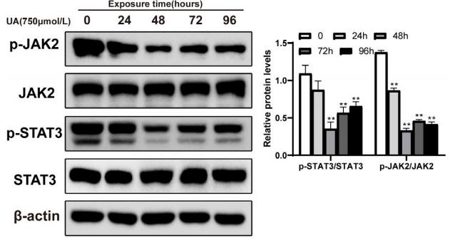
Application: WB    Species: human    Sample: L02 cells

FIGURE 6 |Uric acid induces SREBP-1c activation and JAK2/STAT3 pathway inhibition in vitro.F, p-JAK2 (Tyr1007/1008)/JAK2 and p-STAT3 (Tyr705)/STAT3 protein levels were detected by western blotting.

7). The Food Additive β-Caryophyllene Exerts Its Neuroprotective Effects Through the JAK2-STAT3-BACE1 Pathway. Frontiers in Aging Neuroscience, 2022 (PubMed: 35296033) [IF=4.8]

8). Tert-Butylhydroquinone Mitigates T-2-Toxin-Induced Testicular Dysfunction by Targeting Oxidative Stress, Inflammation, and Apoptosis in Rats. Toxics, 2024 (PubMed: 38787114) [IF=4.6]

Application: WB    Species: Rat    Sample:

Figure 3 tBHQ alleviates the inflammatory response induced by T-2. (A) mRNA expression levels of inflammatory factors; (B,D) protein expression levels and phosphorylation of JAK, represented by grayscale values; (C,E) protein expression levels and phosphorylation of STAT3, represented by grayscale values. n = 8. * represents a significant difference, with p < 0.05. ** represents a significant difference, with p < 0.01.

9). Photobiomodulation promotes repair following spinal cord injury by regulating the transformation of A1/A2 reactive astrocytes. Frontiers in Neuroscience, 2021 (PubMed: 34795557) [IF=4.3]

Application: WB    Species: Rat    Sample:

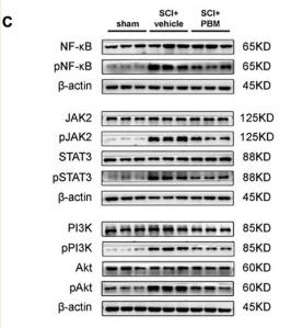
FIGURE 4 Signaling pathways involved in A1/A2 astrocyte activation were regulated by PBM. (A) GO function analysis between the SCI-7d group and sham control group. The inflammatory response and immune system process were dramatically activated. (B) The histogram shows the –log10 (p-value) of the NF-κB pathway, PI3K–Akt pathway, JAK–STAT pathway, and Notch pathway (comparison between multiple groups vs. the sham control group), as determined by KEGG enrichment analysis. (C) Western blot analysis of the expression of proteins related to the NF-κB signaling pathway, JAK2–STAT3 signaling pathway, and PI3K–Akt signaling pathway in the sham control, SCI + vehicle, and SCI + PBM groups at 7 dpi. Quantification of the relative protein levels of pNF-κB, NF-κB, pJAK2, JAK2, pSTAT3, STAT3, pPI3K, PI3K, pAkt, and Akt. n = 3 individuals per group. **p < 0.01, ***p < 0.001, the SCI + vehicle group vs. the sham control group; ##p < 0.01, the SCI + PBM group vs. the SCI + vehicle group. (D) Representative blots and quantification of the relative levels of pNF-κB, NF-κB, Notch1, pJAK2, JAK2, pSTAT3, STAT3, pPI3K, PI3K, pAkt, and Akt in the control, control + PBM, A1, and A1 + PBM groups. Con, control; A1, A1 astrocytes. The experiments were independently repeated three times. **p < 0.01, ***p < 0.001, ****p < 0.0001, the A1 group vs. the control group; ##p < 0.01, ###p < 0.001, the A1 + PBM group vs. the A1 group. (E) Representative blots and quantification of the relative levels of C3 and S100a10 in astrocytes treated with different inhibitors. The experiments were independently repeated three times. **p < 0.01, ***p < 0.001, ****p < 0.0001 vs. the A1 group.

10). The capsaicinoid nonivamide suppresses the inflammatory response and attenuates the progression of steatosis in a NAFLD‐rat model. Journal of Biochemical and Molecular Toxicology, 2023 (PubMed: 36541345) [IF=3.6]

Restrictive clause


Affinity Biosciences tests all products strictly. Citations are provided as a resource for additional applications that have not been validated by Affinity Biosciences. Please choose the appropriate format for each application and consult Materials and Methods sections for additional details about the use of any product in these publications.

For Research Use Only.
Not for use in diagnostic or therapeutic procedures. Not for resale. Not for distribution without written consent. Affinity Biosciences will not be held responsible for patent infringement or other violations that may occur with the use of our products. Affinity Biosciences, Affinity Biosciences Logo and all other trademarks are the property of Affinity Biosciences LTD.