Product: GLUT2 Antibody
Catalog: DF7510
Description: Rabbit polyclonal antibody to GLUT2
Application: WB IHC
Reactivity: Human, Mouse, Rat
Mol.Wt.: 57 kDa.; 57kD(Calculated).
Uniprot: P11168
RRID: AB_2841009

View similar products>>

   Size Price Inventory
 100ul $280 In stock
 200ul $350 In stock

Lead Time: Same day delivery

For pricing and ordering contact:
Local distributors

Product Info

Source:
Rabbit
Application:
WB 1:1000-3000, IHC 1:50-1:200
*The optimal dilutions should be determined by the end user.
*Tips:

WB: For western blot detection of denatured protein samples. IHC: For immunohistochemical detection of paraffin sections (IHC-p) or frozen sections (IHC-f) of tissue samples. IF/ICC: For immunofluorescence detection of cell samples. ELISA(peptide): For ELISA detection of antigenic peptide.

Reactivity:
Human,Mouse,Rat
Clonality:
Polyclonal
Specificity:
GLUT2 Antibody detects endogenous levels of total GLUT2.
RRID:
AB_2841009
Cite Format: Affinity Biosciences Cat# DF7510, RRID:AB_2841009.
Conjugate:
Unconjugated.
Purification:
The antiserum was purified by peptide affinity chromatography using SulfoLink™ Coupling Resin (Thermo Fisher Scientific).
Storage:
Rabbit IgG in phosphate buffered saline , pH 7.4, 150mM NaCl, 0.02% sodium azide and 50% glycerol. Store at -20 °C. Stable for 12 months from date of receipt.
Alias:

Fold/Unfold

liver; Glucose Transporter 2; Glucose Transporter GLUT2; Glucose transporter type 2; Glucose transporter type 2 liver; Glucose transporter, liver/islet; GLUT-2; GLUT2; GTR2_HUMAN; GTT2; SLC2A2; Solute carrier family 2 (facilitated glucose transporter) member 2; Solute carrier family 2 facilitated glucose transporter member 2; Solute carrier family 2, facilitated glucose transporter member 2;

Immunogens

Immunogen:
Uniprot:
Gene(ID):
Expression:
P11168 GTR2_HUMAN:

Liver, insulin-producing beta cell, small intestine and kidney.

Sequence:
MTEDKVTGTLVFTVITAVLGSFQFGYDIGVINAPQQVIISHYRHVLGVPLDDRKAINNYVINSTDELPTISYSMNPKPTPWAEEETVAAAQLITMLWSLSVSSFAVGGMTASFFGGWLGDTLGRIKAMLVANILSLVGALLMGFSKLGPSHILIIAGRSISGLYCGLISGLVPMYIGEIAPTALRGALGTFHQLAIVTGILISQIIGLEFILGNYDLWHILLGLSGVRAILQSLLLFFCPESPRYLYIKLDEEVKAKQSLKRLRGYDDVTKDINEMRKEREEASSEQKVSIIQLFTNSSYRQPILVALMLHVAQQFSGINGIFYYSTSIFQTAGISKPVYATIGVGAVNMVFTAVSVFLVEKAGRRSLFLIGMSGMFVCAIFMSVGLVLLNKFSWMSYVSMIAIFLFVSFFEIGPGPIPWFMVAEFFSQGPRPAALAIAAFSNWTCNFIVALCFQYIADFCGPYVFFLFAGVLLAFTLFTFFKVPETKGKSFEEIAAEFQKKSGSAHRPKAAVEMKFLGATETV

PTMs - P11168 As Substrate

Site PTM Type Enzyme
S491 Phosphorylation
S503 Phosphorylation
S505 Phosphorylation
T523 Phosphorylation

Research Backgrounds

Function:

Facilitative hexose transporter that mediates the transport of glucose and fructose. Likely mediates the bidirectional transfer of glucose across the plasma membrane of hepatocytes and is responsible for uptake of glucose by the beta cells; may comprise part of the glucose-sensing mechanism of the beta cell. May also participate with the Na(+)/glucose cotransporter in the transcellular transport of glucose in the small intestine and kidney. Also able to mediate the transport of dehydroascorbate.

PTMs:

N-glycosylated; required for stability and retention at the cell surface of pancreatic beta cells.

Subcellular Location:

Cell membrane>Multi-pass membrane protein.

Extracellular region or secreted Cytosol Plasma membrane Cytoskeleton Lysosome Endosome Peroxisome ER Golgi apparatus Nucleus Mitochondrion Manual annotation Automatic computational assertionSubcellular location
Tissue Specificity:

Liver, insulin-producing beta cell, small intestine and kidney.

Family&Domains:

Belongs to the major facilitator superfamily. Sugar transporter (TC 2.A.1.1) family. Glucose transporter subfamily.

Research Fields

· Human Diseases > Endocrine and metabolic diseases > Type II diabetes mellitus.

· Human Diseases > Endocrine and metabolic diseases > Insulin resistance.

· Human Diseases > Endocrine and metabolic diseases > Maturity onset diabetes of the young.

· Human Diseases > Cancers: Overview > Central carbon metabolism in cancer.   (View pathway)

· Organismal Systems > Endocrine system > Insulin secretion.   (View pathway)

· Organismal Systems > Endocrine system > Prolactin signaling pathway.   (View pathway)

· Organismal Systems > Endocrine system > Glucagon signaling pathway.

· Organismal Systems > Digestive system > Carbohydrate digestion and absorption.

References

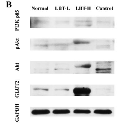
1). Liubao brick tea activates the PI3K-Akt signaling pathway to lower blood glucose, metabolic disorders and insulin resistance via altering the intestinal flora. FOOD RESEARCH INTERNATIONAL, 2021 (PubMed: 34507739) [IF=8.1]

Application: WB    Species: Mice    Sample: liver tissue

Fig. 7. LBT treatment activated the PI3K/Akt pathway in the liver of hyperglycemic and insulin-resistant mice and insulin-resistant HepG2 cells. (A) Hepatic immunohistochemical analysis of PI3K p85, pAkt and Akt protein expressions, (B) Cellular western blotting analysis of GLUT2, PI3K p85 and Akt protein expressions. Different letters represent a significant difference among multiple groups, * represents a significance compared with the Control group, # represents a significance between two groups, ns represents no significance. #,*, p < 0.05; ##, **, p < 0.01.

2). Recurrent moderate hypoglycemia accelerates the progression of cognitive deficits through impairment of TRPC6/GLUT3 pathway in diabetic APP/PS1 mice. JCI Insight, 2022 (PubMed: 35077394) [IF=8.0]

Application: WB    Species: Mice    Sample: hippocampal homogenates

Figure 5 RH reduced brain GLUT3-mediated glucose uptake in STZ-induced APP/PS1-DM mice. (A) Representative PET/CT images showing in vivo brain 18F-FDG uptake (coronal, sagittal, and transaxial sections, n = 6 mice for each group). Cor, Cortex; Hip, hippocampus; Mid, midbrain; HPOA, hypothalamus; BS, brain stem; Cer, cerebellum; OB, Olfactory Bulb; BF, basal forebrain; Tha, thalamus; SC, superior colliculi; CG, central gray; Str, striatum. (B) Quantification for SUV of 18F-FDG in brain region (n = 6 for mice for each group). (C) Western blot and quantitation for GLUT1, GLUT2, GLUT3, GLUT4, GLUT5, SGLT1, and SGLT2 in hippocampal homogenates (n = 3 mice for each group). The data are expressed as the mean ± SEM. Statistical significance was assessed using unpaired Student’s t test. *P < 0.05 and **P < 0.01, APP/PS1-DM versus APP/PS1; #P < 0.05 and ##P < 0.01, APP/PS1-DM-RH versus APP/PS1-DM; WT versus APP/PS1; NS, no significant difference.

3). Network pharmacology-based elucidation of bioactive compounds and experimental exploration of antidiabetic mechanisms of Hydrolea zeylanica. Cellular signalling, 2024 (PubMed: 38052370) [IF=4.8]

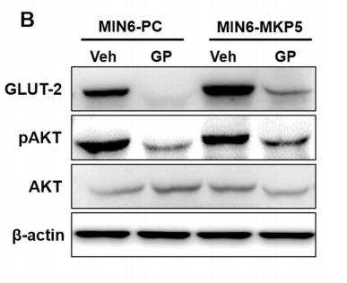
4). The protective role of the MKP-5-JNK/P38 pathway in glucolipotoxicity-induced islet β-cell dysfunction and apoptosis. EXPERIMENTAL CELL RESEARCH, 2019 (PubMed: 31202710) [IF=3.7]

Application: WB    Species: mouse    Sample: MIN6-PC and MIN6-MKP5 cells

Fig. 3. |Cell dysfunction caused by GP is improved in MKP-5 overexpressing MIN6 cells. (B, C) MIN6-PC and MIN6-MKP5 cells were stimulated with GP for 24 h. The phosphorylation of AKT and the expression of GLUT-2 were assessed by western blotting (B). Relative levels of PDX-1 and GCK mRNA were quantified by real-time PCR analysis (C). Data are given as means ± SEM. *, P < 0.05.

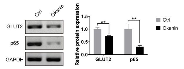
5). Identification of Key Candidate Genes and Pathways of Candida albicans-Infected Human Umbilical Vein Endothelial Cells and Drug Screening. INDIAN JOURNAL OF MICROBIOLOGY, 2020 (PubMed: 32089575) [IF=3.0]

Application: WB    Species: human    Sample:

Fig. 3 Receptor–ligand interactions of compound. a Protocol flowchart of MYC inhibitor discovery strategy, ten candidate inhibitors including okanin were screened at last through HTV, SP,and XP screen methods. b Binding model of MYC–DNA complexes with okanin (PDB code: 1NKP) through molecular docking method.The results showed that okanin interacted with two key amino acid residues in the DNA binding site of MYC. c Okanin could significantly inhibit the transcriptional regulatory activity of MYC by luciferase assay. d Okanin significantly inhibited the expressions of GLUT2 and p65, which were the downstream proteins of MYC(n = 3, **P \0.01)

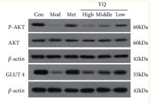
6). YiQi YangYin Decoction Attenuates Nonalcoholic Fatty Liver Disease in Type 2 Diabetes Rats. Evidence-based Complementary and Alternative Medicine, 2021 (PubMed: 34621322)

Application: WB    Species: Rat    Sample: The liver and pancreas tissues

Figure 11 Effect of YQ on insulin secretion-related proteins in pancreas islet. The expressions levels of IRS-2, P-PI3K, PI3K, P-Akt, Akt, and GLUT4 were assessed by western blot analysis. The results are shown as means ± SEM (n = 9–10). ##P < 0.01 compared with the control group; ∗P < 0.05, ∗∗P < 0.01 compared with the model group.

7). Hydrolea Zeylanica Ameliorates Carbohydrate Metabolism Via Modulating GLUTS Expression and Inhibition of Redox-Inflammation in High-Fat Diet Fed. , 2022

Restrictive clause


Affinity Biosciences tests all products strictly. Citations are provided as a resource for additional applications that have not been validated by Affinity Biosciences. Please choose the appropriate format for each application and consult Materials and Methods sections for additional details about the use of any product in these publications.

For Research Use Only.
Not for use in diagnostic or therapeutic procedures. Not for resale. Not for distribution without written consent. Affinity Biosciences will not be held responsible for patent infringement or other violations that may occur with the use of our products. Affinity Biosciences, Affinity Biosciences Logo and all other trademarks are the property of Affinity Biosciences LTD.