
| Product: | Phospho-JAK3 (Tyr981) Antibody |
| Catalog: | AF8160 |
| Description: | Rabbit polyclonal antibody to Phospho-JAK3 (Tyr981) |
| Application: | WB IHC IF/ICC |
| Cited expt.: | WB |
| Reactivity: | Human, Mouse, Rat, Monkey |
| Prediction: | Pig, Bovine, Horse, Sheep, Dog |
| Mol.Wt.: | 125kDa(Observed); 125kD(Calculated). |
| Uniprot: | P52333 |
| RRID: | AB_2840222 |
Control Products

Related Downloads
Protocols
Product Info
*The optimal dilutions should be determined by the end user. For optimal experimental results, antibody reuse is not recommended.
*Tips:
WB: For western blot detection of denatured protein samples. IHC: For immunohistochemical detection of paraffin sections (IHC-p) or frozen sections (IHC-f) of tissue samples. IF/ICC: For immunofluorescence detection of cell samples. ELISA(peptide): For ELISA detection of antigenic peptide.
Cite Format: Affinity Biosciences Cat# AF8160, RRID:AB_2840222.
Fold/Unfold
EC 2.7.10.2; JAK 3; JAK L; JAK-3; Jak3; JAK3 HUMAN; JAK3_HUMAN; JAKL; Janus kinase 3 (a protein tyrosine kinase, leukocyte); Janus kinase 3; Janus Kinase3; L JAK; L-JAK; Leukocyte janus kinase; LJAK; Protein tyrosine kinase leukocyte; Tyrosine protein kinase JAK3; Tyrosine-protein kinase JAK3;
Immunogens
A synthesized peptide derived from human JAK3 around the phosphorylation site of Tyr981.
In NK cells and an NK-like cell line but not in resting T-cells or in other tissues. The S-form is more commonly seen in hematopoietic lines, whereas the B-form is detected in cells both of hematopoietic and epithelial origins.
- P52333 JAK3_HUMAN:
- Protein BLAST With
- NCBI/
- ExPASy/
- Uniprot
MAPPSEETPLIPQRSCSLLSTEAGALHVLLPARGPGPPQRLSFSFGDHLAEDLCVQAAKASGILPVYHSLFALATEDLSCWFPPSHIFSVEDASTQVLLYRIRFYFPNWFGLEKCHRFGLRKDLASAILDLPVLEHLFAQHRSDLVSGRLPVGLSLKEQGECLSLAVLDLARMAREQAQRPGELLKTVSYKACLPPSLRDLIQGLSFVTRRRIRRTVRRALRRVAACQADRHSLMAKYIMDLERLDPAGAAETFHVGLPGALGGHDGLGLLRVAGDGGIAWTQGEQEVLQPFCDFPEIVDISIKQAPRVGPAGEHRLVTVTRTDNQILEAEFPGLPEALSFVALVDGYFRLTTDSQHFFCKEVAPPRLLEEVAEQCHGPITLDFAINKLKTGGSRPGSYVLRRSPQDFDSFLLTVCVQNPLGPDYKGCLIRRSPTGTFLLVGLSRPHSSLRELLATCWDGGLHVDGVAVTLTSCCIPRPKEKSNLIVVQRGHSPPTSSLVQPQSQYQLSQMTFHKIPADSLEWHENLGHGSFTKIYRGCRHEVVDGEARKTEVLLKVMDAKHKNCMESFLEAASLMSQVSYRHLVLLHGVCMAGDSTMVQEFVHLGAIDMYLRKRGHLVPASWKLQVVKQLAYALNYLEDKGLPHGNVSARKVLLAREGADGSPPFIKLSDPGVSPAVLSLEMLTDRIPWVAPECLREAQTLSLEADKWGFGATVWEVFSGVTMPISALDPAKKLQFYEDRQQLPAPKWTELALLIQQCMAYEPVQRPSFRAVIRDLNSLISSDYELLSDPTPGALAPRDGLWNGAQLYACQDPTIFEERHLKYISQLGKGNFGSVELCRYDPLGDNTGALVAVKQLQHSGPDQQRDFQREIQILKALHSDFIVKYRGVSYGPGRQSLRLVMEYLPSGCLRDFLQRHRARLDASRLLLYSSQICKGMEYLGSRRCVHRDLAARNILVESEAHVKIADFGLAKLLPLDKDYYVVREPGQSPIFWYAPESLSDNIFSRQSDVWSFGVVLYELFTYCDKSCSPSAEFLRMMGCERDVPALCRLLELLEEGQRLPAPPACPAEVHELMKLCWAPSPQDRPSFSALGPQLDMLWSGSRGCETHAFTAHPEGKHHSLSFS
Predictions
Score>80(red) has high confidence and is suggested to be used for WB detection. *The prediction model is mainly based on the alignment of immunogen sequences, the results are for reference only, not as the basis of quality assurance.
High(score>80) Medium(80>score>50) Low(score<50) No confidence
Research Backgrounds
Non-receptor tyrosine kinase involved in various processes such as cell growth, development, or differentiation. Mediates essential signaling events in both innate and adaptive immunity and plays a crucial role in hematopoiesis during T-cells development. In the cytoplasm, plays a pivotal role in signal transduction via its association with type I receptors sharing the common subunit gamma such as IL2R, IL4R, IL7R, IL9R, IL15R and IL21R. Following ligand binding to cell surface receptors, phosphorylates specific tyrosine residues on the cytoplasmic tails of the receptor, creating docking sites for STATs proteins. Subsequently, phosphorylates the STATs proteins once they are recruited to the receptor. Phosphorylated STATs then form homodimer or heterodimers and translocate to the nucleus to activate gene transcription. For example, upon IL2R activation by IL2, JAK1 and JAK3 molecules bind to IL2R beta (IL2RB) and gamma chain (IL2RG) subunits inducing the tyrosine phosphorylation of both receptor subunits on their cytoplasmic domain. Then, STAT5A AND STAT5B are recruited, phosphorylated and activated by JAK1 and JAK3. Once activated, dimerized STAT5 translocates to the nucleus and promotes the transcription of specific target genes in a cytokine-specific fashion.
Tyrosine phosphorylated in response to IL-2 and IL-4. Dephosphorylation of Tyr-980 and Tyr-981 by PTPN2 negatively regulates cytokine-mediated signaling (Probable).
Endomembrane system>Peripheral membrane protein. Cytoplasm.
In NK cells and an NK-like cell line but not in resting T-cells or in other tissues. The S-form is more commonly seen in hematopoietic lines, whereas the B-form is detected in cells both of hematopoietic and epithelial origins.
Possesses two phosphotransferase domains. The second one probably contains the catalytic domain (By similarity), while the presence of slight differences suggest a different role for domain 1.
Belongs to the protein kinase superfamily. Tyr protein kinase family. JAK subfamily.
Research Fields
· Cellular Processes > Cell growth and death > Necroptosis. (View pathway)
· Cellular Processes > Cellular community - eukaryotes > Signaling pathways regulating pluripotency of stem cells. (View pathway)
· Environmental Information Processing > Signal transduction > PI3K-Akt signaling pathway. (View pathway)
· Environmental Information Processing > Signal transduction > Jak-STAT signaling pathway. (View pathway)
· Human Diseases > Infectious diseases: Viral > Measles.
· Human Diseases > Infectious diseases: Viral > HTLV-I infection.
· Human Diseases > Infectious diseases: Viral > Epstein-Barr virus infection.
· Human Diseases > Cancers: Overview > Pathways in cancer. (View pathway)
· Human Diseases > Cancers: Overview > Viral carcinogenesis.
· Human Diseases > Cancers: Specific types > Non-small cell lung cancer. (View pathway)
· Human Diseases > Immune diseases > Primary immunodeficiency.
· Organismal Systems > Immune system > Chemokine signaling pathway. (View pathway)
· Organismal Systems > Immune system > Th1 and Th2 cell differentiation. (View pathway)
· Organismal Systems > Immune system > Th17 cell differentiation. (View pathway)
References

Application: WB Species: human Sample:
Application: WB Species: rat Sample: DRG neurons
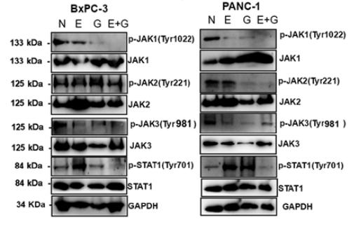
Application: WB Species: human Sample: BxPC-3 and PANC‑1 cells
Restrictive clause
Affinity Biosciences tests all products strictly. Citations are provided as a resource for additional applications that have not been validated by Affinity Biosciences. Please choose the appropriate format for each application and consult Materials and Methods sections for additional details about the use of any product in these publications.
For Research Use Only.
Not for use in diagnostic or therapeutic procedures. Not for resale. Not for distribution without written consent. Affinity Biosciences will not be held responsible for patent infringement or other violations that may occur with the use of our products. Affinity Biosciences, Affinity Biosciences Logo and all other trademarks are the property of Affinity Biosciences LTD.