Product: TFAM Antibody
Catalog: AF0531
Description: Rabbit polyclonal antibody to TFAM
Application: WB IHC IF/ICC
Reactivity: Human, Mouse, Rat
Prediction: Pig, Bovine
Mol.Wt.: 29kDa; 29kD(Calculated).
Uniprot: Q00059
RRID: AB_2834243

View similar products>>

   Size Price Inventory
 100ul $280 In stock
 200ul $350 In stock

Lead Time: Same day delivery

For pricing and ordering contact:
Local distributors

Product Info

Source:
Rabbit
Application:
IHC 1:50-1:200, IF/ICC 1:100-1:500, WB 1:500-1:2000
*The optimal dilutions should be determined by the end user.
*Tips:

WB: For western blot detection of denatured protein samples. IHC: For immunohistochemical detection of paraffin sections (IHC-p) or frozen sections (IHC-f) of tissue samples. IF/ICC: For immunofluorescence detection of cell samples. ELISA(peptide): For ELISA detection of antigenic peptide.

Reactivity:
Human,Mouse,Rat
Prediction:
Pig(82%), Bovine(86%)
Clonality:
Polyclonal
Specificity:
TFAM Antibody detects endogenous levels of total TFAM.
RRID:
AB_2834243
Cite Format: Affinity Biosciences Cat# AF0531, RRID:AB_2834243.
Conjugate:
Unconjugated.
Purification:
The antiserum was purified by peptide affinity chromatography using SulfoLink™ Coupling Resin (Thermo Fisher Scientific).
Storage:
Rabbit IgG in phosphate buffered saline , pH 7.4, 150mM NaCl, 0.02% sodium azide and 50% glycerol. Store at -20 °C. Stable for 12 months from date of receipt.
Alias:

Fold/Unfold

anscription factor 6-like 1; Mitochondrial transcription factor 1; mitochondrial transcription factor A; MtTF1; mtTFA; TCF 6; TCF-6; TCF6; TCF6L1; TCF6L2; TCF6L3; TFAM; TFAM_HUMAN; Transcription factor 6; Transcription factor 6 like 2 (mitochondrial transcription factor); Transcription factor 6 like 2; Transcription factor 6-like 2; transcription factor 6-like 3; Transcription factor A, mitochondrial; Transcription factor A, mitochondrial; Transcription factor A, mitochondrial precursor;

Immunogens

Immunogen:
Uniprot:
Gene(ID):
Description:
This gene encodes a mitochondrial transcription factor that is a key activator of mitochondrial transcription as well as a participant in mitochondrial genome replication. Studies in mice have demonstrated that this gene product is required to regulate the mitochondrial genome copy number and is essential for embryonic development. A mouse model for Kearns-Sayre syndrome was produced when expression of this gene was eliminated by targeted disruption in heart and muscle cells.
Sequence:
MAFLRSMWGVLSALGRSGAELCTGCGSRLRSPFSFVYLPRWFSSVLASCPKKPVSSYLRFSKEQLPIFKAQNPDAKTTELIRRIAQRWRELPDSKKKIYQDAYRAEWQVYKEEISRFKEQLTPSQIMSLEKEIMDKHLKRKAMTKKKELTLLGKPKRPRSAYNVYVAERFQEAKGDSPQEKLKTVKENWKNLSDSEKELYIQHAKEDETRYHNEMKSWEEQMIEVGRKDLLRRTIKKQRKYGAEEC

Predictions

Predictions:

Score>80(red) has high confidence and is suggested to be used for WB detection. *The prediction model is mainly based on the alignment of immunogen sequences, the results are for reference only, not as the basis of quality assurance.

Species
Results
Score
Bovine
86
Pig
82
Xenopus
70
Chicken
64
Zebrafish
60
Horse
0
Sheep
0
Dog
0
Rabbit
0
Model Confidence:
High(score>80) Medium(80>score>50) Low(score<50) No confidence

PTMs - Q00059 As Substrate

Site PTM Type Enzyme
K52 Ubiquitination
S55 Phosphorylation P17612 (PRKACA)
S56 Phosphorylation P17612 (PRKACA)
S61 Phosphorylation P17612 (PRKACA)
K62 Ubiquitination
K69 Methylation
K76 Ubiquitination
R82 Methylation
Y99 Phosphorylation
K111 Acetylation
K111 Ubiquitination
K118 Ubiquitination
T122 Phosphorylation P24941 (CDK2)
S124 Phosphorylation P24941 (CDK2)
S128 Phosphorylation
K146 Methylation
K147 Methylation
S160 Phosphorylation P17612 (PRKACA)
Y165 Phosphorylation
S177 Phosphorylation
K186 Ubiquitination
K190 Ubiquitination
S193 Phosphorylation
S195 Phosphorylation
K197 Ubiquitination
Y200 Phosphorylation
C246 S-Nitrosylation

Research Backgrounds

Function:

Binds to the mitochondrial light strand promoter and functions in mitochondrial transcription regulation. Component of the mitochondrial transcription initiation complex, composed at least of TFB2M, TFAM and POLRMT that is required for basal transcription of mitochondrial DNA. In this complex, TFAM recruits POLRMT to a specific promoter whereas TFB2M induces structural changes in POLRMT to enable promoter opening and trapping of the DNA non-template strand. Required for accurate and efficient promoter recognition by the mitochondrial RNA polymerase. Promotes transcription initiation from the HSP1 and the light strand promoter by binding immediately upstream of transcriptional start sites. Is able to unwind DNA. Bends the mitochondrial light strand promoter DNA into a U-turn shape via its HMG boxes. Required for maintenance of normal levels of mitochondrial DNA. May play a role in organizing and compacting mitochondrial DNA.

PTMs:

Phosphorylation by PKA within the HMG box 1 impairs DNA binding and promotes degradation by the AAA+ Lon protease.

Subcellular Location:

Mitochondrion. Mitochondrion matrix>Mitochondrion nucleoid.

Extracellular region or secreted Cytosol Plasma membrane Cytoskeleton Lysosome Endosome Peroxisome ER Golgi apparatus Nucleus Mitochondrion Manual annotation Automatic computational assertionSubcellular location
Subunit Structure:

Monomer; binds DNA as a monomer. Homodimer. Component of the mitochondrial transcription initiation complex, composed at least of TFB2M, TFAM and POLRMT. In this complex TFAM recruits POLRMT to the promoter whereas TFB2M induces structural changes in POLRMT to enable promoter opening and trapping of the DNA non-template strand. Upon metabolic stress, forms a complex composed of FOXO3, SIRT3, TFAM and POLRMT. Interacts with TFB1M and TFB2M. Interacts with CLPX; this enhances DNA-binding.

Family&Domains:

Binds DNA via its HMG boxes. When bound to the mitochondrial light strand promoter, bends DNA into a U-turn shape, each HMG box bending the DNA by 90 degrees.

Research Fields

· Environmental Information Processing > Signal transduction > Apelin signaling pathway.   (View pathway)

· Human Diseases > Neurodegenerative diseases > Huntington's disease.

References

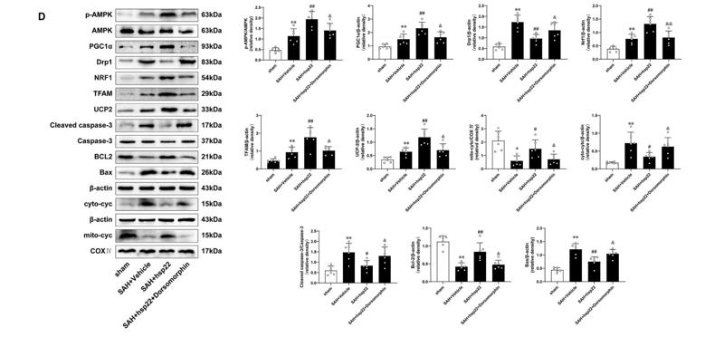
1). Heat shock protein 22 modulates NRF1/TFAM-dependent mitochondrial biogenesis and DRP1-sparked mitochondrial apoptosis through AMPK-PGC1α signaling pathway to alleviate the early brain injury of subarachnoid hemorrhage in rats. Redox Biology, 2021 (PubMed: 33472123) [IF=11.4]

Application: WB    Species: rat    Sample: brain

Fig. 6. Hsp22 regulates PGC1α via AMPK signaling pathway in rats after SAH Beam balance scores, Modified Garcia scores and Brainwater content in various groups. n = 6 per group. (B) Representative photomicrographs of TUNEL staining and quantitative analyses in the indicated groups. n = 4 per group. Scale bar = 100 μm. (C) Typical photomicrographs showing double immunofluorescence staining of PGC1α (green) and NeuN (red) in diverse experimental groups. n = 4 per group. Scale bar = 50 μm. (D) Western blot images and quantitative analyses of p-AMPK/AMPK, PGC1α, Drp1, Nrf1, TFAM, UCP2, Cleaved caspase-3/Caspase-3, Bcl2, Bax, Cytosolic and mitochondrial cytochrome c. n = 6 per group. Bars represent mean ± SD. **P < 0.01, *P < 0.05 vs. Sham group. ##P < 0.01, #P < 0.05 vs. SAH + Vehicle group. &&P < 0.01, &P < 0.05 vs. SAH + hsp22+scramble siRNA. (For interpretation of the references to colour in this figure legend, the reader is referred to the Web version of this article.)

2). Resveratrol and its derivative pterostilbene attenuate oxidative stress-induced intestinal injury by improving mitochondrial redox homeostasis and function via SIRT1 signaling. Free radical biology & medicine, 2021 (PubMed: 34648904) [IF=7.4]

3). Targeting PARK7 Improves Acetaminophen-Induced Acute Liver Injury by Orchestrating Mitochondrial Quality Control and Metabolic Reprogramming. Antioxidants (Basel, Switzerland), 2022 (PubMed: 36358500) [IF=7.0]

4). Sesamol Reverses Myofiber-Type Conversion in Obese States via Activating the SIRT1/AMPK Signal Pathway. JOURNAL OF AGRICULTURAL AND FOOD CHEMISTRY, 2022 (PubMed: 35166533) [IF=6.1]

Application: WB    Species: mouse    Sample:

Figure 2.| Sesamol elevated mitochondrial content and biogenesis reversed myofiber-type conversion in HFD-induced obese mice. (A)Representative Western blot images and quantification of COX2 and COX5A. (B) Representative Western blot images and quantification of PGC1α, TFAM, and NRF1.

5). YY1 inactivated transcription co-regulator PGC-1α to promote mitochondrial dysfunction of early diabetic nephropathy-associated tubulointerstitial fibrosis. Cell Biology and Toxicology, 2022 (PubMed: 35445903) [IF=6.1]

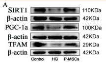
6). Placental Mesenchymal Stem Cells Alleviate Podocyte Injury in Diabetic Kidney Disease by Modulating Mitophagy via the SIRT1-PGC-1alpha-TFAM Pathway. International Journal of Molecular Sciences, 2023 (PubMed: 36902127) [IF=5.6]

Application: WB    Species: Mouse    Sample: MPC5 cell

Figure 5 P-MSCs increased the expressions of SIRT1, PGC-1a, and TFAM in MPC5. (A,B) Representative Western blot analysis of SIRT1, PGC-1a, and TFAM in MPC5. (C) Representative RT-PCR analysis of SIRT1, PGC-1a, and TFAM in MPC5. D-I: Immunofluorescence staining of SIRT1, PGC-1a, and TFAM in MPC5 (Magnification, 400×). Scale bars: 50 μm for (D,F,H). n = 3 for (A–I). Values are expressed as the mean ± SD. Β-actin was used as loading control.

7). Kcnma1 is involved in mitochondrial homeostasis in diabetes-related skeletal muscle atrophy. The FASEB Journal, 2023 (PubMed: 36929614) [IF=4.8]

8). Equisetin inhibits adiposity through AMPK-dependent regulation of brown adipocyte differentiation. Heliyon, 2024 (PubMed: 38327434) [IF=4.0]

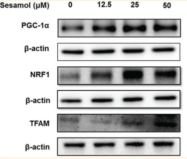
9). Sesamol promotes browning of white adipocytes to ameliorate obesity by inducing mitochondrial biogenesis and inhibition mitophagy via β3-AR/PKA signaling pathway. Food & Nutrition Research, 2023 (PubMed: 34262421) [IF=3.3]

Application: WB    Species: Mouse    Sample: 3T3-L1 adipocytes

Fig. 6 Effects of sesamol on the expressions of mitochondrial biogenesis proteins in mature 3T3-L1 adipocytes. The mature 3T3-L1 adipocytes were treated with different concentrations of sesamol (0, 12.5, 25, and 50 μM) for 48 h. Immunoblotting was used to detect protein expression of PGC-1α, NRF1, TFAM, and densitometric determinations. All values are presented as mean ± SD (n = 3). β-actin and histone were protein loading control. *P < 0.05 versus control group.

10). Mangiferin Alleviates Mitochondrial ROS in Nucleus Pulposus Cells and Protects against Intervertebral Disc Degeneration via Suppression of NF-κB Signaling Pathway. Oxidative Medicine and Cellular Longevity, 2021 (PubMed: 34234886)

Load more

Restrictive clause


Affinity Biosciences tests all products strictly. Citations are provided as a resource for additional applications that have not been validated by Affinity Biosciences. Please choose the appropriate format for each application and consult Materials and Methods sections for additional details about the use of any product in these publications.

For Research Use Only.
Not for use in diagnostic or therapeutic procedures. Not for resale. Not for distribution without written consent. Affinity Biosciences will not be held responsible for patent infringement or other violations that may occur with the use of our products. Affinity Biosciences, Affinity Biosciences Logo and all other trademarks are the property of Affinity Biosciences LTD.