Product: Ubiquitin Antibody
Catalog: AF0289
Description: Rabbit polyclonal antibody to Ubiquitin
Application: WB IHC IF/ICC
Reactivity: Human, Mouse, Rat
Prediction: Pig, Zebrafish, Bovine, Horse, Sheep, Dog, Chicken, Xenopus
Mol.Wt.: 8kDa; 15kD(Calculated).
Uniprot: P62987
RRID: AB_2834166

View similar products>>

   Size Price Inventory
 100ul $280 In stock
 200ul $350 In stock

Lead Time: Same day delivery

For pricing and ordering contact:
Local distributors

Product Info

Source:
Rabbit
Application:
IHC 1:50-1:200, WB 1:500-1:2000, IF/ICC 1:100-1:500
*The optimal dilutions should be determined by the end user.
*Tips:

WB: For western blot detection of denatured protein samples. IHC: For immunohistochemical detection of paraffin sections (IHC-p) or frozen sections (IHC-f) of tissue samples. IF/ICC: For immunofluorescence detection of cell samples. ELISA(peptide): For ELISA detection of antigenic peptide.

Reactivity:
Human,Mouse,Rat
Prediction:
Pig(100%), Zebrafish(100%), Bovine(100%), Horse(100%), Sheep(100%), Dog(100%), Chicken(100%), Xenopus(100%)
Clonality:
Polyclonal
Specificity:
UBA52 Antibody detects endogenous levels of total UBA52.
RRID:
AB_2834166
Cite Format: Affinity Biosciences Cat# AF0289, RRID:AB_2834166.
Conjugate:
Unconjugated.
Purification:
The antiserum was purified by peptide affinity chromatography using SulfoLink™ Coupling Resin (Thermo Fisher Scientific).
Storage:
Rabbit IgG in phosphate buffered saline , pH 7.4, 150mM NaCl, 0.02% sodium azide and 50% glycerol. Store at -20 °C. Stable for 12 months from date of receipt.
Alias:

Fold/Unfold

60S ribosomal protein L40; CEP52; HUBCEP52; L40; MGC126879; MGC57125; RPL40; UBA 52; Ubiquitin 52 amino acid fusion protein; Ubiquitin 60S ribosomal protein L40; Ubiquitin A 52 residue ribosomal protein fusion product 1; Ubiquitin carboxyl extension protein 52; Ubiquitin CEP52; UBA52;

Immunogens

Immunogen:
Uniprot:
Gene(ID):
Description:
ubiquitin a peptide 76 amino acids in length that can be covalently attached to target lysines either as a monomer or as a lysine-linked polymer. Ubiquitin is encoded by 4 different genes. UBA52 and RPS27A genes code for a single copy of ubiquitin fused to the ribosomal proteins L40 and S27a, respectively. UBB and UBC genes code for a polyubiquitin precursor with exact head to tail repeats, the number of repeats differ between species and strains. Only the 76 amino acids of monoubiquitin product are shown in this entry.
Sequence:
MQIFVKTLTGKTITLEVEPSDTIENVKAKIQDKEGIPPDQQRLIFAGKQLEDGRTLSDYNIQKESTLHLVLRLRGGIIEPSLRQLAQKYNCDKMICRKCYARLHPRAVNCRKKKCGHTNNLRPKKKVK

Predictions

Predictions:

Score>80(red) has high confidence and is suggested to be used for WB detection. *The prediction model is mainly based on the alignment of immunogen sequences, the results are for reference only, not as the basis of quality assurance.

Species
Results
Score
Pig
100
Horse
100
Bovine
100
Sheep
100
Dog
100
Xenopus
100
Zebrafish
100
Chicken
100
Rabbit
0
Model Confidence:
High(score>80) Medium(80>score>50) Low(score<50) No confidence

PTMs - P62987 As Substrate

Site PTM Type Enzyme
K6 Acetylation
K6 Methylation
K6 Sumoylation
K6 Ubiquitination
T7 Phosphorylation
K11 Acetylation
K11 Sumoylation
K11 Ubiquitination
T12 Phosphorylation
T14 Phosphorylation
S20 Phosphorylation
T22 Phosphorylation
K27 Sumoylation
K27 Ubiquitination
K29 Sumoylation
K29 Ubiquitination
K33 Sumoylation
K33 Ubiquitination
K48 Acetylation
K48 Sumoylation
K48 Ubiquitination
S57 Phosphorylation
Y59 Phosphorylation
K63 Methylation
K63 Sumoylation
K63 Ubiquitination
S65 Phosphorylation
T66 Phosphorylation
R72 Methylation
S81 Phosphorylation
K88 Acetylation
K88 Ubiquitination
C91 S-Nitrosylation
K93 Acetylation
K93 Ubiquitination
K98 Methylation
C115 S-Nitrosylation

Research Backgrounds

Function:

Exists either covalently attached to another protein, or free (unanchored). When covalently bound, it is conjugated to target proteins via an isopeptide bond either as a monomer (monoubiquitin), a polymer linked via different Lys residues of the ubiquitin (polyubiquitin chains) or a linear polymer linked via the initiator Met of the ubiquitin (linear polyubiquitin chains). Polyubiquitin chains, when attached to a target protein, have different functions depending on the Lys residue of the ubiquitin that is linked: Lys-6-linked may be involved in DNA repair; Lys-11-linked is involved in ERAD (endoplasmic reticulum-associated degradation) and in cell-cycle regulation; Lys-29-linked is involved in lysosomal degradation; Lys-33-linked is involved in kinase modification; Lys-48-linked is involved in protein degradation via the proteasome; Lys-63-linked is involved in endocytosis, DNA-damage responses as well as in signaling processes leading to activation of the transcription factor NF-kappa-B. Linear polymer chains formed via attachment by the initiator Met lead to cell signaling. Ubiquitin is usually conjugated to Lys residues of target proteins, however, in rare cases, conjugation to Cys or Ser residues has been observed. When polyubiquitin is free (unanchored-polyubiquitin), it also has distinct roles, such as in activation of protein kinases, and in signaling.

Component of the 60S subunit of the ribosome. Ribosomal protein L40 is essential for translation of a subset of cellular transcripts, and especially for cap-dependent translation of vesicular stomatitis virus mRNAs.

PTMs:

Phosphorylated at Ser-65 by PINK1 during mitophagy. Phosphorylated ubiquitin specifically binds and activates parkin (PRKN), triggering mitophagy. Phosphorylation does not affect E1-mediated E2 charging of ubiquitin but affects discharging of E2 enzymes to form polyubiquitin chains. It also affects deubiquitination by deubiquitinase enzymes such as USP30.

Mono-ADP-ribosylated at the C-terminus by PARP9, a component of the PPAR9-DTX3L complex. ADP-ribosylation requires processing by E1 and E2 enzymes and prevents ubiquitin conjugation to substrates such as histones.

Subcellular Location:

Cytoplasm. Nucleus.

Cytoplasm.

Extracellular region or secreted Cytosol Plasma membrane Cytoskeleton Lysosome Endosome Peroxisome ER Golgi apparatus Nucleus Mitochondrion Manual annotation Automatic computational assertionSubcellular location
Subunit Structure:

Ribosomal protein L40 is part of the 60S ribosomal subunit. Interacts with UBQLN1 (via UBA domain).

Family&Domains:

In the N-terminal section; belongs to the ubiquitin family.

In the C-terminal section; belongs to the eukaryotic ribosomal protein eL40 family.

Research Fields

· Genetic Information Processing > Translation > Ribosome.

References

1). Salidroside can target both P4HB-mediated inflammation and melanogenesis of the skin. Theranostics (PubMed: 33042273) [IF=12.4]

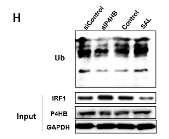
Application: WB    Species: human    Sample: A375 cells

Figure 4. P4HB regulates the ubiquitination degradation of IRF1. A. Changes in TYR activity in P4HB overexpression or knockdown A375 cells. B. Changes in TYR mRNA expression in Sal treated, P4HB overexpression, and P4HB knockdown A375 cells. C. Western blot analysis of TYR expression in P4HB overexpression or knockdown A375 cells. D. Proteins have interactions with P4HB and USF1 analyzed by FpClass. E. Interaction of P4HB and IRF1 in A375 cells detected by PLA. F. Western blot analysis of the expression of IRF1 and P4HB in P4HB overexpression or knockdown A375 cells. G. Western blot analysis of IRF1 expression in the nucleus of A375 cells after P4HB overexpression or knockdown. H. Effect of SAL on the ubiquitination of IRF1 as determined by Western blot. I. Western blot analysis of IRF1 expression in whole cells and nucleus of SAL-treated A375 cells. Data are expressed as mean ± SD (*P < 0.05, **P < 0.01).

2). Natural isoflavone formononetin inhibits IgE-mediated mast cell activation and allergic inflammation by increasing IgE receptor degradation. Food & Function (PubMed: 36880662) [IF=6.1]

3). Forensic Application of Epidermal Ubiquitin Expression to Determination of Wound Vitality in Human Compressed Neck Skin. Frontiers in Medicine (PubMed: 35492347) [IF=3.9]

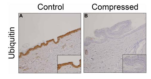
Application: IHC    Species: human    Sample: skin

FIGURE 1 | Immunohistochemical analysis. Immunohistochemical analysis were performed by using anti-ubiquitin pAb in the human skin samples. (A) control; (B) compressed neck skin. Original magnification, × 200;inset, × 400.

4). RACK1 degrades MAVS to promote bovine ephemeral fever virus replication via upregulating E3 ubiquitin ligase STUB1. VETERINARY MICROBIOLOGY (PubMed: 33940459) [IF=3.3]

Application: WB    Species: Bovine    Sample: BHK-21 cells

Fig. 5. RACK1 up-regulates E3 ubiquitin ligase STUB1 to promote MAVS ubiquitination and degradation. (A) Immunoblot analysis of MAVS, STUB1, and Flag in RACK1-overexpressing and negative control BHK-21 cell lines infected with BEFV of 0.1MOI for 24 h. Antiβ-tubulin was used as an internal reference control. (B) BHK-21 cells were co-transfected with pcDNA3.1-MAVS-HA and pcDNA3.1-STUB1-Flag (0.6, 0.8, and 1.2 μg) for 24 h. Cells were pretreated with the proteasome inhibitor MG132 (0.5 μM) for 4 h and then infected with BEFV (MOI of 0.1) for 24 h. The cells were harvested to detect the expression of MAVS by Western blotting. Anti-β-actin was used as an internal reference control. (C) BHK-21 cells were co-transfected with the indicated plasmids. After 24 h post-transfection, cells were infected with BEFV at an MOI of 0.1 for 24 h. Immunoblotting was performed with the indicated antibodies. (D) BHK-21 cells were co-transfected with the indicated plasmids. After 24 h post-transfection, cells were pretreated with the proteasome inhibitor MG132 (0.5 μM) for 4 h and then infected with BEFV at an MOI of 0.1 for 24 h. Ubiquitination assays and immunoblotting were performed with the indicated antibodies. Data were confirmed by three independent experiments.

Restrictive clause


Affinity Biosciences tests all products strictly. Citations are provided as a resource for additional applications that have not been validated by Affinity Biosciences. Please choose the appropriate format for each application and consult Materials and Methods sections for additional details about the use of any product in these publications.

For Research Use Only.
Not for use in diagnostic or therapeutic procedures. Not for resale. Not for distribution without written consent. Affinity Biosciences will not be held responsible for patent infringement or other violations that may occur with the use of our products. Affinity Biosciences, Affinity Biosciences Logo and all other trademarks are the property of Affinity Biosciences LTD.