
 and mouse anti-beta tubulin Ab(T0023) for 1 hour at 37°C. An AlexaFluor594 conjugated goat anti-rabbit IgG(H+L) Ab(Red) and an AlexaFluor488 conjugated goat anti-mouse IgG(H+L) Ab(Green) were used as the secondary antibody.
The nuclear counter stain is DAPI(blue).")
 and mouse anti-beta tubulin Ab(#T0023) for 1 hour at 37°C. An AlexaFluor594 conjugated goat anti-rabbit IgG Ab(Red) and an AlexaFluor488 conjugated goat anti-mouse IgG Ab(Green) were used as the secondary antibody.
The nuclear counter stain is DAPI (blue).")
| Product: | ITGA1 Antibody |
| Catalog: | DF2538 |
| Description: | Rabbit polyclonal antibody to ITGA1 |
| Application: | WB IF/ICC |
| Cited expt.: | WB |
| Reactivity: | Human, Mouse, Rat |
| Prediction: | Pig, Bovine, Sheep, Rabbit, Dog |
| Mol.Wt.: | 131 kDa(Observed); 131kD(Calculated). |
| Uniprot: | P56199 |
| RRID: | AB_2839744 |
Control Products

Related Downloads
Protocols
Product Info
*The optimal dilutions should be determined by the end user. For optimal experimental results, antibody reuse is not recommended.
*Tips:
WB: For western blot detection of denatured protein samples. IHC: For immunohistochemical detection of paraffin sections (IHC-p) or frozen sections (IHC-f) of tissue samples. IF/ICC: For immunofluorescence detection of cell samples. ELISA(peptide): For ELISA detection of antigenic peptide.
Cite Format: Affinity Biosciences Cat# DF2538, RRID:AB_2839744.
Fold/Unfold
CD 49a; CD49 antigen-like family member A; CD49a; CD49a antigen; Integrin alpha-1; ITA1_HUMAN; Itga 1; ITGA1; Laminin and collagen receptor; Very late activation protein 1; VLA 1; VLA-1; VLA1;
Immunogens
A synthesized peptide derived from human ITGA1, corresponding to a region within the internal amino acids.
- P56199 ITA1_HUMAN:
- Protein BLAST With
- NCBI/
- ExPASy/
- Uniprot
MAPRPRARPGVAVACCWLLTVVLRCCVSFNVDVKNSMTFSGPVEDMFGYTVQQYENEEGKWVLIGSPLVGQPKNRTGDVYKCPVGRGESLPCVKLDLPVNTSIPNVTEVKENMTFGSTLVTNPNGGFLACGPLYAYRCGHLHYTTGICSDVSPTFQVVNSIAPVQECSTQLDIVIVLDGSNSIYPWDSVTAFLNDLLERMDIGPKQTQVGIVQYGENVTHEFNLNKYSSTEEVLVAAKKIVQRGGRQTMTALGIDTARKEAFTEARGARRGVKKVMVIVTDGESHDNHRLKKVIQDCEDENIQRFSIAILGSYNRGNLSTEKFVEEIKSIASEPTEKHFFNVSDELALVTIVKTLGERIFALEATADQSAASFEMEMSQTGFSAHYSQDWVMLGAVGAYDWNGTVVMQKASQIIIPRNTTFNVESTKKNEPLASYLGYTVNSATASSGDVLYIAGQPRYNHTGQVIIYRMEDGNIKILQTLSGEQIGSYFGSILTTTDIDKDSNTDILLVGAPMYMGTEKEEQGKVYVYALNQTRFEYQMSLEPIKQTCCSSRQHNSCTTENKNEPCGARFGTAIAAVKDLNLDGFNDIVIGAPLEDDHGGAVYIYHGSGKTIRKEYAQRIPSGGDGKTLKFFGQSIHGEMDLNGDGLTDVTIGGLGGAALFWSRDVAVVKVTMNFEPNKVNIQKKNCHMEGKETVCINATVCFDVKLKSKEDTIYEADLQYRVTLDSLRQISRSFFSGTQERKVQRNITVRKSECTKHSFYMLDKHDFQDSVRITLDFNLTDPENGPVLDDSLPNSVHEYIPFAKDCGNKEKCISDLSLHVATTEKDLLIVRSQNDKFNVSLTVKNTKDSAYNTRTIVHYSPNLVFSGIEAIQKDSCESNHNITCKVGYPFLRRGEMVTFKILFQFNTSYLMENVTIYLSATSDSEEPPETLSDNVVNISIPVKYEVGLQFYSSASEYHISIAANETVPEVINSTEDIGNEINIFYLIRKSGSFPMPELKLSISFPNMTSNGYPVLYPTGLSSSENANCRPHIFEDPFSINSGKKMTTSTDHLKRGTILDCNTCKFATITCNLTSSDISQVNVSLILWKPTFIKSYFSSLNLTIRGELRSENASLVLSSSNQKRELAIQISKDGLPGRVPLWVILLSAFAGLLLLMLLILALWKIGFFKRPLKKKMEK
Predictions
Score>80(red) has high confidence and is suggested to be used for WB detection. *The prediction model is mainly based on the alignment of immunogen sequences, the results are for reference only, not as the basis of quality assurance.
High(score>80) Medium(80>score>50) Low(score<50) No confidence
Research Backgrounds
Integrin alpha-1/beta-1 is a receptor for laminin and collagen. It recognizes the proline-hydroxylated sequence G-F-P-G-E-R in collagen. Involved in anchorage-dependent, negative regulation of EGF-stimulated cell growth.
Membrane>Single-pass type I membrane protein.
The integrin I-domain (insert) is a VWFA domain. Integrins with I-domains do not undergo protease cleavage.
Belongs to the integrin alpha chain family.
Research Fields
· Cellular Processes > Cellular community - eukaryotes > Focal adhesion. (View pathway)
· Cellular Processes > Cell motility > Regulation of actin cytoskeleton. (View pathway)
· Environmental Information Processing > Signal transduction > PI3K-Akt signaling pathway. (View pathway)
· Environmental Information Processing > Signaling molecules and interaction > ECM-receptor interaction. (View pathway)
· Human Diseases > Infectious diseases: Viral > Human papillomavirus infection.
· Human Diseases > Cardiovascular diseases > Hypertrophic cardiomyopathy (HCM).
· Human Diseases > Cardiovascular diseases > Arrhythmogenic right ventricular cardiomyopathy (ARVC).
· Human Diseases > Cardiovascular diseases > Dilated cardiomyopathy (DCM).
· Organismal Systems > Immune system > Hematopoietic cell lineage. (View pathway)
References
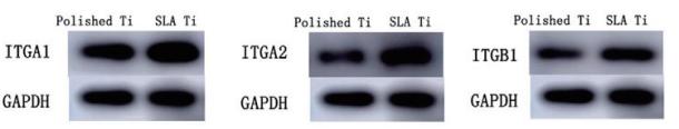
Application: WB Species: mouse Sample: MC3T3-E1 cells
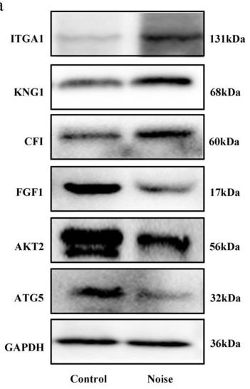
Application: WB Species: Mice Sample:
Restrictive clause
Affinity Biosciences tests all products strictly. Citations are provided as a resource for additional applications that have not been validated by Affinity Biosciences. Please choose the appropriate format for each application and consult Materials and Methods sections for additional details about the use of any product in these publications.
For Research Use Only.
Not for use in diagnostic or therapeutic procedures. Not for resale. Not for distribution without written consent. Affinity Biosciences will not be held responsible for patent infringement or other violations that may occur with the use of our products. Affinity Biosciences, Affinity Biosciences Logo and all other trademarks are the property of Affinity Biosciences LTD.