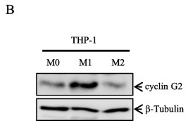
Product: CCNG2 Antibody
Catalog: DF2284
Description: Rabbit polyclonal antibody to CCNG2
Application: WB IF/ICC
Reactivity: Human, Mouse
Prediction: Bovine, Horse, Sheep, Rabbit, Dog, Chicken
Mol.Wt.: 39 kD; 39kD(Calculated).
Uniprot: Q16589
RRID: AB_2839511

View similar products>>

   Size Price Inventory
 100ul $280 In stock
 200ul $350 In stock

Lead Time: Same day delivery

For pricing and ordering contact:
Local distributors

Product Info

Source:
Rabbit
Application:
WB 1:500-1:2000, IF/ICC 1:100-1:500
*The optimal dilutions should be determined by the end user.
*Tips:

WB: For western blot detection of denatured protein samples. IHC: For immunohistochemical detection of paraffin sections (IHC-p) or frozen sections (IHC-f) of tissue samples. IF/ICC: For immunofluorescence detection of cell samples. ELISA(peptide): For ELISA detection of antigenic peptide.

Reactivity:
Human,Mouse
Prediction:
Bovine(100%), Horse(88%), Sheep(100%), Rabbit(100%), Dog(100%), Chicken(100%)
Clonality:
Polyclonal
Specificity:
CCNG2 Antibody detects endogenous levels of total CCNG2.
RRID:
AB_2839511
Cite Format: Affinity Biosciences Cat# DF2284, RRID:AB_2839511.
Conjugate:
Unconjugated.
Purification:
The antiserum was purified by peptide affinity chromatography using SulfoLink™ Coupling Resin (Thermo Fisher Scientific).
Storage:
Rabbit IgG in phosphate buffered saline, pH 7.4, 150mM NaCl, 0.02% sodium azide and 50% glycerol. Store at -20 °C. Stable for 12 months from date of receipt.
Alias:

Fold/Unfold

CCNG 2; Ccng2; CCNG2 protein; CCNG2_HUMAN; Cyclin-G2; CyclinG2;

Immunogens

Immunogen:
Uniprot:
Gene(ID):
Expression:
Q16589 CCNG2_HUMAN:

High levels in cerebellum, thymus, spleen and prostate. Low levels in skeletal muscle.

Description:
May play a role in growth regulation and in negative regulation of cell cycle progesssion
Sequence:
MKDLGAEHLAGHEGVQLLGLLNVYLEQEERFQPREKGLSLIEATPENDNTLCPGLRNAKVEDLRSLANFFGSCTETFVLAVNILDRFLALMKVKPKHLSCIGVCSFLLAARIVEEDCNIPSTHDVIRISQCKCTASDIKRMEKIISEKLHYELEATTALNFLHLYHTIILCHTSERKEILSLDKLEAQLKACNCRLIFSKAKPSVLALCLLNLEVETLKSVELLEILLLVKKHSKINDTEFFYWRELVSKCLAEYSSPECCKPDLKKLVWIVSRRTAQNLHNSYYSVPELPTIPEGGCFDESESEDSCEDMSCGEESLSSSPPSDQECTFFFNFKVAQTLCFPS

Predictions

Predictions:

Score>80(red) has high confidence and is suggested to be used for WB detection. *The prediction model is mainly based on the alignment of immunogen sequences, the results are for reference only, not as the basis of quality assurance.

Species
Results
Score
Bovine
100
Sheep
100
Dog
100
Chicken
100
Rabbit
100
Horse
88
Pig
0
Xenopus
0
Zebrafish
0
Model Confidence:
High(score>80) Medium(80>score>50) Low(score<50) No confidence

PTMs - Q16589 As Substrate

Site PTM Type Enzyme
K36 Ubiquitination
T134 Phosphorylation
S136 Phosphorylation
K184 Ubiquitination
S220 Phosphorylation
S234 Phosphorylation

Research Backgrounds

Function:

May play a role in growth regulation and in negative regulation of cell cycle progression.

Subcellular Location:

Cytoplasm.

Extracellular region or secreted Cytosol Plasma membrane Cytoskeleton Lysosome Endosome Peroxisome ER Golgi apparatus Nucleus Mitochondrion Manual annotation Automatic computational assertionSubcellular location
Tissue Specificity:

High levels in cerebellum, thymus, spleen and prostate. Low levels in skeletal muscle.

Family&Domains:

Belongs to the cyclin family. Cyclin G subfamily.

Research Fields

· Cellular Processes > Cell growth and death > p53 signaling pathway.   (View pathway)

· Environmental Information Processing > Signal transduction > FoxO signaling pathway.   (View pathway)

References

1). LncRNA AK023391 promotes tumorigenesis and invasion of gastric cancer through activation of the PI3K/Akt signaling pathway. Journal of Experimental & Clinical Cancer Research (PubMed: 29282102) [IF=11.3]

Application: WB    Species: human    Sample: Gastric cancer cells

Fig. 8 | LncRNA AK023391 was involved in the regulation of the PI3K/Akt signaling pathway.e-f Western blotting validation of the effects of AK023391 knockdown on the expression of PI3K/Akt, NF-κB, p53, and FOXO3a pathways, and their downstream transcription factors c-myb, cyclinB1/G2, and BCL-6 in HGC 27, AGS, and SGC-7901 cells

2). Cyclin G2 suppresses Wnt/β-catenin signaling and inhibits gastric cancer cell growth and migration through Dapper1. JOURNAL OF EXPERIMENTAL & CLINICAL CANCER RESEARCH (PubMed: 30547803) [IF=11.3]

Application: WB    Species: human    Sample: three gastric cell lines and human normal gastric epithelial cell

Fig. 1| Cyclin G2 expression is down-regulated in gastric cancer..e Comparison of the protein expression levels of cyclin G2 in three gastric cell lines and human normal gastric epithelial cell line

Application: IHC    Species: human    Sample: SGC-7901 or MGC-803 gastric cancer cells

Fig. 2| Cyclin G2 inhibits the proliferation and migration of SGC-7901 or MGC-803 gastric cancer cells.. i and j Representative Transwell® migration assays in cyclin G2 overexpression SGC-7901 or MGC-803 cells determined by stained with Giemsa. The migration ratio is expressed as a percentage of the control cells at the indicated time-points (n = 3). **P < 0.01

Application: IF/ICC    Species: monkey    Sample: COS-7 cells

Fig. 5| Cyclin G2 inhibits Wnt/β-catenin signaling through DPR1. a Cell lysates from COS-7 cells were immunoprecipitated with Dpr1 or IgG control antibody, followed by western blot analysis with a cyclin G2 antibody (upper panel). Total cellular protein from COS-7 cells was immunoprecipitated with an anti-cyclin G2 or IgG control antibody, then blotted with anti-DPR1 and anti-Dvl2 antibody, respectively (middle panel). Western blot analysis of endogenous Dpr1 and cyclin G2 protein level in COS-7 cells (lower panel). b Direct interaction of Dpr1 and cyclin G2 was detected using Duolink in situ PLA. Red spots represent the interaction of Dpr1 and cyclin G2. The nuclei were stained using DAPI and are shown in blue. Images were acquired using a confocal microscopy with a 40× objective.

3). Cyclin G2 in macrophages triggers CTL-mediated antitumor immunity and antiangiogenesis via interferon-gamma. Journal of Experimental & Clinical Cancer Research (PubMed: 36566226) [IF=11.3]

Application: IHC    Species: Mouse    Sample:

Fig. 1 Knockout of cyclin G2 in macrophages attenuates the tumor suppressive effects of IFN-γ. A CCNG2 mRNA expression in THP-1 cells treated with IFN-γ, LPS, IL-4 and IL-13 was detected RT–qPCR using β-actin as an internal control. Data are presented as the mean ± SD (Data of CCNG2 mRNA expression in THP-1 cells treated with IFN-γ representing 3 independent experiments). B Cyclin G2 protein levels in THP-1 cells treated with IFN-γ, IL-4 and IL-13 were evaluated by western blotting using β-tubulin as a loading control (representing 3 independent experiments). C Cyclin G2 protein levels in the human peripheral blood monocytes were determined by western blotting following IFN-γ, IL-4 and IL-13 treatment. β-tubulin was used as a loading control (representing 3 independent experiments). D After IFN-γ (100 ng/mL) treatment of BMDMs, the cyclin G2 protein expression was detected by western blotting. β-tubulin was used as a loading control (representing 3 independent experiments). E BMDMs were isolated from WT and Ccng2−/− C57BL/6 mice and identified by western blotting. β-tubulin was used as a loading control. F LLC cells were mixed with BMDMs from WT and Ccng2−/− C57BL/6 mice at a ratio of 5:1 and injected subcutaneously into C57BL/6 mice, which were then treated with IFN-γ at specific times. G–J Gross tumors (G), tumor growth curves (H), and tumor weights (I) and volumes (J) measured at the study endpoint. (n = 5). K Representative Ki-67 immunohistochemical staining of tumors from mice in the WT and Ccng2−/− groups. 10× scale bar = 200 μm; 40× scale bar = 50 μm. (L) Graph showing the number of Ki-67-positive cells in each field (n = 5). The data in H, I, J, and L are presented as the mean ± SEM. Data presented in A, I, J, and L were analyzed with the unpaired Student’s t-test. Data in H were analyzed using two-way ANOVA. *p 

Restrictive clause


Affinity Biosciences tests all products strictly. Citations are provided as a resource for additional applications that have not been validated by Affinity Biosciences. Please choose the appropriate format for each application and consult Materials and Methods sections for additional details about the use of any product in these publications.

For Research Use Only.
Not for use in diagnostic or therapeutic procedures. Not for resale. Not for distribution without written consent. Affinity Biosciences will not be held responsible for patent infringement or other violations that may occur with the use of our products. Affinity Biosciences, Affinity Biosciences Logo and all other trademarks are the property of Affinity Biosciences LTD.