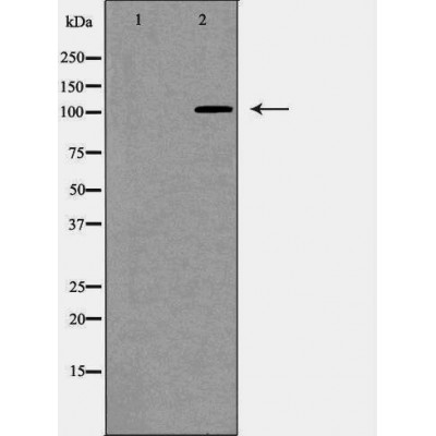
ANPEP/CD13 Antibody - #DF7445
| Product: | ANPEP/CD13 Antibody |
| Catalog: | DF7445 |
| Description: | Rabbit polyclonal antibody to ANPEP/CD13 |
| Application: | WB IHC |
| Reactivity: | Human, Mouse, Rat |
| Prediction: | Pig, Bovine, Horse, Sheep, Rabbit, Dog, Chicken |
| Mol.Wt.: | 109kDa; 110kD(Calculated). |
| Uniprot: | P15144 |
| RRID: | AB_2839383 |
Related Downloads
Protocols
Product Info
*The optimal dilutions should be determined by the end user. For optimal experimental results, antibody reuse is not recommended.
*Tips:
WB: For western blot detection of denatured protein samples. IHC: For immunohistochemical detection of paraffin sections (IHC-p) or frozen sections (IHC-f) of tissue samples. IF/ICC: For immunofluorescence detection of cell samples. ELISA(peptide): For ELISA detection of antigenic peptide.
Cite Format: Affinity Biosciences Cat# DF7445, RRID:AB_2839383.
Fold/Unfold
Alanyl (membrane) aminopeptidase; Alanyl aminopeptidase; Aminopeptidase M; Aminopeptidase N; AMPN_HUMAN; ANPEP; AP M; AP N; AP-M; AP-N; APN; CD 13; CD13; CD13 antigen; gp150; hAPN; LAP 1; LAP1; Microsomal aminopeptidase; Myeloid plasma membrane glycoprotein CD13; p150; PEPN;
Immunogens
A synthesized peptide derived from human ANPEP/CD13, corresponding to a region within the internal amino acids.
Expressed in epithelial cells of the kidney, intestine, and respiratory tract; granulocytes, monocytes, fibroblasts, endothelial cells, cerebral pericytes at the blood-brain barrier, synaptic membranes of cells in the CNS. Also expressed in endometrial stromal cells, but not in the endometrial glandular cells. Found in the vasculature of tissues that undergo angiogenesis and in malignant gliomas and lymph node metastases from multiple tumor types but not in blood vessels of normal tissues. A soluble form has been found in plasma. It is found to be elevated in plasma and effusions of cancer patients.
- P15144 AMPN_HUMAN:
- Protein BLAST With
- NCBI/
- ExPASy/
- Uniprot
MAKGFYISKSLGILGILLGVAAVCTIIALSVVYSQEKNKNANSSPVASTTPSASATTNPASATTLDQSKAWNRYRLPNTLKPDSYRVTLRPYLTPNDRGLYVFKGSSTVRFTCKEATDVIIIHSKKLNYTLSQGHRVVLRGVGGSQPPDIDKTELVEPTEYLVVHLKGSLVKDSQYEMDSEFEGELADDLAGFYRSEYMEGNVRKVVATTQMQAADARKSFPCFDEPAMKAEFNITLIHPKDLTALSNMLPKGPSTPLPEDPNWNVTEFHTTPKMSTYLLAFIVSEFDYVEKQASNGVLIRIWARPSAIAAGHGDYALNVTGPILNFFAGHYDTPYPLPKSDQIGLPDFNAGAMENWGLVTYRENSLLFDPLSSSSSNKERVVTVIAHELAHQWFGNLVTIEWWNDLWLNEGFASYVEYLGADYAEPTWNLKDLMVLNDVYRVMAVDALASSHPLSTPASEINTPAQISELFDAISYSKGASVLRMLSSFLSEDVFKQGLASYLHTFAYQNTIYLNLWDHLQEAVNNRSIQLPTTVRDIMNRWTLQMGFPVITVDTSTGTLSQEHFLLDPDSNVTRPSEFNYVWIVPITSIRDGRQQQDYWLIDVRAQNDLFSTSGNEWVLLNLNVTGYYRVNYDEENWRKIQTQLQRDHSAIPVINRAQIINDAFNLASAHKVPVTLALNNTLFLIEERQYMPWEAALSSLSYFKLMFDRSEVYGPMKNYLKKQVTPLFIHFRNNTNNWREIPENLMDQYSEVNAISTACSNGVPECEEMVSGLFKQWMENPNNNPIHPNLRSTVYCNAIAQGGEEEWDFAWEQFRNATLVNEADKLRAALACSKELWILNRYLSYTLNPDLIRKQDATSTIISITNNVIGQGLVWDFVQSNWKKLFNDYGGGSFSFSNLIQAVTRRFSTEYELQQLEQFKKDNEETGFGSGTRALEQALEKTKANIKWVKENKEVVLQWFTENSK
Predictions
Score>80(red) has high confidence and is suggested to be used for WB detection. *The prediction model is mainly based on the alignment of immunogen sequences, the results are for reference only, not as the basis of quality assurance.
High(score>80) Medium(80>score>50) Low(score<50) No confidence
Research Backgrounds
Broad specificity aminopeptidase which plays a role in the final digestion of peptides generated from hydrolysis of proteins by gastric and pancreatic proteases. Also involved in the processing of various peptides including peptide hormones, such as angiotensin III and IV, neuropeptides, and chemokines. May also be involved the cleavage of peptides bound to major histocompatibility complex class II molecules of antigen presenting cells. May have a role in angiogenesis and promote cholesterol crystallization. May have a role in amino acid transport by acting as binding partner of amino acid transporter SLC6A19 and regulating its activity (By similarity).
(Microbial infection) Acts as a receptor for human coronavirus 229E/HCoV-229E. In case of human coronavirus 229E (HCoV-229E) infection, serves as receptor for HCoV-229E spike glycoprotein.
(Microbial infection) Mediates as well Human cytomegalovirus (HCMV) infection.
Sulfated.
N- and O-glycosylated.
May undergo proteolysis and give rise to a soluble form.
Cell membrane>Single-pass type II membrane protein.
Note: Also found as a soluble form.
Expressed in epithelial cells of the kidney, intestine, and respiratory tract; granulocytes, monocytes, fibroblasts, endothelial cells, cerebral pericytes at the blood-brain barrier, synaptic membranes of cells in the CNS. Also expressed in endometrial stromal cells, but not in the endometrial glandular cells. Found in the vasculature of tissues that undergo angiogenesis and in malignant gliomas and lymph node metastases from multiple tumor types but not in blood vessels of normal tissues. A soluble form has been found in plasma. It is found to be elevated in plasma and effusions of cancer patients.
Belongs to the peptidase M1 family.
Research Fields
· Metabolism > Metabolism of other amino acids > Glutathione metabolism.
· Metabolism > Global and overview maps > Metabolic pathways.
· Organismal Systems > Endocrine system > Renin-angiotensin system. (View pathway)
· Organismal Systems > Immune system > Hematopoietic cell lineage. (View pathway)
References
Restrictive clause
Affinity Biosciences tests all products strictly. Citations are provided as a resource for additional applications that have not been validated by Affinity Biosciences. Please choose the appropriate format for each application and consult Materials and Methods sections for additional details about the use of any product in these publications.
For Research Use Only.
Not for use in diagnostic or therapeutic procedures. Not for resale. Not for distribution without written consent. Affinity Biosciences will not be held responsible for patent infringement or other violations that may occur with the use of our products. Affinity Biosciences, Affinity Biosciences Logo and all other trademarks are the property of Affinity Biosciences LTD.
