Product: ZEB1 Antibody
Catalog: DF7414
Description: Rabbit polyclonal antibody to ZEB1
Application: WB IHC IF/ICC IHC-F
Reactivity: Human, Mouse, Rat
Prediction: Pig, Bovine, Sheep, Rabbit, Dog, Chicken, Xenopus
Mol.Wt.: 124kDa,180kDa; 124kD(Calculated).
Uniprot: P37275
RRID: AB_2839352

View similar products>>

   Size Price Inventory
 100ul $280 In stock
 200ul $350 In stock

Lead Time: Same day delivery

For pricing and ordering contact:
Local distributors

Product Info

Source:
Rabbit
Application:
WB 1:500-1:2000, IHC 1:50-1:200, IF/ICC 1:100
*The optimal dilutions should be determined by the end user.
*Tips:

WB: For western blot detection of denatured protein samples. IHC: For immunohistochemical detection of paraffin sections (IHC-p) or frozen sections (IHC-f) of tissue samples. IF/ICC: For immunofluorescence detection of cell samples. ELISA(peptide): For ELISA detection of antigenic peptide.

Reactivity:
Human,Mouse,Rat
Prediction:
Pig(100%), Bovine(100%), Sheep(100%), Rabbit(100%), Dog(100%), Chicken(100%), Xenopus(100%)
Clonality:
Polyclonal
Specificity:
ZEB1 Antibody detects endogenous levels of total ZEB1.
RRID:
AB_2839352
Cite Format: Affinity Biosciences Cat# DF7414, RRID:AB_2839352.
Conjugate:
Unconjugated.
Purification:
The antiserum was purified by peptide affinity chromatography using SulfoLink™ Coupling Resin (Thermo Fisher Scientific).
Storage:
Rabbit IgG in phosphate buffered saline , pH 7.4, 150mM NaCl, 0.02% sodium azide and 50% glycerol. Store at -20 °C. Stable for 12 months from date of receipt.
Alias:

Fold/Unfold

AREB 6; BZP; Delta crystallin enhancer binding factor 1; DELTA EF1; FECD6; MGC133261; Negative regulator of IL 2; Negative regulator of IL2; NIL 2 A; NIL 2 A zinc finger protein; NIL 2A; NIL-2-A zinc finger protein; NIL2A; Posterior polymorphous corneal dystrophy 3; PPCD3; Represses interleukin 2 expression; TCF 8; TCF-8; TCF8; Transcription factor 8 (represses interleukin 2 expression); Transcription factor 8; ZEB 1; ZEB; ZEB1; ZEB1_HUMAN; ZFHEP; ZFHX 1A; ZFHX1A; Zinc finger E box binding homeobox 1; Zinc finger E-box-binding homeobox 1; Zinc finger homeodomain enhancer binding protein;

Immunogens

Immunogen:
Uniprot:
Gene(ID):
Expression:
P37275 ZEB1_HUMAN:

Colocalizes with SMARCA4/BRG1 in E-cadherin-negative cells from established lines, and stroma of normal colon as well as in de-differentiated epithelial cells at the invasion front of colorectal carcinomas (at protein level). Expressed in heart and skeletal muscle, but not in liver, spleen, or pancreas.

Description:
This gene encodes a zinc finger transcription factor. The encoded protein likely plays a role in transcriptional repression of interleukin 2. Mutations in this gene have been associated with posterior polymorphous corneal dystrophy-3 and late-onset Fuchs endothelial corneal dystrophy. Alternatively spliced transcript variants encoding different isoforms have been described.
Sequence:
MADGPRCKRRKQANPRRNNVTNYNTVVETNSDSDDEDKLHIVEEESVTDAADCEGVPEDDLPTDQTVLPGRSSEREGNAKNCWEDDRKEGQEILGPEAQADEAGCTVKDDECESDAENEQNHDPNVEEFLQQQDTAVIFPEAPEEDQRQGTPEASGHDENGTPDAFSQLLTCPYCDRGYKRFTSLKEHIKYRHEKNEDNFSCSLCSYTFAYRTQLERHMTSHKSGRDQRHVTQSGCNRKFKCTECGKAFKYKHHLKEHLRIHSGEKPYECPNCKKRFSHSGSYSSHISSKKCISLIPVNGRPRTGLKTSQCSSPSLSASPGSPTRPQIRQKIENKPLQEQLSVNQIKTEPVDYEFKPIVVASGINCSTPLQNGVFTGGGPLQATSSPQGMVQAVVLPTVGLVSPISINLSDIQNVLKVAVDGNVIRQVLENNQANLASKEQETINASPIQQGGHSVISAISLPLVDQDGTTKIIINYSLEQPSQLQVVPQNLKKENPVATNSCKSEKLPEDLTVKSEKDKSFEGGVNDSTCLLCDDCPGDINALPELKHYDLKQPTQPPPLPAAEAEKPESSVSSATGDGNLSPSQPPLKNLLSLLKAYYALNAQPSAEELSKIADSVNLPLDVVKKWFEKMQAGQISVQSSEPSSPEPGKVNIPAKNNDQPQSANANEPQDSTVNLQSPLKMTNSPVLPVGSTTNGSRSSTPSPSPLNLSSSRNTQGYLYTAEGAQEEPQVEPLDLSLPKQQGELLERSTITSVYQNSVYSVQEEPLNLSCAKKEPQKDSCVTDSEPVVNVIPPSANPINIAIPTVTAQLPTIVAIADQNSVPCLRALAANKQTILIPQVAYTYSTTVSPAVQEPPLKVIQPNGNQDERQDTSSEGVSNVEDQNDSDSTPPKKKMRKTENGMYACDLCDKIFQKSSSLLRHKYEHTGKRPHECGICKKAFKHKHHLIEHMRLHSGEKPYQCDKCGKRFSHSGSYSQHMNHRYSYCKREAEERDSTEQEEAGPEILSNEHVGARASPSQGDSDERESLTREEDEDSEKEEEEEDKEMEELQEEKECEKPQGDEEEEEEEEEVEEEEVEEAENEGEEAKTEGLMKDDRAESQASSLGQKVGESSEQVSEEKTNEA

Predictions

Predictions:

Score>80(red) has high confidence and is suggested to be used for WB detection. *The prediction model is mainly based on the alignment of immunogen sequences, the results are for reference only, not as the basis of quality assurance.

Species
Results
Score
Pig
100
Bovine
100
Sheep
100
Dog
100
Xenopus
100
Chicken
100
Rabbit
100
Horse
0
Zebrafish
0
Model Confidence:
High(score>80) Medium(80>score>50) Low(score<50) No confidence

PTMs - P37275 As Substrate

Site PTM Type Enzyme
S31 Phosphorylation
S33 Phosphorylation
T151 Phosphorylation
S155 Phosphorylation
T162 Phosphorylation
S184 Phosphorylation
K186 Ubiquitination
T243 Phosphorylation
S263 Phosphorylation
S282 Phosphorylation
S285 Phosphorylation
S288 Phosphorylation
S289 Phosphorylation
S309 Phosphorylation
S312 Phosphorylation
S313 Phosphorylation
S315 Phosphorylation
S317 Phosphorylation
S319 Phosphorylation
S322 Phosphorylation
T324 Phosphorylation
K335 Ubiquitination
S342 Phosphorylation
K347 Sumoylation
T443 Phosphorylation
S447 Phosphorylation
S455 Phosphorylation
K493 Sumoylation
K504 Sumoylation
K504 Ubiquitination
K507 Ubiquitination
K515 Sumoylation
K515 Ubiquitination
S516 Phosphorylation
S521 Phosphorylation
S529 Phosphorylation
S583 Phosphorylation
S585 Phosphorylation
K613 Ubiquitination
S641 Phosphorylation
S642 Phosphorylation
S645 Phosphorylation
S646 Phosphorylation
S679 Phosphorylation
S686 Phosphorylation
S693 Phosphorylation
S698 Phosphorylation
S700 Phosphorylation
S701 Phosphorylation
T702 Phosphorylation
S704 Phosphorylation
S706 Phosphorylation
T716 Phosphorylation
S850 Phosphorylation
S887 Phosphorylation
S889 Phosphorylation
T890 Phosphorylation
T927 Phosphorylation
S955 Phosphorylation
S995 Phosphorylation
T996 Phosphorylation
S1016 Phosphorylation
S1018 Phosphorylation
S1022 Phosphorylation
S1036 Phosphorylation
S1100 Phosphorylation
K1108 Ubiquitination

Research Backgrounds

Function:

Acts as a transcriptional repressor. Inhibits interleukin-2 (IL-2) gene expression. Enhances or represses the promoter activity of the ATP1A1 gene depending on the quantity of cDNA and on the cell type. Represses E-cadherin promoter and induces an epithelial-mesenchymal transition (EMT) by recruiting SMARCA4/BRG1. Represses BCL6 transcription in the presence of the corepressor CTBP1. Positively regulates neuronal differentiation. Represses RCOR1 transcription activation during neurogenesis. Represses transcription by binding to the E box (5'-CANNTG-3'). Promotes tumorigenicity by repressing stemness-inhibiting microRNAs.

Subcellular Location:

Nucleus.

Extracellular region or secreted Cytosol Plasma membrane Cytoskeleton Lysosome Endosome Peroxisome ER Golgi apparatus Nucleus Mitochondrion Manual annotation Automatic computational assertionSubcellular location
Tissue Specificity:

Colocalizes with SMARCA4/BRG1 in E-cadherin-negative cells from established lines, and stroma of normal colon as well as in de-differentiated epithelial cells at the invasion front of colorectal carcinomas (at protein level). Expressed in heart and skeletal muscle, but not in liver, spleen, or pancreas.

Subunit Structure:

Interacts (via N-terminus) with SMARCA4/BRG1.

Family&Domains:

Belongs to the delta-EF1/ZFH-1 C2H2-type zinc-finger family.

Research Fields

· Human Diseases > Cancers: Overview > Transcriptional misregulation in cancer.

· Human Diseases > Cancers: Overview > MicroRNAs in cancer.

· Human Diseases > Cancers: Specific types > Prostate cancer.   (View pathway)

References

1). Salidroside improves the hypoxic tumor microenvironment and reverses the drug resistance of platinum drugs via HIF-1α signaling pathway. EBioMedicine, 2018 (PubMed: 30396856) [IF=11.1]

Application: WB    Species: human    Sample: PLC/PRF/5 cells

Fig. 3.|Sal reversed the drug resistance and inhibited HIF-1α signaling pathway. (I) Protein expression level of Twist1, Zeb1 and E-cadherin in PLC/PRF/5 cells treated hypoxia and hypoxia combined with Sal. Error bars represent the standard deviation of experiments performed in triplicate (*P b .05, **P b .01).

2). Hypoxia-induced ZEB1 promotes cervical cancer progression via CCL8-dependent tumour-associated macrophage recruitment. Cell Death & Disease, 2019 (PubMed: 31263103) [IF=9.0]

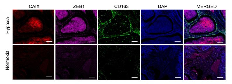
Application: IF/ICC    Species: human    Sample: human cervical cancer tissues

Fig. 1| The number of TAMs is correlated with ZEB1 expression in the hypoxic regions of cervical cancer. a Immunofluorescence staining of CAIX, ZEB1, and CD163+ TAMs in normoxic and hypoxic areas of human cervical cancer tissues (scale bar, 100 μm).

Application: WB    Species: human    Sample: hypoxic cancer cells

Fig. 2| Hypoxia-induced ZEB1 in cancer cells enhances TAM migration in vitro. A Immunofluorescence revealed that hypoxic SiHa and C33a cells exhibited upregulated expression of CAIX (green) and ZEB1 (red). Nuclei were stained with DAPI (blue) (scale bar, 50 μm). b Western blotting shows that the protein levels of both ZEB1 (upper panel) and CAIX (middle panel) were higher in hypoxic cancer cells than in normoxic cancer cells (left panel shows SiHa cells; right panel shows C33a cells).

3). d-Borneol enhances cisplatin sensitivity via autophagy dependent EMT signaling and NCOA4-mediated ferritinophagy. PHYTOMEDICINE, 2022 (PubMed: 36030746) [IF=7.9]

4). MicroRNA-181a regulates epithelial-mesenchymal transition by targeting PTEN in drug-resistant lung adenocarcinoma cells. INTERNATIONAL JOURNAL OF ONCOLOGY, 2015 (PubMed: 26323677) [IF=5.2]

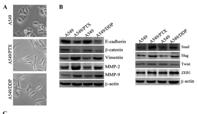
Application: WB    Species: human    Sample: A549 cells

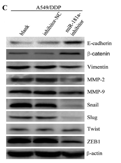
Figure 3. A549/DDP and A549/PTX cells showed molecular and morphological changes that were consistent with EMT. (A) microscopy at x200 magnification was used to assess cell morphology. The A549 cells (parental cells) had an epithelioid, rounded cobblestone appearance and there was limited formation of pseudopodia. A549/PTX and A549/DDP cells exhibited a spindle-shaped morphology and an increased formation of pseudopodia, indicating a loss of cell polarity. (B) E-cadherin, β-catenin, vimentin, MMP-2 and MMP-9 which are EMT-related proteins, were assessed in terms of expression levels. EMT-related transcription factors (Snail, Slug, Twist and ZEB1) were measured in A549/PTX and A549/DDP cells using western blot analysis. (C) The expression changes were confirmed at the mRNA level by qRT-PCR. Expression was standardized to the expression of GAPDH and normalized to 1.0 in the parental cells (compared with the parental A549 cells, means ± SEM, n=3, * P<0.05)

Application: WB    Species: human    Sample: A549/PTX cells

Figure 8.| miR-181a downregulation in A549/DDP cells reverses both drug resistance and morphological and molecular changes. (C) Expression of E-cadherin, β-catenin, vimentin, MMP-2, MMP-9, Snail, Slug, Twist and ZEB1 in A549/DDP cells was measured with western blotting.

5). The regulatory effects of metformin on the [SNAIL/miR-34]:[ZEB/miR-200] system in the epithelial-mesenchymal transition(EMT) for colorectal cancer(CRC). European Journal of Pharmacology, 2018 (PubMed: 30017802) [IF=5.0]

Application: WB    Species: human    Sample: SW480 and HCT116 cells

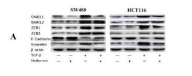
Fig. 3. |Western-blot for SW480 and HCT116 cells (A)(B) Protein strips and quantitative analysis: significant decreases in the SNAIL1, ZEB1 and vimentin and a significant increase in the E-cadherin of metformin and TGF-β-treated samples compared with only TGF-β-treated samples for both SW480 and HCT116 cells; a significant increase in the ZEB2 of metformin and TGF-β-treated samples compared with only TGF-β-treated samples for SW480 cells; significant decreases in the SNAIL2, ZEB1 and ZEB2 of metformin-treated samples compared with negative control samples for HCT116 cells. *P<0.05

Application: IF/ICC    Species: human    Sample: SW480 and HCT116 cells

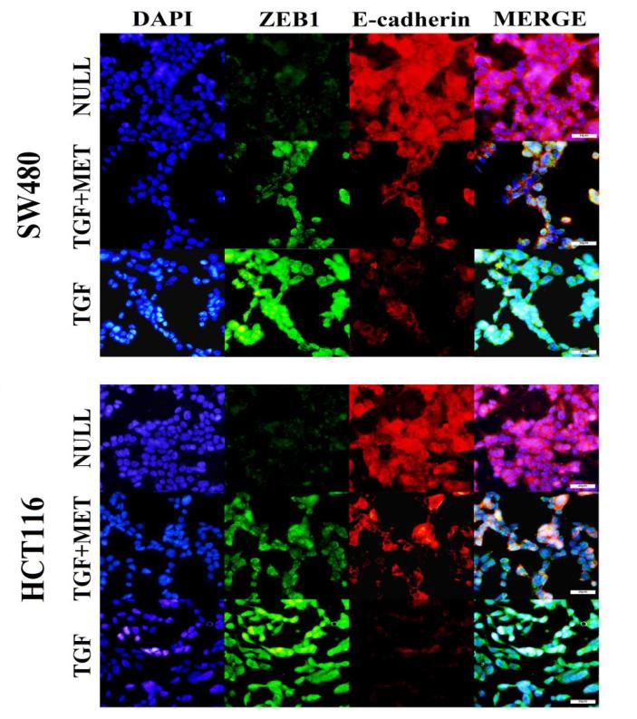
Fig. 5.| Immunofluorescence detection for SW480 and HCT116 cells: (A) The coverglass with metformin and TGF-β-treated cells showed stronger E-cadherin(red) signals and weaker ZEB1(green) signals than with only TGF-β-treated cells; there were more hybrid cells(stacked signal) on the coverglass with metformin and TGF-βtreated cells.

6). MicroRNA-145 promotes the epithelial-mesenchymal transition in peritoneal dialysis-associated fibrosis by suppressing fibroblast growth factor 10. Journal of Biological Chemistry, 2019 (PubMed: 31431501) [IF=4.8]

7). Value of Pyruvate Carboxylase in Thyroid Fine-Needle Aspiration Wash-Out Fluid for Predicting Papillary Thyroid Cancer Lymph Node Metastasis. Frontiers in Oncology, 2021 (PubMed: 34136384) [IF=4.7]

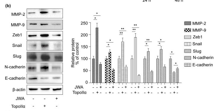
8). EGCG regulates the cross-talk between JWA and topoisomerase IIα in non-small-cell lung cancer (NSCLC) cells. Scientific Reports, 2015 (PubMed: 26046674) [IF=4.6]

Application: WB    Species: human    Sample: NCI-H460 cells


9). Estradiol promotes EMT in endometriosis via MALAT1/miR200s sponge function. REPRODUCTION, 2019 (PubMed: 30500775) [IF=3.8]

Application: WB    Species: human    Sample: EECs

Figure 2.| (A and B) Expression levels of EMT-associated markers (Zeb1, Zeb2, E-cadherin and Vimentin) with E2 treatment for different doses (0, 10−12, 10−10, 10−8, 10−6 mol/L) and time (0, 24, 48 or 72 h) in EECs detected by Western blot.(M: mol/L); (C and D) Expression levels of MALAT1 and miR200s under E2 for various doses (0, 10−12, 10−10, 10−8, 10−6 mol/L) for 24 h in EECs measured by qRT-PCR. Data were evaluated by one-way ANOVA analysis (*P < 0.05, **P < 0.01, ***P < 0.001 compared with untreated group).

10). MicroRNA-139-5p affects cisplatin sensitivity in human nasopharyngeal carcinoma cells by regulating the epithelial-to-mesenchymal transition. GENE, 2018 (PubMed: 29427737) [IF=3.5]

Load more

Restrictive clause


Affinity Biosciences tests all products strictly. Citations are provided as a resource for additional applications that have not been validated by Affinity Biosciences. Please choose the appropriate format for each application and consult Materials and Methods sections for additional details about the use of any product in these publications.

For Research Use Only.
Not for use in diagnostic or therapeutic procedures. Not for resale. Not for distribution without written consent. Affinity Biosciences will not be held responsible for patent infringement or other violations that may occur with the use of our products. Affinity Biosciences, Affinity Biosciences Logo and all other trademarks are the property of Affinity Biosciences LTD.