Product: PLAU Antibody
Catalog: DF6904
Description: Rabbit polyclonal antibody to PLAU
Application: WB IHC
Reactivity: Human, Mouse, Rat, Monkey
Prediction: Horse, Rabbit
Mol.Wt.: 49kDa, 57 kDa; 49kD(Calculated).
Uniprot: P00749
RRID: AB_2838863

View similar products>>

   Size Price Inventory
 100ul $280 In stock
 200ul $350 In stock

Lead Time: Same day delivery

For pricing and ordering contact:
Local distributors

Product Info

Source:
Rabbit
Application:
WB 1:500-1:2000, IHC 1:50-1:200
*The optimal dilutions should be determined by the end user.
*Tips:

WB: For western blot detection of denatured protein samples. IHC: For immunohistochemical detection of paraffin sections (IHC-p) or frozen sections (IHC-f) of tissue samples. IF/ICC: For immunofluorescence detection of cell samples. ELISA(peptide): For ELISA detection of antigenic peptide.

Reactivity:
Human,Mouse,Rat,Monkey
Prediction:
Horse(82%), Rabbit(82%)
Clonality:
Polyclonal
Specificity:
PLAU Antibody detects endogenous levels of total PLAU.
RRID:
AB_2838863
Cite Format: Affinity Biosciences Cat# DF6904, RRID:AB_2838863.
Conjugate:
Unconjugated.
Purification:
The antiserum was purified by peptide affinity chromatography using SulfoLink™ Coupling Resin (Thermo Fisher Scientific).
Storage:
Rabbit IgG in phosphate buffered saline , pH 7.4, 150mM NaCl, 0.02% sodium azide and 50% glycerol. Store at -20 °C. Stable for 12 months from date of receipt.
Alias:

Fold/Unfold

ATF; ATF uPA; BDPLT5; Plasminogen activator; Plasminogen activator urinary; Plasminogen activator urokinase; PLAU; QPD; u PA; U plasminogen activator; u-PA; U-plasminogen activator; uPA; URK; UROK_HUMAN; Urokinase plasminogen activator; Urokinase type plasminogen activator; Urokinase type plasminogen activator precursor; Urokinase-type plasminogen activator chain B;

Immunogens

Immunogen:
Uniprot:
Gene(ID):
Expression:
P00749 UROK_HUMAN:

Expressed in the prostate gland and prostate cancers.

Description:
The human urokinase-type plasminogen activator receptor (uPAR) is a 55-65 kDa, highly glycosylated, GPI-anchored cell surface receptor (the deglycosylated protein is 35 kDa) (1-3). It is a central player in the plasminogen activation pathway. uPAR binds with high affinity to a serine protease urokinase-type plasminogen activator (uPA) and converts plasminogen to its active form plasmin in a spatially restricted manner on the cell surface (4). Plasmin further carries out the activation of uPA, which is inhibited by serpins, such as plasminogen activator inhibitors (5). Therefore, uPAR plays a key role in regulating extracellular proteolysis. In addition, uPAR plays an important role in regulating cell proliferation, adhesion, and mobility (6,7). Research studies have shown that overexpression of uPAR is found in various cancer cells and tissues (8,9).
Sequence:
MRALLARLLLCVLVVSDSKGSNELHQVPSNCDCLNGGTCVSNKYFSNIHWCNCPKKFGGQHCEIDKSKTCYEGNGHFYRGKASTDTMGRPCLPWNSATVLQQTYHAHRSDALQLGLGKHNYCRNPDNRRRPWCYVQVGLKPLVQECMVHDCADGKKPSSPPEELKFQCGQKTLRPRFKIIGGEFTTIENQPWFAAIYRRHRGGSVTYVCGGSLISPCWVISATHCFIDYPKKEDYIVYLGRSRLNSNTQGEMKFEVENLILHKDYSADTLAHHNDIALLKIRSKEGRCAQPSRTIQTICLPSMYNDPQFGTSCEITGFGKENSTDYLYPEQLKMTVVKLISHRECQQPHYYGSEVTTKMLCAADPQWKTDSCQGDSGGPLVCSLQGRMTLTGIVSWGRGCALKDKPGVYTRVSHFLPWIRSHTKEENGLAL

Predictions

Predictions:

Score>80(red) has high confidence and is suggested to be used for WB detection. *The prediction model is mainly based on the alignment of immunogen sequences, the results are for reference only, not as the basis of quality assurance.

Species
Results
Score
Rabbit
82
Horse
82
Bovine
64
Sheep
64
Pig
0
Dog
0
Xenopus
0
Zebrafish
0
Chicken
0
Model Confidence:
High(score>80) Medium(80>score>50) Low(score<50) No confidence

PTMs - P00749 As Substrate

Site PTM Type Enzyme
T38 O-Glycosylation
T38 Phosphorylation
S41 Phosphorylation
S158 O-Glycosylation
S158 Phosphorylation P17252 (PRKCA)
S323 Phosphorylation P17252 (PRKCA)
K424 Ubiquitination

Research Backgrounds

Function:

Specifically cleaves the zymogen plasminogen to form the active enzyme plasmin.

PTMs:

Phosphorylation of Ser-158 and Ser-323 abolishes proadhesive ability but does not interfere with receptor binding.

Subcellular Location:

Secreted.

Extracellular region or secreted Cytosol Plasma membrane Cytoskeleton Lysosome Endosome Peroxisome ER Golgi apparatus Nucleus Mitochondrion Manual annotation Automatic computational assertionSubcellular location
Tissue Specificity:

Expressed in the prostate gland and prostate cancers.

Subunit Structure:

Found in high and low molecular mass forms. Each consists of two chains, A and B. The high molecular mass form contains a long chain A which is cleaved to yield a short chain A. Forms heterodimer with SERPINA5. Binds LRP1B; binding is followed by internalization and degradation. Interacts with MRC2. Interacts with PLAUR. In complex with SERPINE1, interacts with PLAUR/uPAR. Interacts with SORL1 and LRP1, either alone or in complex with SERPINE1; these interactions are abolished in the presence of LRPAP1/RAP. The ternary complex composed of PLAUR-PLAU-PAI1 also interacts with SORLA.

Family&Domains:

Belongs to the peptidase S1 family.

Research Fields

· Environmental Information Processing > Signal transduction > NF-kappa B signaling pathway.   (View pathway)

· Human Diseases > Cancers: Overview > Transcriptional misregulation in cancer.

· Human Diseases > Cancers: Overview > Proteoglycans in cancer.

· Human Diseases > Cancers: Overview > MicroRNAs in cancer.

· Human Diseases > Cancers: Specific types > Prostate cancer.   (View pathway)

· Organismal Systems > Immune system > Complement and coagulation cascades.   (View pathway)

References

1). Fucoxanthin Activates Apoptosis via Inhibition of PI3K/Akt/mTOR Pathway and Suppresses Invasion and Migration by Restriction of p38-MMP-2/9 Pathway in Human Glioblastoma Cells. NEUROCHEMICAL RESEARCH (PubMed: 27394418) [IF=4.4]

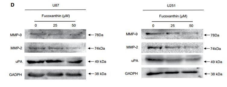
Application: WB    Species: human    Sample:

d Cell lysates were electrophoresed and MMP- 2/9 and uPA proteins were detected by their respective specific antibodies in indicated concentrations

2). Loureirin B Alleviates Myocardial Ischemia/Reperfusion Injury via Inhibiting PAI-1/TGF-β1/Smad Signaling Pathway. Evidence-based Complementary and Alternative Medicine (PubMed: 35535157)

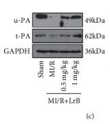
Application: WB    Species: Mice    Sample:

Figure 4 LrB suppressed the expression of PAI-1 and the activation of TGF-β1/Smad signaling pathway in MI/R mice. (a) Representative images showing the distribution of PAI-1 in cardiac tissues after immunofluorescence staining. Scar bar = 50 μm. (b, c) Relative mRNA and protein expression of urokinase plasminogen activator (uPA) and tissue-type plasminogen activator (tPA) in the cardiac tissues. (d) Western blot analysis for TGF-β1R, TGF-β1, p-Smad2/3, and Smad2/3. Relative protein expression of (e) PAI-1, (f) TGF-β1R, (g) TGF-β1, (h) p-Smad2/3, and (i) Smad2/3 in the cardiac tissues. Values are expressed as mean ± SD (n = 6). +++p < 0.001vs. sham group. ∗p < 0.05, ∗∗p < 0.01, ∗∗∗p < 0.001vs. MI/R group.

Restrictive clause


Affinity Biosciences tests all products strictly. Citations are provided as a resource for additional applications that have not been validated by Affinity Biosciences. Please choose the appropriate format for each application and consult Materials and Methods sections for additional details about the use of any product in these publications.

For Research Use Only.
Not for use in diagnostic or therapeutic procedures. Not for resale. Not for distribution without written consent. Affinity Biosciences will not be held responsible for patent infringement or other violations that may occur with the use of our products. Affinity Biosciences, Affinity Biosciences Logo and all other trademarks are the property of Affinity Biosciences LTD.