Product: Wnt5a Antibody
Catalog: DF6856
Description: Rabbit polyclonal antibody to Wnt5a
Application: WB IHC
Reactivity: Human, Mouse, Rat
Prediction: Pig, Bovine, Horse, Sheep, Rabbit, Dog, Chicken, Xenopus
Mol.Wt.: 42kDa; 42kD(Calculated).
Uniprot: P41221
RRID: AB_2838815

View similar products>>

   Size Price Inventory
 100ul $280 In stock
 200ul $350 In stock

Lead Time: Same day delivery

For pricing and ordering contact:
Local distributors

Product Info

Source:
Rabbit
Application:
WB 1:500-1:2000, IHC 1:50-1:200
*The optimal dilutions should be determined by the end user.
*Tips:

WB: For western blot detection of denatured protein samples. IHC: For immunohistochemical detection of paraffin sections (IHC-p) or frozen sections (IHC-f) of tissue samples. IF/ICC: For immunofluorescence detection of cell samples. ELISA(peptide): For ELISA detection of antigenic peptide.

Reactivity:
Human,Mouse,Rat
Prediction:
Pig(100%), Bovine(100%), Horse(100%), Sheep(100%), Rabbit(100%), Dog(100%), Chicken(82%), Xenopus(82%)
Clonality:
Polyclonal
Specificity:
Wnt5a Antibody detects endogenous levels of total Wnt5a.
RRID:
AB_2838815
Cite Format: Affinity Biosciences Cat# DF6856, RRID:AB_2838815.
Conjugate:
Unconjugated.
Purification:
The antiserum was purified by peptide affinity chromatography using SulfoLink™ Coupling Resin (Thermo Fisher Scientific).
Storage:
Rabbit IgG in phosphate buffered saline , pH 7.4, 150mM NaCl, 0.02% sodium azide and 50% glycerol. Store at -20 °C. Stable for 12 months from date of receipt.
Alias:

Fold/Unfold

hWNT 5A; hWNT5A; Protein Wnt 5a; Protein Wnt-5a; Protein Wnt5a; Wingless type MMTV integration site family member 5A; Wnt 5a; WNT 5A protein; WNT 5A protein precursor; WNT5A; WNT5A protein precursor; WNT5A_HUMAN;

Immunogens

Immunogen:
Uniprot:
Gene(ID):
Expression:
P41221 WNT5A_HUMAN:

Expression is increased in differentiated thyroid carcinomas compared to normal thyroid tissue and anaplastic thyroid tumors where expression is low or undetectable. Expression is found in thyrocytes but not in stromal cells (at protein level) (PubMed:15735754). Detected in neonate heart and lung (PubMed:8288227).

Description:
The Wnt family includes several secreted glycoproteins that play important roles in animal development (1). There are 19 Wnt genes in the human genome that encode functionally distinct Wnt proteins (2). Wnt members bind to the Frizzled family of seven-pass transmembrane proteins and activate several signaling pathways (3). The canonical Wnt/β-catenin pathway also requires a coreceptor from the low-density lipoprotein receptor family (4). Aberrant activation of Wnt signaling pathways is involved in several types of cancers (5).
Sequence:
MKKSIGILSPGVALGMAGSAMSSKFFLVALAIFFSFAQVVIEANSWWSLGMNNPVQMSEVYIIGAQPLCSQLAGLSQGQKKLCHLYQDHMQYIGEGAKTGIKECQYQFRHRRWNCSTVDNTSVFGRVMQIGSRETAFTYAVSAAGVVNAMSRACREGELSTCGCSRAARPKDLPRDWLWGGCGDNIDYGYRFAKEFVDARERERIHAKGSYESARILMNLHNNEAGRRTVYNLADVACKCHGVSGSCSLKTCWLQLADFRKVGDALKEKYDSAAAMRLNSRGKLVQVNSRFNSPTTQDLVYIDPSPDYCVRNESTGSLGTQGRLCNKTSEGMDGCELMCCGRGYDQFKTVQTERCHCKFHWCCYVKCKKCTEIVDQFVCK

Predictions

Predictions:

Score>80(red) has high confidence and is suggested to be used for WB detection. *The prediction model is mainly based on the alignment of immunogen sequences, the results are for reference only, not as the basis of quality assurance.

Species
Results
Score
Pig
100
Horse
100
Bovine
100
Sheep
100
Dog
100
Rabbit
100
Xenopus
82
Chicken
82
Zebrafish
0
Model Confidence:
High(score>80) Medium(80>score>50) Low(score<50) No confidence

PTMs - P41221 As Substrate

Site PTM Type Enzyme
Y86 Phosphorylation
Y92 Phosphorylation
T99 Phosphorylation
S116 Phosphorylation
T117 Phosphorylation
T121 Phosphorylation
S122 Phosphorylation
K194 Ubiquitination
K208 Ubiquitination
K261 Ubiquitination
Y270 Phosphorylation
S272 Phosphorylation
S280 Phosphorylation
K283 Ubiquitination
K348 Ubiquitination

Research Backgrounds

Function:

Ligand for members of the frizzled family of seven transmembrane receptors. Can activate or inhibit canonical Wnt signaling, depending on receptor context. In the presence of FZD4, activates beta-catenin signaling. In the presence of ROR2, inhibits the canonical Wnt pathway by promoting beta-catenin degradation through a GSK3-independent pathway which involves down-regulation of beta-catenin-induced reporter gene expression (By similarity). Suppression of the canonical pathway allows chondrogenesis to occur and inhibits tumor formation. Stimulates cell migration. Decreases proliferation, migration, invasiveness and clonogenicity of carcinoma cells and may act as a tumor suppressor. Mediates motility of melanoma cells. Required during embryogenesis for extension of the primary anterior-posterior axis and for outgrowth of limbs and the genital tubercle. Inhibits type II collagen expression in chondrocytes (By similarity).

PTMs:

Glycosylation is necessary for secretion but not for activity.

Palmitoleoylation is required for efficient binding to frizzled receptors. Depalmitoleoylation leads to Wnt signaling pathway inhibition.

Proteolytic processing by TIKI1 and TIKI2 promotes oxidation and formation of large disulfide-bond oligomers, leading to inactivation of WNT5A.

Subcellular Location:

Secreted>Extracellular space>Extracellular matrix. Secreted.

Extracellular region or secreted Cytosol Plasma membrane Cytoskeleton Lysosome Endosome Peroxisome ER Golgi apparatus Nucleus Mitochondrion Manual annotation Automatic computational assertionSubcellular location
Tissue Specificity:

Expression is increased in differentiated thyroid carcinomas compared to normal thyroid tissue and anaplastic thyroid tumors where expression is low or undetectable. Expression is found in thyrocytes but not in stromal cells (at protein level). Detected in neonate heart and lung.

Subunit Structure:

Forms a soluble 1:1 complex with AFM; this prevents oligomerization and is required for prolonged biological activity. The complex with AFM may represent the physiological form in body fluids. Homooligomer; disulfide-linked, leading to inactivation (in vitro). Interacts with PORCN. Interacts with WLS (By similarity). Interacts with glypican GCP3. Interacts with PKD1 (via extracellular domain).

Family&Domains:

Belongs to the Wnt family.

Research Fields

· Cellular Processes > Cellular community - eukaryotes > Signaling pathways regulating pluripotency of stem cells.   (View pathway)

· Environmental Information Processing > Signal transduction > mTOR signaling pathway.   (View pathway)

· Environmental Information Processing > Signal transduction > Wnt signaling pathway.   (View pathway)

· Environmental Information Processing > Signal transduction > Hippo signaling pathway.   (View pathway)

· Human Diseases > Infectious diseases: Viral > Human papillomavirus infection.

· Human Diseases > Infectious diseases: Viral > HTLV-I infection.

· Human Diseases > Cancers: Overview > Pathways in cancer.   (View pathway)

· Human Diseases > Cancers: Overview > Proteoglycans in cancer.

· Human Diseases > Cancers: Specific types > Basal cell carcinoma.   (View pathway)

· Human Diseases > Cancers: Specific types > Breast cancer.   (View pathway)

· Human Diseases > Cancers: Specific types > Hepatocellular carcinoma.   (View pathway)

· Human Diseases > Cancers: Specific types > Gastric cancer.   (View pathway)

· Organismal Systems > Development > Axon guidance.   (View pathway)

· Organismal Systems > Endocrine system > Melanogenesis.

References

1). Ginsenoside Rd promotes omentin secretion in adipose through TBK1-AMPK to improve mitochondrial biogenesis via WNT5A/Ca2+ pathways in heart failure. Redox biology, 2023 (PubMed: 36652744) [IF=11.4]

Application: WB    Species: Mouse    Sample:

Fig. 5 Adipose tissue-specific omentin overexpression inhibited WNT5A/Ca2+ signaling pathway and improved mitochondrial biogenesis to ameliorate myocardial injury in HF mice. (A) Serum omentin level of HF patients and healthy subjects. A total of 58 HF patients and 38 healthy subjects were enrolled. (B) The omentin serum content of adipose tissue-specific omentin overexpression by intravenous injection of AAV-omentin and negative control by intravenous injection of AAV-NC (n = 8). (C) The expression of omentin in the inguinal fat of mice with adipose tissue-specific omentin overexpression was detected by immunohistochemistry (n = 4). Scale bar = 200 μm. (D) Representative echocardiographs of mice with AAV-omentin overexpression and the statistical results of (E) LV EF, (F) LV FS, (G) stroke volume, (H) LVPW; d, (I) IVS; d, (J) RWT and (K) LV Mass were presented (n = 8). (L) Representative TTC staining images of heart tissues of mice with AAV-omentin overexpression (n = 6). (M) Representative images of H&E and Masson staining of heart tissues of mice with AAV-omentin overexpression (n = 3), scale bar = 250 μm. (N) The serum content of BNP in mice with AAV-omentin overexpression (n = 8). (O) The serum CK activity in mice with AVV-omentin overexpression (n = 8). (P) The protein expression level of WNT5A, Frizzled2 and p-CAMKII/CAMKII were determined by Western blot. β-actin was used as a loading control (n = 5). (Q) The protein expression level of PGC-1α, NRF1, NRF2 and TFAM were determined by Western blot in mice with AAV-omentin overexpression. β-actin was used as a loading control (n = 5). Values are expressed as the means ± SD. (A) Unpaired Student's two-tailed t-test; (B–Q) One-way ANOVA followed by the Dunnett's post hoc test. #P < 0.05, ##P < 0.01, ###P < 0.001 vs. Sham; *P < 0.05, **P < 0.01, ***P < 0.001 vs. CAL.

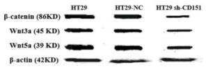
2). CD151 promotes Colorectal Cancer progression by a crosstalk involving CEACAM6, LGR5 and Wnt signaling via TGFβ1. International Journal of Biological Sciences, 2021 (PubMed: 33767593) [IF=9.2]

Application: WB    Species: Mouse    Sample: HT29 and HCT116 cells

Figure 8 CEACAM6, TGFβ1, LGR5, β-catenin, Wnt3a and Wnt5a are downregulated by CD151 silencing in HT29 and HCT116 cells. A and B. Western blot and qRT-PCR showed that CEACAM6, TGFβ1, LGR5 (A), β-catenin, Wnt3a and Wnt5a (B) were downregulated at both protein and mRNA levels by CD151 silencing in HT29 cells, as compared with controls. C and D. Western blot and qRT-PCR demonstrated CEACAM6, TGFβ1, LGR5 (C), and β-catenin, Wnt3a and Wnt5a (D) were downregulated at both protein and mRNA levels by CD151 silencing in HCT116 cells, as compared with controls. *P < 0.05, **P < 0.01.

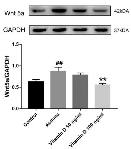
3). Vitamin D alleviates airway remodeling in asthma by down-regulating the activity of Wnt/β-catenin signaling pathway. International Immunopharmacology, 2019 (PubMed: 30616171) [IF=5.6]

Application: WB    Species: rat    Sample: lung

Fig. 5. |Vitamin D reduced the mRNA and protein expression of Wnt5a and β-catenin.The expression of Wnt5a and β-catenin mRNAs in the lung was determined by RT-qPCR (A and B); the expression of Wnt5a and β-catenin was determined by Western blot, which was normalized to the levels of GAPDH, including the quantitation of the ratio between Wnt5a/GAPDH and β-catenin/GAPDH (C and D). The results are expressed as mean ± SEM based on 6 random individual rats. #P < 0.05 and ##P < 0.01 compared with the control group, *P < 0.05, **P < 0.01 compared to the asthma group.

4). 10‐Gingerol alleviates hypoxia/reoxygenation‐induced cardiomyocyte injury through inhibition of the Wnt5a/Frizzled‐2 pathway. Food Science & Nutrition, 2021 (PubMed: 34262748) [IF=3.9]

Application: WB    Species: Rat    Sample: H9c2 cells

FIGURE 10 Effects of 10- Gin on the Wnt5a (a, c, e) and Frizzled- 2 (b, d, f) gene and protein expressions. Data are represented as mean ± SEM. ** p <.01 versus. CONT; # p <.05, ## p <.01 versus. H/R group, n = 3

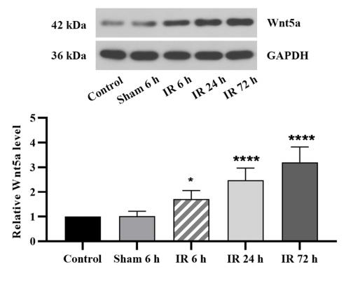
5). miR-374 improves cerebral ischemia reperfusion injury by targeting Wnt5a. EXPERIMENTAL ANIMALS, 2021 (PubMed: 33116025) [IF=2.4]

Application: WB    Species: rat    Sample: brain

Fig. 1.| Cerebral IR caused neurological deficit, cerebral infarct, downregulation of miR-374, and upregulation of Wnt5a.(E) After 6 h, 24 h and 72 h of reperfusion, the protein expression of WNT5A was determined by Western blot analysis.

Restrictive clause


Affinity Biosciences tests all products strictly. Citations are provided as a resource for additional applications that have not been validated by Affinity Biosciences. Please choose the appropriate format for each application and consult Materials and Methods sections for additional details about the use of any product in these publications.

For Research Use Only.
Not for use in diagnostic or therapeutic procedures. Not for resale. Not for distribution without written consent. Affinity Biosciences will not be held responsible for patent infringement or other violations that may occur with the use of our products. Affinity Biosciences, Affinity Biosciences Logo and all other trademarks are the property of Affinity Biosciences LTD.