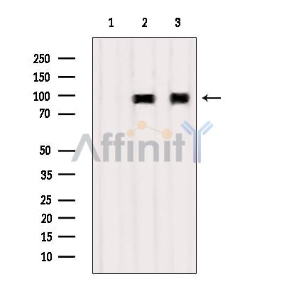
A20/TNFAIP3 Antibody - #DF6850
| Product: | A20/TNFAIP3 Antibody |
| Catalog: | DF6850 |
| Description: | Rabbit polyclonal antibody to A20/TNFAIP3 |
| Application: | WB IHC IF/ICC |
| Reactivity: | Human, Mouse, Rat |
| Prediction: | Pig, Bovine, Horse, Rabbit, Dog |
| Mol.Wt.: | 90kDa; 90kD(Calculated). |
| Uniprot: | P21580 |
| RRID: | AB_2838809 |
Related Downloads
Protocols
Product Info
*The optimal dilutions should be determined by the end user. For optimal experimental results, antibody reuse is not recommended.
*Tips:
WB: For western blot detection of denatured protein samples. IHC: For immunohistochemical detection of paraffin sections (IHC-p) or frozen sections (IHC-f) of tissue samples. IF/ICC: For immunofluorescence detection of cell samples. ELISA(peptide): For ELISA detection of antigenic peptide.
Cite Format: Affinity Biosciences Cat# DF6850, RRID:AB_2838809.
Fold/Unfold
A20; AISBL; MGC104522; MGC138687; MGC138688; OTU domain containing protein 7C; OTU domain-containing protein 7C; OTUD7C; Putative DNA binding protein A20; Putative DNA-binding protein A20; TNAP3_HUMAN; TNF alpha-induced protein 3; TNFA1P2; TNFAIP 3; TNFAIP3 (A20); TNFAIP3; Tumor necrosis factor alpha induced protein 3; Tumor necrosis factor alpha-induced protein 3; Tumor necrosis factor induced protein 3; Tumor necrosis factor inducible protein A20; tumor necrosis factor, alpha-induced protein 3; Zinc finger protein A20;
Immunogens
A synthesized peptide derived from human A20/TNFAIP3, corresponding to a region within the internal amino acids.
- P21580 TNAP3_HUMAN:
- Protein BLAST With
- NCBI/
- ExPASy/
- Uniprot
MAEQVLPQALYLSNMRKAVKIRERTPEDIFKPTNGIIHHFKTMHRYTLEMFRTCQFCPQFREIIHKALIDRNIQATLESQKKLNWCREVRKLVALKTNGDGNCLMHATSQYMWGVQDTDLVLRKALFSTLKETDTRNFKFRWQLESLKSQEFVETGLCYDTRNWNDEWDNLIKMASTDTPMARSGLQYNSLEEIHIFVLCNILRRPIIVISDKMLRSLESGSNFAPLKVGGIYLPLHWPAQECYRYPIVLGYDSHHFVPLVTLKDSGPEIRAVPLVNRDRGRFEDLKVHFLTDPENEMKEKLLKEYLMVIEIPVQGWDHGTTHLINAAKLDEANLPKEINLVDDYFELVQHEYKKWQENSEQGRREGHAQNPMEPSVPQLSLMDVKCETPNCPFFMSVNTQPLCHECSERRQKNQNKLPKLNSKPGPEGLPGMALGASRGEAYEPLAWNPEESTGGPHSAPPTAPSPFLFSETTAMKCRSPGCPFTLNVQHNGFCERCHNARQLHASHAPDHTRHLDPGKCQACLQDVTRTFNGICSTCFKRTTAEASSSLSTSLPPSCHQRSKSDPSRLVRSPSPHSCHRAGNDAPAGCLSQAARTPGDRTGTSKCRKAGCVYFGTPENKGFCTLCFIEYRENKHFAAASGKVSPTASRFQNTIPCLGRECGTLGSTMFEGYCQKCFIEAQNQRFHEAKRTEEQLRSSQRRDVPRTTQSTSRPKCARASCKNILACRSEELCMECQHPNQRMGPGAHRGEPAPEDPPKQRCRAPACDHFGNAKCNGYCNECFQFKQMYG
Predictions
Score>80(red) has high confidence and is suggested to be used for WB detection. *The prediction model is mainly based on the alignment of immunogen sequences, the results are for reference only, not as the basis of quality assurance.
High(score>80) Medium(80>score>50) Low(score<50) No confidence
Research Backgrounds
Ubiquitin-editing enzyme that contains both ubiquitin ligase and deubiquitinase activities. Involved in immune and inflammatory responses signaled by cytokines, such as TNF-alpha and IL-1 beta, or pathogens via Toll-like receptors (TLRs) through terminating NF-kappa-B activity. Essential component of a ubiquitin-editing protein complex, comprising also RNF11, ITCH and TAX1BP1, that ensures the transient nature of inflammatory signaling pathways. In cooperation with TAX1BP1 promotes disassembly of E2-E3 ubiquitin protein ligase complexes in IL-1R and TNFR-1 pathways; affected are at least E3 ligases TRAF6, TRAF2 and BIRC2, and E2 ubiquitin-conjugating enzymes UBE2N and UBE2D3. In cooperation with TAX1BP1 promotes ubiquitination of UBE2N and proteasomal degradation of UBE2N and UBE2D3. Upon TNF stimulation, deubiquitinates 'Lys-63'-polyubiquitin chains on RIPK1 and catalyzes the formation of 'Lys-48'-polyubiquitin chains. This leads to RIPK1 proteasomal degradation and consequently termination of the TNF- or LPS-mediated activation of NF-kappa-B. Deubiquitinates TRAF6 probably acting on 'Lys-63'-linked polyubiquitin. Upon T-cell receptor (TCR)-mediated T-cell activation, deubiquitinates 'Lys-63'-polyubiquitin chains on MALT1 thereby mediating disassociation of the CBM (CARD11:BCL10:MALT1) and IKK complexes and preventing sustained IKK activation. Deubiquitinates NEMO/IKBKG; the function is facilitated by TNIP1 and leads to inhibition of NF-kappa-B activation. Upon stimulation by bacterial peptidoglycans, probably deubiquitinates RIPK2. Can also inhibit I-kappa-B-kinase (IKK) through a non-catalytic mechanism which involves polyubiquitin; polyubiquitin promotes association with IKBKG and prevents IKK MAP3K7-mediated phosphorylation. Targets TRAF2 for lysosomal degradation. In vitro able to deubiquitinate 'Lys-11'-, 'Lys-48'- and 'Lys-63' polyubiquitin chains. Inhibitor of programmed cell death. Has a role in the function of the lymphoid system. Required for LPS-induced production of proinflammatory cytokines and IFN beta in LPS-tolerized macrophages.
Proteolytically cleaved by MALT1 upon TCR stimulation; disrupts NF-kappa-B inhibitory function and results in increased IL-2 production. It is proposed that only a fraction of TNFAIP3 colocalized with TCR and CBM complex is cleaved, leaving the main TNFAIP3 pool intact.
Cytoplasm. Nucleus. Lysosome.
Cytoplasm.
The A20-type zinc fingers mediate the ubiquitin ligase activity. The A20-type zinc finger 4 selectively recognizes 'Lys-63'-linked polyubiquitin. The A20-type zinc finger 4-7 are sufficient to bind polyubiquitin.
The OTU domain mediates the deubiquitinase activity.
Belongs to the peptidase C64 family.
Research Fields
· Cellular Processes > Cell growth and death > Necroptosis. (View pathway)
· Environmental Information Processing > Signal transduction > NF-kappa B signaling pathway. (View pathway)
· Environmental Information Processing > Signal transduction > TNF signaling pathway. (View pathway)
· Human Diseases > Infectious diseases: Viral > Measles.
· Human Diseases > Infectious diseases: Viral > Epstein-Barr virus infection.
· Organismal Systems > Immune system > NOD-like receptor signaling pathway. (View pathway)
· Organismal Systems > Immune system > IL-17 signaling pathway. (View pathway)
References
Restrictive clause
Affinity Biosciences tests all products strictly. Citations are provided as a resource for additional applications that have not been validated by Affinity Biosciences. Please choose the appropriate format for each application and consult Materials and Methods sections for additional details about the use of any product in these publications.
For Research Use Only.
Not for use in diagnostic or therapeutic procedures. Not for resale. Not for distribution without written consent. Affinity Biosciences will not be held responsible for patent infringement or other violations that may occur with the use of our products. Affinity Biosciences, Affinity Biosciences Logo and all other trademarks are the property of Affinity Biosciences LTD.


