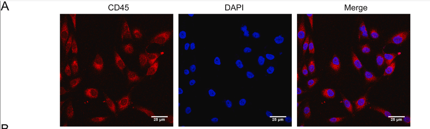
 and mouse anti-beta tubulin Ab(T0023) for 1 hour at 37°C. An AlexaFluor594 conjugated goat anti-rabbit IgG(H+L) Ab(Red) and an AlexaFluor488 conjugated goat anti-mouse IgG(H+L) Ab(Green) were used as the secondary antibody.
The nuclear counter stain is DAPI (blue).")
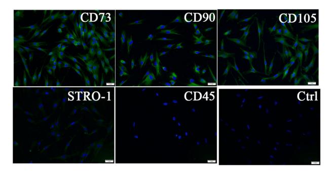
的表达情况(荧光显微镜,x400)Figure 1 | Expression ofspecific markers(CD29, cD90 and CD45) onthe surface ofrat bone marrow mesenchymal stem cells (fluorescencemicroscope, x400)")

| Product: | CD45 Antibody |
| Catalog: | DF6839 |
| Description: | Rabbit polyclonal antibody to CD45 |
| Application: | WB IHC IF/ICC |
| Cited expt.: | IF/ICC |
| Reactivity: | Human, Mouse, Rat |
| Prediction: | Zebrafish, Bovine, Sheep, Rabbit, Dog |
| Mol.Wt.: | 147kDa(Observed); 147kD(Calculated). |
| Uniprot: | P08575 |
| RRID: | AB_2838798 |
Control Products

Related Downloads
Protocols
Product Info
*The optimal dilutions should be determined by the end user. For optimal experimental results, antibody reuse is not recommended.
*Tips:
WB: For western blot detection of denatured protein samples. IHC: For immunohistochemical detection of paraffin sections (IHC-p) or frozen sections (IHC-f) of tissue samples. IF/ICC: For immunofluorescence detection of cell samples. ELISA(peptide): For ELISA detection of antigenic peptide.
Cite Format: Affinity Biosciences Cat# DF6839, RRID:AB_2838798.
Fold/Unfold
B220; CD 45; CD45; CD45 antigen; CD45R; GP180; L-CA; LCA; Leukocyte common antigen; loc; Ly-5; LY5; Ly5, homolog of; Lyt-4; OTTHUMP00000033813; OTTHUMP00000033816; OTTHUMP00000033817; OTTHUMP00000038574; Protein tyrosine phosphatase receptor type c polypeptide; Protein tyrosine phosphatase, receptor type C; protein tyrosine phosphatase, receptor type, C; Protein tyrosine phosphatase, receptor type, c polypeptide; Ptprc; PTPRC_HUMAN; Receptor-type tyrosine-protein phosphatase C; T200; T200 glycoprotein; T200 leukocyte common antigen;
Immunogens
A synthesized peptide derived from human CD45, corresponding to a region within C-terminal amino acids.
Isoform 1: Detected in thymocytes. Isoform 2: Detected in thymocytes. Isoform 3: Detected in thymocytes. Isoform 4: Not detected in thymocytes. Isoform 5: Detected in thymocytes. Isoform 6: Not detected in thymocytes. Isoform 7: Detected in thymocytes. Isoform 8: Not detected in thymocytes.
- P08575 PTPRC_HUMAN:
- Protein BLAST With
- NCBI/
- ExPASy/
- Uniprot
MTMYLWLKLLAFGFAFLDTEVFVTGQSPTPSPTGLTTAKMPSVPLSSDPLPTHTTAFSPASTFERENDFSETTTSLSPDNTSTQVSPDSLDNASAFNTTGVSSVQTPHLPTHADSQTPSAGTDTQTFSGSAANAKLNPTPGSNAISDVPGERSTASTFPTDPVSPLTTTLSLAHHSSAALPARTSNTTITANTSDAYLNASETTTLSPSGSAVISTTTIATTPSKPTCDEKYANITVDYLYNKETKLFTAKLNVNENVECGNNTCTNNEVHNLTECKNASVSISHNSCTAPDKTLILDVPPGVEKFQLHDCTQVEKADTTICLKWKNIETFTCDTQNITYRFQCGNMIFDNKEIKLENLEPEHEYKCDSEILYNNHKFTNASKIIKTDFGSPGEPQIIFCRSEAAHQGVITWNPPQRSFHNFTLCYIKETEKDCLNLDKNLIKYDLQNLKPYTKYVLSLHAYIIAKVQRNGSAAMCHFTTKSAPPSQVWNMTVSMTSDNSMHVKCRPPRDRNGPHERYHLEVEAGNTLVRNESHKNCDFRVKDLQYSTDYTFKAYFHNGDYPGEPFILHHSTSYNSKALIAFLAFLIIVTSIALLVVLYKIYDLHKKRSCNLDEQQELVERDDEKQLMNVEPIHADILLETYKRKIADEGRLFLAEFQSIPRVFSKFPIKEARKPFNQNKNRYVDILPYDYNRVELSEINGDAGSNYINASYIDGFKEPRKYIAAQGPRDETVDDFWRMIWEQKATVIVMVTRCEEGNRNKCAEYWPSMEEGTRAFGDVVVKINQHKRCPDYIIQKLNIVNKKEKATGREVTHIQFTSWPDHGVPEDPHLLLKLRRRVNAFSNFFSGPIVVHCSAGVGRTGTYIGIDAMLEGLEAENKVDVYGYVVKLRRQRCLMVQVEAQYILIHQALVEYNQFGETEVNLSELHPYLHNMKKRDPPSEPSPLEAEFQRLPSYRSWRTQHIGNQEENKSKNRNSNVIPYDYNRVPLKHELEMSKESEHDSDESSDDDSDSEEPSKYINASFIMSYWKPEVMIAAQGPLKETIGDFWQMIFQRKVKVIVMLTELKHGDQEICAQYWGEGKQTYGDIEVDLKDTDKSSTYTLRVFELRHSKRKDSRTVYQYQYTNWSVEQLPAEPKELISMIQVVKQKLPQKNSSEGNKHHKSTPLLIHCRDGSQQTGIFCALLNLLESAETEEVVDIFQVVKALRKARPGMVSTFEQYQFLYDVIASTYPAQNGQVKKNNHQEDKIEFDNEVDKVKQDANCVNPLGAPEKLPEAKEQAEGSEPTSGTEGPEHSVNGPASPALNQGS
Predictions
Score>80(red) has high confidence and is suggested to be used for WB detection. *The prediction model is mainly based on the alignment of immunogen sequences, the results are for reference only, not as the basis of quality assurance.
High(score>80) Medium(80>score>50) Low(score<50) No confidence
Research Backgrounds
Protein tyrosine-protein phosphatase required for T-cell activation through the antigen receptor. Acts as a positive regulator of T-cell coactivation upon binding to DPP4. The first PTPase domain has enzymatic activity, while the second one seems to affect the substrate specificity of the first one. Upon T-cell activation, recruits and dephosphorylates SKAP1 and FYN. Dephosphorylates LYN, and thereby modulates LYN activity (By similarity).
(Microbial infection) Acts as a receptor for human cytomegalovirus protein UL11 and mediates binding of UL11 to T-cells, leading to reduced induction of tyrosine phosphorylation of multiple signaling proteins upon T-cell receptor stimulation and impaired T-cell proliferation.
Heavily N- and O-glycosylated.
Cell membrane>Single-pass type I membrane protein. Membrane raft.
Note: Colocalized with DPP4 in membrane rafts.
Isoform 1: Detected in thymocytes. Isoform 2: Detected in thymocytes. Isoform 3: Detected in thymocytes. Isoform 4: Not detected in thymocytes. Isoform 5: Detected in thymocytes. Isoform 6: Not detected in thymocytes. Isoform 7: Detected in thymocytes. Isoform 8: Not detected in thymocytes.
The first PTPase domain interacts with SKAP1.
Belongs to the protein-tyrosine phosphatase family. Receptor class 1/6 subfamily.
Research Fields
· Environmental Information Processing > Signaling molecules and interaction > Cell adhesion molecules (CAMs). (View pathway)
· Human Diseases > Immune diseases > Primary immunodeficiency.
· Organismal Systems > Immune system > T cell receptor signaling pathway. (View pathway)
· Organismal Systems > Immune system > Fc gamma R-mediated phagocytosis. (View pathway)
References

Application: IF/ICC Species: canine Sample: cDPSCs
Application: IF/ICC Species: beagles Sample: cDPSCs
Application: IF/ICC Species: Human Sample:
Restrictive clause
Affinity Biosciences tests all products strictly. Citations are provided as a resource for additional applications that have not been validated by Affinity Biosciences. Please choose the appropriate format for each application and consult Materials and Methods sections for additional details about the use of any product in these publications.
For Research Use Only.
Not for use in diagnostic or therapeutic procedures. Not for resale. Not for distribution without written consent. Affinity Biosciences will not be held responsible for patent infringement or other violations that may occur with the use of our products. Affinity Biosciences, Affinity Biosciences Logo and all other trademarks are the property of Affinity Biosciences LTD.