- WB
- IHC
- IF/ICC
- Cites


, using E-cadherin Antibody. The lane on the left was treated with blocking peptide.")


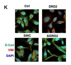
 by IF/ICC. The samples were fixed with PFA and permeabilized in 0.1% Triton X-100,then blocked in 10% serum for 45 minutes at 25°C. Samples were then incubated with primary Ab(AF0131) and mouse anti-beta tubulin Ab(T0023) for 1 hour at 37°C. An AlexaFluor594 conjugated goat anti-rabbit IgG(H+L) Ab(Red) and an AlexaFluor488 conjugated goat anti-mouse IgG(H+L) Ab(Green) were used as the secondary antibody.
The nuclear counter stain is DAPI(blue).")
 microscopy at x200 magnification was used to assess cell morphology. The A549 cells (parental cells) had an epithelioid, rounded cobblestone appearance and there was limited formation of pseudopodia. A549/PTX and A549/DDP cells exhibited a spindle-shaped morphology and an increased formation of pseudopodia, indicating a loss of cell polarity. (B) E-cadherin, β-catenin, vimentin, MMP-2 and MMP-9 which are EMT-related proteins, were assessed in terms of expression levels. EMT-related transcription factors (Snail, Slug, Twist and ZEB1) were measured in A549/PTX and A549/DDP cells using western blot analysis. (C) The expression changes were confirmed at the mRNA level by qRT-PCR. Expression was standardized to the expression of GAPDH and normalized to 1.0 in the parental cells (compared with the parental A549 cells, means ± SEM, n=3, * P<0.05)")
 Overexpression of miR-1271 could inhibit MGC-803 cell migration and invasion, whereas its downregulation in SGC-7901 cells increased the cell migration and invasion processes. (B) Western blots showed that overexpression of miR-1271 could upregulate E-cadherin and downregulate N-cadherin and vimentin expression in MGC-803 cells, whereas its downregulation had the opposite effect in SGC- 7901 cells. Three independent experiments were conducted. *P < 0.05, **P < 0.01.")

 By human cancer pathway PCR array, ectopic expression of BCL2L10 up- or down-regulated several genes related to tumor proliferation, apoptosis, metastasis and angiogenesis. (B) Western blot was performed to confirm the downstream gene expression regulated by BCL2L10 in HepG2 cells. GAPDH was used as an internal control. (C) Schematic diagram of the molecular events for BCL2L10 function as a tumor suppressor through regulating cell cycle, proliferation, apoptosis metastasis and angiogenesis effectors.")
 Crystal violet staining of the shNS and control group cells that crossed the polycarbonate membrane of the Transwell chamber to detect cell migration. (B) The number of cells that crossed the Transwell migration chamber in different groups. (C) Crystal violet staining of the shNS and control group cells that crossed the Matrigel-coated polycarbonate membrane of the Transwell chamber to detect cell invasion. (D) The number of cells that crossed the Transwell invasion chamber in different groups. (E) Representative Western blotting results indicate the EMT marker expressions in the different groups. The results are presented as the means ± SD, as based on three independent experiments. Statistical significance was determined using Student's t-test. *P < 0.05. Scale: 100 mm.")
 inhibited JAM-A-transfected cell invasion (B) and EMT (C).")
 The EMT markers in MKN45 and MKN74 cells were analyzed using western blotting after being co-cultured with THP-1 cells. (B and C) The EMT markers in MKN45 and MKN74 cells were analyzed by RT-PCR after being co-cultured with THP-1 cells; * P")
 miR-93-5p overexpression suppressed and increased E-cadherin and N-cadherin expression, respectively. The opposite result was observed in response to miR-93-5p downregulation.")

 Results of IHC assays. The expression levels of E-cadherin were significantly upregulated, whereas those of vimentin, MMP2, and MMP9 were downregulated by OA or regorafenib treatment, and OA enhanced the effects of regorafenib. The expression levels of iNOS and NT were upregulated by OA but not by regorafenib. (G) Staining indexes of IHC assays. Data are represented as mean ± standard error of the mean (*P < 0.05, **P < 0.01).")
 Immunofluorescence assay conducted in the HepG2 cancer cell line demonstrated that Downloaded from mct.aacrjournals.org on October 10, 2018. © 2018 American Association for Cancer Research. Author manuscripts have been peer reviewed and accepted for publication but have not yet been edited. Author Manuscript Published OnlineFirst on October 8, 2018; DOI: 10.1158/1535-7163.MCT-18-0448 22 OA can enhance fluorescence intensity of E-cadherin and attenuate that of vimentin, whereas treatment of NO scavenger (C-PTIO) or transfection of siRNA iNOS exhibits the opposite effect. This outcome is consistent with the finding of Western blot assay. The results were obtained from three independent experiments, each performed in triplicate. Data are represented as mean ± standard error of the mean (*P < 0.05, **P < 0.01).")
 Protein expression level of Twist1, Zeb1 and E-cadherin in PLC/PRF/5 cells treated hypoxia and hypoxia combined with Sal. Error bars represent the standard deviation of experiments performed in triplicate (*P b .05, **P b .01).")
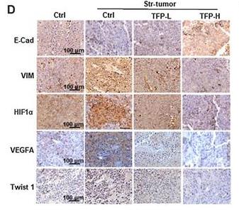
 Cell migration was measured after treatment with various drugs for 48 h. (C–D)Representative images of Transwell cell invasion assays were obtained at 200× magnification. (E–F) Double immunofluorescence staining for E-cadherin and vimentin after treated with Sal. (G-H) The expression levels of HIF-1α in tumor tissues of subcutaneously transplanted tumor mice. Error bars represent the standard deviation of experiments performed intriplicate (*P b .05, **P b .01).")
 Cell migration was measured after treatment with various drugs for 48 h. (C–D)Representative images of Transwell cell invasion assays were obtained at 200× magnification. (E–F) Double immunofluorescence staining for E-cadherin and vimentin after treated with Sal. (G-H) The expression levels of HIF-1α in tumor tissues of subcutaneously transplanted tumor mice. Error bars represent the standard deviation of experiments performed intriplicate (*P b .05, **P b .01).")
 Effect of Pyr or MTX on the migration of NCI-H460 at 24 and 48 h. (B) Effects of Pyr and MTX on the migration of A549 cells at 24 and 48 h. (C) Transwell chambers were utilized for the invasion assay, and images were obtained under 200× magnification. NCI-H460 and A549 cells were treated with Pyr or MTX. (D) Changes of E-cadherin and vimentin expression in NCI-H460 cells treated with Pyr or MTX (Western blot assay). β-actin was used as the loading control. (E) Changes of E-cadherin and vimentin expression in NCI-H460 cells treated with Pyr or MTX (immunofluorescence assay). Each experiment was performed in triplicate. Results are shown as means ± SD (*P < 0.05, **P < 0.01).")
 Effect of Pyr or MTX on the migration of NCI-H460 at 24 and 48 h. (B) Effects of Pyr and MTX on the migration of A549 cells at 24 and 48 h. (C) Transwell chambers were utilized for the invasion assay, and images were obtained under 200× magnification. NCI-H460 and A549 cells were treated with Pyr or MTX. (D) Changes of E-cadherin and vimentin expression in NCI-H460 cells treated with Pyr or MTX (Western blot assay). β-actin was used as the loading control. (E) Changes of E-cadherin and vimentin expression in NCI-H460 cells treated with Pyr or MTX (immunofluorescence assay). Each experiment was performed in triplicate. Results are shown as means ± SD (*P < 0.05, **P < 0.01).")


")
 E-cadherin, β-catenin, vimentin, MMP-2 and MMP-9 which are EMT-related proteins, were assessed in terms of expression levels. EMT-related transcription factors (Snail, Slug, Twist and ZEB1) were measured in A549/PTX and A549/DDP cells using western blot analysis.")
 The levels of cyclin A, cyclin B, cyclin D1, and CDK2 were reduced in A375 cells transfected with OVOS2-shRNA; (b) The downregulated expression of N-cadherin accompanied with the upregulated expression of E-cadherin and β-catenin were observed in A375 cells transfected with OVOS2-shRNA; (c) The expression of p-FAK, p-AKT, and p-ERK were reduced in A375 cells transfected with OVOS2-shRNA; (d) The increased production of MMP-2 was observed in A375 transfected with OVOS2-shRNA; (e) GAPDH was used as the reference.")
 and Vimentin (red) was performed. The nucleus was staining with DAPI.")

 and Western blot (B and C) indicated that transient
transfection of HTR8/SVneo cells with miR-210 mimic increases E-cadherin expression and decreases N-cadherin and vimentin expression at the transcriptional and
protein levels compared to those in the cells transfected with miR-210 mimic NC. qRT-PCR (E and H) and Western blot (F and G) indicated that silencing of miR-210
in HTR8/SVneo cells got the opposite results. β-actin served as the loading control. (*P < 0.05, **P < 0.01, ***P < 0.001). n = 3.")

 Overexpression of KRT7 increased Snail, vimentin and N-cadherin expression, and decreased E-cadherin expression in HEY cells as detected by western blotting. (C and D) Knockdown of KRT7 resulted in increased E-cadherin expression and reduced vimentin and Snail expression in OVCAR433 cells as detected by western blotting. All experiments were performed at least three times. Results are presented as the mean ± standard deviation. **P<0.01. KRT7, keratin 7; sh, short hairpin RNA; NC, negative control.")
.")
 Cell migration was assayed by Transwell assay in HG-induced MPC5 cells treated with miR-215-5p mimic. (b) The number of migratory cells was analyzed by ImageJ software. (c) The expression of miR215-5p was measured by qRT-PCR in HG-induced MPC5 cells treated with miR-215-5p mimic. (d) The expression of E-cadherin and a-SMA was measured by western blotting under miR-215-5p mimic treatment.")
 and α-SMA (red), and quantitative analysis of the staining intensity. Scale bar, 50 μm. Statistically significant differences are presented as the mean ± SD (n = 3)by one-way ANOVA with Tukey’s test for multiple comparisons.")
 DDTC increased the protein expression level of E-cadherin and decreased the protein expression level of N-cadherin and Vimentin in SKOV3 cells.")
 The protein expression of c-Myc, vimentin, E-cadherin, HIF-1α, CXCR4, and SDF-1 in PANC-1 and SW1990 pancreatic cancer cells under different treatments was detected via western blot. (B) Quantification results of protein expressions of c-Myc, vimentin, E-cadherin, HIF-1α, CXCR4, and SDF-1 in PANC-1 pancreatic cancer cells. (C) Quantification results of protein expression of c-Myc, vimentin, E-cadherin, HIF-1α, CXCR4, and SDF-1 in SW1990 pancreatic cancer cells (* p < 0.05, ** p < 0.01 vs control, ▲ p < 0.05, ▲▲ p < 0.01 vs bufalin treatment group, n = 3).")
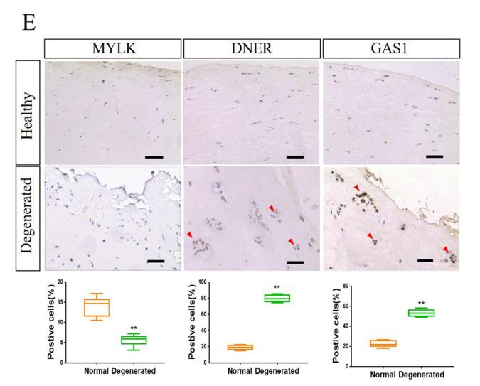
 N-cadherin, Vimentin and E-cadherin expression levels detected by immunohistochemistry. Scale bar, 100 µm. (B) mRNA expression levels detected via reverse transcription-quantitative PCR. (C) Protein expression levels detected via western blotting. *P<0.05 vs. control. Huc-MSCs-exo, human umbilical cord mesenchymal stem cell-derived exosomes.")
 N-cadherin, Vimentin and E-cadherin expression levels detected by immunohistochemistry. Scale bar, 100 µm. (B) mRNA expression levels detected via reverse transcription-quantitative PCR. (C) Protein expression levels detected via western blotting. *P<0.05 vs. control. Huc-MSCs-exo, human umbilical cord mesenchymal stem cell-derived exosomes.")
 The downregulation of NEAT1 in si-NEAT1 cell lines (A549 and H460) detected by qRT-PCR. (B, C) The
cell proliferation of A549 and H460 cells with si-NEAT1 measured by CCK8 assay. (D) The regulation of si-NEAT1 on
cell apoptosis tested by flow cytometry. (E, F) Transwell experiments on cell migration, invasion of si-NEAT1 cell lines
(A549 and H460). (G) The expression of EMT marker protein by Western blot normalized to GAPDH. *p < 0.05. CCK8,
Cell Counting Kit-8; EMT, epithelial–mesenchymal transition; qRT-PCR, quantitative real-time polymerase chain
reaction.")
 Relative LINP1 expression in EC9706 sh1, sh2, sh3, and sh4 was verified by qRT-PCR. The LINP1 sh2 cell line had the most significant knockdown effect compared with the control cell line (61.75%, P<0.001). (B) The Alamar Blue proliferation assay indicated that the proliferation of shRNA-LINP1 cells was significantly inhibited compared with that of the corresponding control cells and shRNA-scr cells (P<0.001). (C,D) Colony formation assays showing that the number of colonies formed was significantly lower in shRNA-LINP1 cells than in NC and shRNA-scr cells in macroscopic view (71.7 vs. 73.3 vs. 24.7, P<0.001). (E,F) Wound healing assays showed that the migration rate was slower in shRNA-LINP1 cells than in NC and shRNA-scr cells after 48 h (19.1% vs. 46.4% vs. 46.6%; P<0.001) (magnification 100×). (G,H) Transwell migration assays demonstrated that the number of migratory cells was lower in shRNA-LINP1 than in the two control cells (60.3 vs. 236.3 vs. 238.7; P<0.001) (magnification 100×). (I,J,K) qRT-PCR and western blot analyses of the expression of EMT markers. E-cadherin was significantly upregulated, whereas N-cadherin, vimentin, snail and slug were significantly downregulated in shRNA-LINP1 cells compared with NC and shRNA-scr cells (all P<0.05). **, P<0.01; ***, P<0.001.")
 qRT-PCR showing the knock down of lncRNA AL161431.1, and the corresponding changes in E-cadherin, N-cadherin, and vimentin in SW1990 and BxPC-3 cells; (B) Western blots showing the changes in protein level in E-cadherin, N-cadherin, and vimentin in SW1990 and BxPC-3 cells after transfection of scrambled siRNA or siRNA1; (C) Immunofluorescence analysis showing the change of expression of E-cadherin, N-cadherin, and vimentin in SW1990 and BxPC-3 cells after transfection of scrambled siRNA or siRNA1 (200x). *p < 0.05, **p < 0.01, ***p < 0.001.")
 qRT-PCR showing the knock down of lncRNA AL161431.1, and the corresponding changes in E-cadherin, N-cadherin, and vimentin in SW1990 and BxPC-3 cells; (B) Western blots showing the changes in protein level in E-cadherin, N-cadherin, and vimentin in SW1990 and BxPC-3 cells after transfection of scrambled siRNA or siRNA1; (C) Immunofluorescence analysis showing the change of expression of E-cadherin, N-cadherin, and vimentin in SW1990 and BxPC-3 cells after transfection of scrambled siRNA or siRNA1 (200x). *p < 0.05, **p < 0.01, ***p < 0.001.")
 Wound-healing assay in LINC01554-overexpressing SK-Hep1 and HCCLM9 cells. (B) Invasion ability of LINC01554-overexpressing SK-Hep1 and HCCLM9 cells. (C) Expression levels of EMT markers (ZO-1, E-cadherin, N-cadherin and vimentin) in LINC01554-overexpressing HCCLM9 and SK-Hep1 cells are analysed by western blotting. (D) FISH analysis of LINC01554 in the SK-Hep1 and HCCLM9 cells. The U6 and 18S rRNA probes are labelled red, the LINC01554 RNA probe is labelled green, and nuclear DNA with DAPI staining is labelled blue. Scale bar, 50 µm. All results are from at least three independent experiments. *P<0.05, **P<0.01, comparison with the Lv-NC group. HCC, hepatocellular carcinoma; EMT, epithelial-mesenchymal transition.")
 were determined by western blot (a, b). Compared with the LOVO, *, P < 0.05; **, P < 0.01; compared with the LOVO+NFs, #, P < 0.05; ##, P < 0.01. The induction of the FAK pathway, which is involved in EMT in colon cancer cells, by CAFs, was analyzed by western blot (c) and (d).
EMT, epithelial–mesenchymal transition; NFs, normal fibroblasts; FAK, focal adhesion kinase; CAFs, cancer-associated fibroblasts.")
 NSE overexpression promoted the EMT process of SCLC. (A) Western blot assay was performed to measure the protein expression levels of the EMT-related markers (Snail, N-cadherin and E-cadherin).")
immunofluorescence analysis showing the change of expression of E-cadherin, N-cadherin, and vimentin in SW1990 and BxPC-3 cells after transfection of scrambled siRNA or siRNA1 (200×). *P<0.05, **P<0.01.")
 western blots showing the changes in protein level in E-cadherin, N-cadherin, and vimentin in SW1990 and BxPC-3 cells after transfection of scrambled siRNA or siRNA1")
 NUDCD1 knockdown decreased the expression of Ncadherin and vimentin and upregulated the expression of E-cadherin in PANC-1 cells.")
 NSE overexpression promoted the EMT process of SCLC. (A) Western blot assay was performed to measure the protein expression levels of the EMT-related markers (Snail, N-cadherin and E-cadherin). (B) qRT-PCR was performed to measure the mRNA expression levels of the EMT markers. (C, D) NSE knockdown represses the EMT process of SCLC. Protein levels (C) and mRNA levels (D) of EMT-related markers were measured using western blot or qRT-PCR, respectively. These results were repeated of three independent experiments.")
 Protein levels of the EMT markers E-cadherin, N-cadherin, Vimentin, and Snail were detected in Hep3B
cells transfected with NC mimics or miR-4458 mimics by western blot analysis and immunofluorescence assay. (B,D) Huh7 cells were transfected with NC
inhibitor or miR-4458 inhibitor, and E-cadherin, N-cadherin, Vimentin, and Snail protein levels were detected by western blot analysis and immunofluorescence
assay. Data are presented as the mean±SD. ∗P<0.05, ∗∗P<0.01.")
 NSE overexpression promoted the EMT process of SCLC. (A) Western blot assay was performed to measure the protein expression levels of the EMT-related markers (Snail, N-cadherin and E-cadherin). (B) qRT-PCR was performed to measure the mRNA expression levels of the EMT markers. (C, D) NSE knockdown represses the EMT process of SCLC. Protein levels (C) and mRNA levels (D) of EMT-related markers were measured using western blot or qRT-PCR, respectively. These results were repeated of three independent experiments.")
 EMT-related protein expression; representative blots of the related proteins. (B) Quantitative data of the related protein expression. (C) EMT-related gene expression at mRNA level. *P<0.05 vs. control; #P<0.05 vs. AOPPs. AOPPs; EMT, epithelial-mesenchymal transition.")
 qRT-PCR showing the knock down of lncRNA ELFN1-AS1, and the corresponding changes in E-cadherin, N-cadherin, and vimentin in SW1990 and BxPC-3 cells; (B) western blots showing the changes in protein level in E-cadherin, N-cadherin, and vimentin in SW1990 and BxPC-3 cells after transfection of scrambled siRNA or siRNA1; (C) immunofluorescence analysis showing the change of expression of E-cadherin, N-cadherin, and vimentin in SW1990 and BxPC-3 cells after transfection of scrambled siRNA or siRNA1 (200×). *P<0.05, **P<0.01.")
 in colon tissue. The bar graph of the relative intensities of ZO-1 (b), occludin (c), claudin-1 (d), claudin-2 (e), and E-cadherin (f) Western Blotting bands. The mRNA expression of occludin (g), ZO-1 (h), and claudin-1 (i) and claudin-2 (j) in colon tissue. All values are presented as the mean ± SEM. ##P < 0.01 and #P < 0.05 versus normal group; ∗P < 0.05, ∗∗P < 0.01, and ∗∗∗P < 0.001 versus DSS group.")
 NUDCD1 knockdown decreased the expression of N-cadherin and vimentin and upregulated the expression of E-cadherin in PANC-1 cells. (E–H) NUDCD1 knockdown decreased the expression of N-cadherin and vimentin, and upregulated the expression of E-cadherin in Patu8988 cells. ***p<0.001, ****p<0.0001.")
 Glioblastoma cells transfected with siRNA control and siLDHA and U87 cells were treated with TGF-β1 for 24 hours before being adhered to microplates, and extracellular acidification rate (ECAR) was determined over time and analyzed as bar graphs. (b) Glioblastoma cells transfected with siRNA control and siPDK1 and U87 cells were treated with TGF-β1 for 24 hours before being adhered to microplates, and oxygen consumption rate (OCR) was determined over time and analyzed as bar graphs. (c) EMT protein marker expression in glioblastoma cells treated with the siLDHA and siPDK1 in the presence of TGF-β1 for 24 hours. (d) EMT protein marker expression in glioblastoma cells when treated with LDHA inhibitor GSK2837808A (10 μM) and PDK1 inhibitor Dichloroacetate (20 μM) combined with TGF-β1 for 24 hours. (e) Immunofluorescences observed Vimentin expression in glioblastoma cells after treated with siLDHA and siPDK1 under treatment of TGF-β1. Scale bars = 20 μm. (f) Migration and invasion ability of glioblastoma cells transfected with siLDHA and siPDK1 were determined by transwell assay under treatment of TGF-β1. Scale bars = 100 μm. (g) Glioblastoma cells were treated with rotenone (50 nM) for 24 hours before staining for reactive oxygen species with CellROX Deep Red Reagents. Scale bars = 100 μm. (h) EMT protein marker expression when inhibited mitochondrial respiration with rotenone (50 nM) for 24 hours in glioblastoma cells. Data represent mean and SD of three independent experiments. ∗P < 0.05; ∗∗P < 0.01; ∗∗∗P < 0.001.")
 in vitro. B Western blot analysis of E-cadherin, Snail1, ZEB2, and Vimentin expression in TC cells compared with PC-knockdown TC cells; the full blot is provided in the supplementary file")
 were performed with bar plot by gray analysis (B). For qRT-PCR assay (C), relative quantification of tight junction proteins were calculated by 2– △△Ct method. Error bars indicate standard deviations. *p < 0.05, **p < 0.01, and ***p < 0.001 were compared by Student’s t-test to evaluate differences between every two groups.")
. ADCY7 expression in FLSs derived from the HFD-OA model. Cells were transfected with siRNA-ADCY7 and pCDNA-ADCY7 in vitro (n ¼ 3). (b). Expression of epithelial-mesenchymal transition (EMT)-related markers in FLSs (n¼3).")
; E, Scatterplot showing the correlation between plasma levels of ALB and uPAR. The vertical position represents the expression levels of uPAR (lg pg/mL)")
, vimentin (B), α-SMA (C) and Collagen I (F)
were determined by RT-qPCR. (D) The protein expression level of E-cadherin analyzed by Western blot. (E) The protein expression of E-cadherin relative to GADPH
protein expression. (G) The protein expression levels of vimentin and α-SMA analyzed by Western blot. The protein expression of vimentin (H) and α-SMA (I) relative
to GADPH protein expression. Data are expressed as the mean ± S.E.M. (n = 3–7), *P < 0.05 vs Sham; **P < 0.01 vs Sham; ***P < 0.001 vs Sham; ##P < 0.01 vs 5/6
Nx; ###P < 0.001 vs 5/6 Nx; ns, no significance.")
 HE staining and Masson staining of lung tissue in the sham-operated, memantine, BLM, and BLM + memantine groups (×100, n=5); (B) ashcroft scores of four groups of lung tissue pathological specimens; (C) determination of hydroxyproline content in the four groups of lung tissue samples; (D,E) changes in the protein expression levels of α-SMA, vimentin, E-cadherin, and EpCAM in the four groups of lung tissues. **P<0.01 vs. Sham; ##P<0.01 vs. BLM (n=5). BLM, bleomycin; α-SMA, alpha-smooth muscle actin; EpCAM, epithelial cell adhesion molecule.")

 The representative band of Western blot. (b)–(h) The quantitative result of Western blot. Data were expressed as mean ± SD. #P < 0.05/##p < 0.01 vs. control; ∗p < 0.05/∗∗p < 0.01 vs. the BLM group. n = 3.")
 Cell morphology of A2780 and SKOV3 after CA and EGF treatment. (B) Expression of E-cadherin, N-cadherin, vimentin, and Snail were detected by Western blotting in A2780 and SKOV3 cells after CA and EGF treatment. Representative fluorescence images of E-cadherin (C) and N-cadherin (D) in A2780 and SKOV3 cells. At least three independent experiments were performed.")
 Representative images of Masson's trichrome stained kidney tissues of mice in the sham and ALD groups. Scale bar, 50 µm. (B) RT-qPCR analysis of miR-26a expression levels in the kidney tissues of mice in the sham and ALD groups; U6 was used for normalization. (C) RT-qPCR analysis of collagen I, α-SMA and LCN2 mRNA expression levels in the kidney tissues of mice from the sham and ALD groups; β-actin was used for normalization. (D) Representative western blotting images and semi-quantitative analysis of E-cadherin, collagen I, α-SMA, CTGF and LCN2 protein expression levels in the kidney tissue of mice in the sham and ALD groups. (E) Immunohistochemical analysis of E-cadherin (green), α-SMA (red) and fibronectin (green) in the kidney tissue of mice in the sham and ALD groups; DAPI (blue) was used to stain the nuclei. Scale bar, 50 µm. Data are presented as the mean ± SD; n=5 mice/group; *P<0.05 vs. sham. α-SMA, α-smooth muscle actin2; CTGF, connective tissue growth factor; LCN2, lipocalin; RT-qPCR, reverse transcription-quantitative PCR.")
 Representative images of Masson's trichrome stained kidney tissues of mice in the sham and ALD groups. Scale bar, 50 µm. (B) RT-qPCR analysis of miR-26a expression levels in the kidney tissues of mice in the sham and ALD groups; U6 was used for normalization. (C) RT-qPCR analysis of collagen I, α-SMA and LCN2 mRNA expression levels in the kidney tissues of mice from the sham and ALD groups; β-actin was used for normalization. (D) Representative western blotting images and semi-quantitative analysis of E-cadherin, collagen I, α-SMA, CTGF and LCN2 protein expression levels in the kidney tissue of mice in the sham and ALD groups. (E) Immunohistochemical analysis of E-cadherin (green), α-SMA (red) and fibronectin (green) in the kidney tissue of mice in the sham and ALD groups; DAPI (blue) was used to stain the nuclei. Scale bar, 50 µm. Data are presented as the mean ± SD; n=5 mice/group; *P<0.05 vs. sham. α-SMA, α-smooth muscle actin2; CTGF, connective tissue growth factor; LCN2, lipocalin; RT-qPCR, reverse transcription-quantitative PCR.")
 Cell migratory abilities were assessed via the wound-healing assay (scale bar = 200 μm). (b, c) Migratory and invasive abilities was examined using Transwell assay (scale bar = 50 μm). (d) Protein levels were estimated by Western blot. ∗A significant difference compared with the sh-NC group. ∗∗P < 0.01.")
 Overexpression of LAMC2 promoted the expression of N-cadherin and vimentin proteins in TU177 cells and inhibited the expression of E-cadherin protein. Western blot was performed to detect the effect of LAMC2 overexpression on the EMT in LSCC cells. (b) LAMC2 knockdown inhibited N-cadherin and vimentin protein expression and promoted E-cadherin protein expression in AMC-HN-8 cells. Western blot was performed to detect the effect of LAMC2 knockdown on the EMT in LSCC cells. Data were expressed as mean ± SD, n = 3. Compared to the vector group, ##P < 0.01. Compared to the shNC group, ∗∗P < 0.01.")
 and a transwell assay with matrigel for cell invasion (C) were performed on 786-O and ACHN cells following SSE treatment for 24 h. The images shown are representatives (Scale bar = 100 μm). Following SSE treatment for 6 h, E-cadherin, MMP-9 and VEGF were determined in cells using real-time RT PCR (D) and Western blot (E). β-Actin mRNA and β-actin were used as loading controls. The blots shown are representatives. The black bars and white bars denote 786-O cells and ACHN cells respectively. Data are presented as means ± SD (n ≥ 3). *P < 0.05 and **P < 0.01 vs the control group")
 and (b). The BHLHE41, hypoxia-inducible factor-1alpha (HIF-1α), and epithelial-mesenchymal transition- (EMT-) related factor levels in hypoxia-induced CC cells were tested by Western blot. All experiments have been performed in triplicate, and data were expressed as mean ± SD. ∗∗P < 0.01 vs. hypoxia;")
 and those cultured in regular media for the same course (Ctrl) were analyzed. (A) CCK-8 assay was applied to assess cell viability for 6 days. (B) The migration of cells was studied and quantified by transwell assay. (C) Cytotoxicity assay was used to examine apoptosis after 3 days of growth factor deprivation stress (Q3). (D) Western blot analysis showed altered expression of apoptotic markers in cells subjected to 5 days of growth factor deprivation (Q5). (E) Flow cytometry analysis was performed to study cell death in response to 48 h treatment of 5-FU (6 μM). (F) Protein levels of E-cadherin, N-cadherin, and vimentin were determined by Western blot analysis. Data were representatives of at least three independent experiments, shown as mean ± SD. Significance threshold determined by one-way ANOVA or Student's t-test:")
 and body weight (B) were measured at the indicated weeks (n = 6). 24-h urine volume (C), 24-h urinary albumin (D), ACR (E), and urinary NAG levels (F) were detected at 0 and 10 weeks (n = 6). Histopathological changes were determined by H&E staining, PAS staining, and Masson staining (n = 6) (G). The mesangial scores and the degree of fibrosis were quantified (n = 6) (H). The qRT-PCR results of E-cadherin, Vimentin, and α-SMA in each group (n = 3) (I). The WB analysis of the indicated molecules among groups (n = 3) (J - K). The immunohistochemistry analysis of the EMT-related molecules in the renal tissues of the indicated groups (n = 3) (L). Data are expressed as the mean ± SEM.")
 and body weight (B) were measured at the indicated weeks (n = 6). 24-h urine volume (C), 24-h urinary albumin (D), ACR (E), and urinary NAG levels (F) were detected at 0 and 10 weeks (n = 6). Histopathological changes were determined by H&E staining, PAS staining, and Masson staining (n = 6) (G). The mesangial scores and the degree of fibrosis were quantified (n = 6) (H). The qRT-PCR results of E-cadherin, Vimentin, and α-SMA in each group (n = 3) (I). The WB analysis of the indicated molecules among groups (n = 3) (J - K). The immunohistochemistry analysis of the EMT-related molecules in the renal tissues of the indicated groups (n = 3) (L). Data are expressed as the mean ± SEM.")
, collagen I (b), vimentin (c), N-cadherin (d), and a-SMA (e) protein and their protein band (f) in the lung tissue of mice in each group. NC, normal control group; BLM, bleomycin-induced systemic sclerosis model group; PESV-L, low-dose PESV intervention group; PESV-M, medium-dose PESV intervention group; PESV-H, high-dose PESV intervention group; DXM, dexamethasone intervention group.")
 miR-22-5p inhibitor could inhibit the content of miR-22-5p in NCI-H1299 cells, and miR-22-5p mimics could increase the content of miR-22-5p in A549 cells. (b) miR-22-5p downregulation promoted the proliferation of NCI-H1299 cells, and miR-22-5p upregulation inhibited the proliferation of A549 cells. (c) miR-22-5p inhibitor promoted the wound healing speed of NCI-H1299 cells, and miR-22-5p upregulation decreased the migration ability of A549 cells. (d) miR-22-5p inhibitor promoted the invasion of NCI-H1299 cells, and miR-22-5p upregulation decreased the invasion ability of A549 cells. (e) miR-22-5p inhibitor promoted vimentin expression and inhibited E-cadherin expression in NCI-H1299 cells, whereas miR-22-5p upregulation inhibited vimentin expression and increased E-cadherin expression in A549 cells")
 Flow cytometry assay for the analysis of cell apoptosis. (B) Cell migration via Transwell assay (scale bar, 50 µm). (C) CCK-8 cell viability assay. (D) Expression level of vimentin and E-cadherin via western blotting. *P")
 Phase image of cells (100×). (B) Immunofluorescence staining of FaDu cells (treated with CM of OSF for 30 days) with Texas-568 phalloidin to visualize the actin cytoskeleton. The nuclei were labelled using DAPI stain. The results from three repeated and separated experiments were similar. The cell lysates were then subjected to western blot analysis. (C) Fibronectin and vimentin with β-actin as an internal control. (D) E-cadherin and N-cadherin with β-actin as an internal control. (E) t-FAK and p-FAK, (F) Rho A and Rac-1/CDC42 with β-actin as an internal control. The results from three repeated and separated experiments were similar")
 NSE overexpression promoted the EMT process of SCLC. (A) Western blot assay was performed to measure the protein expression levels of the EMT-related markers (Snail, N-cadherin and E-cadherin). (B) qRT-PCR was performed to measure the mRNA expression levels of the EMT markers. (C, D) NSE knockdown represses the EMT process of SCLC. Protein levels (C) and mRNA levels (D) of EMT-related markers were measured using western blot or qRT-PCR, respectively. These results were repeated of three independent experiments.")
 The mRNA and protein expression levels of FOSL2 in mice were analyzed using qRT-PCR and western blotting. (B, C) Representative images show the kidney tissues subjected to H&E and Masson staining. The arrows point to renal tubular injury (B) and deposition of collagen and fibers (C). Magnification: 200×. Scale bar: 100 μm. (D) Semi-quantitative assessment of tubulointerstitial fibrosis. (E) The protein expression levels of α-SMA, fibronectin, and E-cadherin were examined using western blotting. (F) qRT-PCR analysis of mRNA expression levels of Col1a1, Col1a2, and Col5a1. The values are expressed as mean ± standard deviation. **P < 0.01 versus sham group; ##P < 0.01 versus UUO + shNC group. UUO: unilateral ureteral obstruction; H&E: hematoxylin–eosin; α-SMA: α-smooth muscle actin; qRT-PCR: quantitative real-time polymerase chain reaction; FOSL2: fos-related antigen 2.")
 Immunofluorescence staining showed that BV-LPS could abolish EC-LPS-induced EMT in A549 cells (magnification, ×600). (C) Reverse transcription-quantitative PCR assay showed that BV-LPS could abolish EC-LPS-induced inflammation in A549 cells. (D and E) Western blot analysis showed that BV-LPS could abolish EC-LPS-induced EMT in A549 cells. **P")
 Immunofluorescence staining showed that BV-LPS could abolish EC-LPS-induced EMT in A549 cells (magnification, ×600). (C) Reverse transcription-quantitative PCR assay showed that BV-LPS could abolish EC-LPS-induced inflammation in A549 cells. (D and E) Western blot analysis showed that BV-LPS could abolish EC-LPS-induced EMT in A549 cells. **P")
 HEp-2 cells were infected with HSV-1 at an MOI of 5. Cells were fixed at 0.5, 1, 1.5, and 2 hpi, and the intranuclear HSV-1 genomes were stained by FISH and scanned under a confocal microscope. Representative image of the 2 hpi sample and its 3D reconstruction are shown in the left panel. The percentage of the nuclear edge-located HSV-1 genomes vs total intranuclear stains at each time point was calculated, plotted and shown in the right panel. The total number of counted dots is shown as n on the top of the right panel. (B) The cross-section of a representative image from A is shown in the left panel. The while line measures the diameter of an HSV-1 FISH spot. The yellow line measures the center of an HSV-1 FISH spot to the edge of DAPI. The red arrow points to a nuclear edge-localized HSV-1 FISH spot. Scale bars, 2 μm. In the right panel, the distance of more than 100 nuclear edge-HSV-1 spots to the margin of the DAPI-stained area was measured and plotted. (C) Sequence, position, and number of targeting sites of 5 HSV-1 sgRNAs in the viral genome (GenBank accession number GU734771). Note that the figure is not drawn to scale as the sgRNA targeting site’s sequence is relatively short compared to the entire HSV-1 genome. (D) dCas9-emerin cells expressing a ctrl sgRNA or each of the 5 HSV-1 sgRNAs were infected with HSV-1 at an MOI of 1. Virus yield at 24 hpi was titrated by plaque assay. Data is shown as mean ± SD, n = 3, and P values are calculated using the Student's t-test. ‘***’ represents ≤0.001 and ‘****’ represents p≤0.0001. (E) GFP-expressing HEp-2 cells (GFP), dCas9-expressing cells or dCas9-emerin cells were transfected with plasmids expressing HSV-1 sgRNA2 or ctrl sgRNA for 24 hr and infected with HSV-1 at an MOI of 1. Cell-associated viruses at 24 hpi were titrated by plaque assay. Data is shown as mean ± SD, n = 3. (F) dCas9 and dCas9-emerin cells were transfected with plasmids expressing HSV-1 sgRNA1 or sgRNA2 for 24 hr and infected with HSV-1 at an MOI of 1. Cell-associated viruses at 24 hpi were titrated by plaque assay. Data is shown as mean ± SD, n = 3, and P values are calculated using the Student's t-test. ‘****’ represents p≤0.0001. (G) dCas9 and dCas9-emerin cells were transfected with plasmids expressing HSV-1 sgRNA2 and infected with R8515 (a GFP expressing recombinant HSV-1) at an MOI of 1. Cells were imaged at 24 hpi. (H) dCas9-emerin cells transfected with plasmids expressing ctrl sgRNA or HSV-1 sgRNA2 for 24 hr were infected with HSV-1 (2 hr on ice) at different MOIs. The mRNA level of ICP27 at the indicated time points was quantified by qPCR. Data is shown as mean ± SD, n = 3. (I) dCas9-emerin cells were transfected with plasmids expressing HSV-1 sgRNA2 or ctrl sgRNA for 24 hr and infected (2 hr on ice) with HSV-1 at an MOI of 1. Cells were collected at the indicated times and separated into cytosol (C), membranous organelles (M) and nuclear compartments (N). Successful separation of the subcellular compartments was confirmed by immunoblotting with marker antibodies (I upper panel). The viral DNA content in the nuclear compartment was quantified by qPCR targeting the ICP27 region of the HSV-1 genome and normalized to total cellular viral DNA load. Data is shown as mean ± SD, n = 3, and P values are calculated using the Student's t-test. n.s. represents not significant, p>0.05, ‘*’ represents p≤0.05 and ‘**’ represents p≤0.01. Two independent experimental results are shown at the bottom panel of I. (J–K) dCas9-emerin cells with (+DOX) or without DOX (-DOX) were transfected with plasmids expressing HSV-1 sgRNA or ctrl sgRNA for 24 hr and then infected with HSV-1 at an MOI of 5. At the indicated time point post infection, cells were fixed and stained with anti-ICP8 (green) and anti-Flag (red) antibodies. Two representative images at each time point are shown in J. White arrows indicate nuclei with diffuse ICP8 (inefficient replication compartment formation), and red arrows indicate nuclei with aggregated/condensed ICP8 (replication compartment formation). (K) More than 200 nuclei in each sampling group were counted, and the HSV-1 replication center formation efficiency (nuclei of aggregated/condensed ICP8 vs total ICP8-positive nuclei) was quantified and plotted. Data is shown as mean ± SD, n = 3. (L) The fraction of viral DNA (targeting the ICP27 region; top panel) or host genomic DNA (targeting GAPDH) (bottom panel) immunoprecipitated with the indicated antibodies was quantified by qPCR and compared to their respective levels immunoprecipitated by a nonspecific antibody (ab) (IgG) and plotted. Data is shown as mean ± SD, n = 3. (M) ChIP assay examining the histone packaging level on the promoters of ICP27, ICP8 and VP16 of HSV-1 in HSV-1 sgRNA- or ctrl sgRNA-expressing dCas9-emerin cells at the indicated time points post infection (2 hr on ice) was performed using anti-H3 antibody. The results were displayed as enrichment (fold) by comparing the fraction of viral promoters immunoprecipitated by anti-H3 to the fraction of GAPDH immunoprecipitated in the same reaction. Data is shown as mean ± SD, n = 3, and P values are calculated using the Student's t-test. n.s. represents not significant, p>0.05 and ‘**’ represents p≤0.01. (N) HSV-1 sgRNA- or ctrl sgRNA-expressing dCas9-emerin cells were treated with DMSO or different HDACi: valproic acid (VPA), trichostatin A (TSA), romidepsin (FK 228) or sodium butyrate (NaB) and infected with HSV-1 (2 hr on ice) at an MOI of 1 (drug treatment started 5 hr before the infection). The mRNA level of ICP27 at 3 hpi was quantified by qPCR, and the ICP27 level in ctrl sgRNA-expressing cells was set as 100%. Data is shown as mean ± SD, n = 3, and P values are calculated using the Student's t-test.")
 The protein expression of CA9, IKBKB, NF-κB/p65, MMP-2, MMP-9, E-cadherin, and GAPDH in AGS cells was estimated by western blotting. (c, d) The protein expression of CA9, IKBKB, NF-κB/p65, MMP-2, MMP-9, E-cadherin, and GAPDH in MKN-45 cells was estimated by western blotting. (e, f) The mRNA expression of CA9, IKBKB, NF-κB/p65, MMP-2, MMP-9, and E-cadherin in AGS and MKN-45 cells treated with the control group, ArBu group (40 nM), CDDP group (40 μM), and ArBu combination with CDDP group (24.61 nM plus 28.37 μM). After treatment for 24 h, the mRNA level was detected by RT-PCR analysis. The data were presented as the mean (SD) of three independent experiments. *P < 0.05 and **P < 0.01, significantly different compared with the control treatment.")
 A schematic overview of ectopic tumor model using female nude mice (6 mice for each group). Metformin treatment was performed in the second week. (B) Images of xenograft tumors, (C) tumor weight, and (D) tumor volume, were recorded at the indicated times. (E) Representative IHC images using light microscope (X10 objective) showed the expression of Ki-67 in xenografted tumor tissues from the NC and metformin groups. (F) Measurement of Ki-67 levels. (G) The expression of FN1, N-cadherin, vimentin, and E-cadherin was detected in the tumor samples. (H) Quantification of protein expression levels. (I) qPCR analysis of genes related to DNA replication in nude mice. (J) Abundance of aspartic acid, glutathione, citric acid, L-glutamic acid, and pyruvic acid following metformin treatment in vivo (BCPAP cell lines). Data are presented as the mean ± SEM; data were compared using a two-tailed unpaired Student’s t-test, one-way ANOVA, and Tukey's multiple comparison test. *P")
 Immune Score predicted by SangerBox (http://vip.sangerbox.com/login.html). (B) T cell infiltration predicted by SangerBox (http://vip.sangerbox.com/login.html). (C) Immune cell infiltration predicted by TIMER2.0 (http://timer.comp-genomics.org/timer/). (D) Anti-PD1 prognosis in TJP3 low and high expression groups. (E) Anti-PD1 response differences between TJP3 low and high expression groups. (F) Si-RNA down-regulates TJP3 expression, and the corresponding down-stream targets expression features. (G) Statistical analysis, normalized by GAPDH. (H) Recombinant plasmid up-regulation TJP3 expression, and the corresponding down-stream targets expression features. (I) Statistical analysis, normalized by GAPDH.")
 and Transwell assays (scale = 50 μm), respectively. 2 F: Changes in the levels of proteins related to the proliferation, apoptosis, and migration of HCT116 and SW480 cells after lncRNA RP11-197K6.1 knockdown, as analyzed by western blotting assay (**P")
. Scale bar: 200 μm. B Cell invasion was determined by transwell assays (× 100 magnification). Scale bar: 200 μm. C Immunoblotting was used to detect EMT-related proteins, including E-cadherin, ZO-1, and Vimentin. D Phalloidin staining was performed to detect actin cytoskeleton (× 600 magnification). Scale bar: 50 μm. Data are expressed as the mean ± SD. CCDC88C coiled-coil domain containing 88C. ev empty vectors. oe, overexpression. shRNA short hairpin RNA. shnc negative control shRNA. EMT epithelial-mesenchymal transition.")
 reduced the migration of MHCC97H cells when co-cultured with M2 macrophages by knocking down CSF-1. A. Transwell assay was used to measure the migration ability of MHCC97H cancer cells. B. Western blotting was used to determine the expression of CSF-1 protein in MHCC-97H cells. C. qPCR detected the expression of CSF-1 mRNA in M2 macrophages. D. The concentration of CSF-1 in the lower chamber culture system was determined by ELISA. E. Immunofluorescence detection of CSF-1R+ M2 macrophages. The original blots for the protein immunoblotting were provided in Supplementary Fig. S5. **P < 0.01 vs. Scramble; #P < 0.05 and ##P < 0.01 vs. shCSF-1; ^ P < 0.05 and ^^ P < 0.01 vs. JDF.")
.")
 cells. (A) Transwell assays for cell migration and invasion capacity in ccRCC cells. Scale bar, 100 μm. (B) ELISA for MMP‐2 and MMP‐9 levels in ccRCC cells. (C) Immunofluorescence of E‐cadherin (an epithelial marker) and N‐cadherin (a mesenchymal marker) in ccRCC cells. Scale bar, 50 μm. Data are expressed as the mean ± SD.")
. Expression levels of E-cadherin and c-Met proteins in uterine tissues (E,F). Expression levels of E-cadherin and c-Met mRNA in uterine tissues (G). The brown area indicates the location of the target protein.*p")
 Immunofluorescence showing the expression of N-cadherin (green), E-cadherin (white) in different groups in the epidermis, dermis and subcutaneous tissue. The nuclear is labeled by DAPI (blue). Scale: 50 μm. (D–F) Representative images and analysis of western blots of N-cadherin and E-cadherin in the indicated groups. GAPDH was used as a reference. * represents P < 0.05. ** represents P < 0.01. Data are represented as mean ± SD (n = 3).")
 Immunofluorescence detection of E-cadherin and Vimentin in cattle RTECs under hypoxia at 0 h, 24 h, 48 h, and 72 h. Magnification: ×200, scale bar = 100 μm. (B) Relative expression analysis of E-cadherin and Vimentin in cattle RTECs. (C) Immunofluorescence detection of E-cadherin and Vimentin in yak RTECs under hypoxia at 0 h, 24 h, 48 h, and 72 h. Magnification: ×200, scale bar = 100 μm. (D) Relative expression analysis of E-cadherin and Vimentin in yak RTECs. (E,F) Protein expression levels of α-SMA and Collagen I in cattle RTECs after 0 h, 24 h, 48 h, and 72 h of hypoxia induction. The original WB images are shown in Figure S2. (G,H) Protein expression levels of α-SMA and Collagen I in yak RTECs after 0 h, 24 h, 48 h, and 72 h of hypoxia induction. The original WB images are shown in Figure S3. All data are presented as the mean ± SEM, ns = not significant, * p < 0.05; ** p < 0.01; *** p < 0.001.")
 GO and KEGG enrichment analyses showed that SULF1 was involved in CRC progression via the FAK/PI3K/AKT/mTor pathway. (B) Western blotting showed that SULF1 knockdown could decrease the protein expression of p-FAK, p-PI3K, p-AKT, and p-mTor. (C) Western blotting showed that SULF1 knockdown could decrease the protein expression of EMT markers, N-cadherin and Snail, but increase E-cadherin. All experiments were repeated three times and the data are represented as mean ± SD. *p")
 and epithelial-mesenchymal transition (EMT) signaling through CBX4. (A) We used Western blotting to measure the levels of MMP-associated proteins (MMP2 and MMP9) and EMT-associated proteins (E-Cadherin and N-Cadherin) in four groups of cells (HOS cells and MG-63 cells): shRNA-NC, shRNA-METTL3-1, shRNA-METTL3-1+OE-CBX4, and shRNA-METTL3-1+OE-NC. The differences in protein (MMP2, MMP9, E-cadherin, N-cadherin) expression in the four groups of cells were analyzed. The results are expressed as the mean ±SD of three independent experiments. (B) shows the calculated protein gray value. **p < 0.01, ***p < 0.001 using two-tailed Student’s t test.")
 Representative images of wound healing experiments and quantitative data of migration of HCC cells with upregulated or downregulated HOXB4 expression. Scale bar, 200 μm; (e-f) Invasion ability of HCC cells with upregulated or downregulated HOXB4 expression was determined by Transwell assays. Scale bar, 100 μm; (g) Immunoblots analysis of active MMP-2, active MMP-9, E-cadherin, N-cadherin, and Vimentin in Li-7 cells with upregulated or downregulated HOXB4 expression; (h) Representative immunofluorescence images of E-cadherin expression in Li-7 cells with upregulated or downregulated HOXB4 expression. Scale bar, 50 μm. Data are expressed as mean ± SD, N = 3. **p")
 Representative images of HE-stained esophageal tissue slices. Scale bars: 50 μm. (B) Immunofluorescence assay showing E-cadherin and claudin-1 expression levels in esophageal tissues. Scale bars: 50 μm. Transmission electron microscopy images illustrating the ultrastructure of intercellular spaces (C) and desmosome counts (D) in esophageal tissues. Scale bars: 10 μm (C) and 1 μm (D). (E) Immunohistochemistry and immunofluorescence detection of CD206-positive, iNOS-positive, and CD86-positive macrophages in esophageal tissue. Scale bars: 50 μm. (F) Western blot analysis of p65, JNK, ERK, and their phosphorylated forms in the MAPK and NF-κB pathways in esophageal tissues (n = 3). Data are presented as mean ± SD. *P < 0.05, **P < 0.01, ***P < 0.001.")
 Representative blots. Full images of the Western blot are in the Supplementary Material (Figures S2–S12). (b,c) Western blot analysis of the intercellular junction proteins levels. The intensity of bends was normalized to the intensity of β-actin bends in the corresponding samples. One-way ANOVA followed by Tukey’s multiple comparisons test,")


 and the corresponding statistical graphs of sphere numbers and diameters (bottom), comparing the sphere-forming abilities of the three cell lines, Bar = 100 μm. D, E Western blot analysis showing differences in EMT-related protein expression among the cell lines. F Transwell invasion assay (top, Bar = 50 μm) and wound healing assay (bottom, Bar = 100 μm) results demonstrate the invasion and migration abilities of the different cell lines. The number of invasive cells and the percentage of unhealed areas are compared among the three treatments; ① indicates Tu177/CDDP cells, ② indicates Tu177/CDDPTNFAIP2-/- cells, and ③ indicates Tu177/CDDPTNFAIP2-/-+oe-NRF2 cells. * represents p")
 Western blot and RT-qPCR were used to validate the knockdown efficiency of TRIML2 in HNSC cell lines. (C-F) The proliferative ability was assessed by CCK-8 and EdU assays. (G-H) Migration and invasion ability were measured by wound-healing and transwell assays. (I) the canonical wnt and EMT-related proteins were detected by Western blot after TRIML2 knockdown. All in vitro experiments were performed in at least three independent replicates. The data are presented as the mean ± SD.")
 Representative images of Masson-stained lung sections (scale bar: 100 μm) with quantitative analysis of collagen deposition. Green dashed lines outline areas of collagen accumulation, and green arrows indicate airway wall thickening caused by extracellular matrix remodeling. Collagen deposition is a percentage of the total tissue area (mean ± SD). (C–F) Western blot analysis of EMT-associated proteins (Vimentin, E-Cadherin, α-SMA) in lung tissue samples. Protein expression levels were normalized to GAPDH and are shown as fold changes relative to sham controls (mean ± SD). (G) Immunofluorescence staining of E-cadherin and α-SMA in lung tissues (scale bar: 100 μm). (H–I) Quantification of E-cadherin intensity and α-SMA intensity (RFU). N = 8 mice group. ###p")
 Results from proteomic analysis indicated that MDK affected the Wnt/β-catenin signalling pathway and protein ubiquitination. The raw data are shown in Table S8. (A) Volcano plot analysis results showed differentially expressed proteins (Table S9). The red dots represent upregulated expression in the siMDK group compared with the control group, and the yellow dots represent downregulated expression in the siMDK group compared with the control group. (B) Cluster analysis results showed similarities and differences among the groups (control vs. siMDK) and the stability of the mass spectrometry results of the three repeated experiments (Table S10). (C) KEGG pathway enrichment analysis revealed the signalling pathway affected by MDK knockdown (Table S11). (D) After treatment with 10 mM MG132 for 4 h, siMDK-U118MG and siMDK-SF126 cell lysates were immunoprecipitated with c-Myc and immunoblotted with ubiquitination antibodies. (E) After treatment with 10 mM MG132 for 4 h, OE-MDK-SHG44 and OE-MDK-U87 cell lysates were immunoprecipitated with c-Myc and immunoblotted with ubiquitination antibodies. (F–G) 50 mg/mL cycloheximide was added (at different time points: 0, 2, 4, 6, and 8 h) to glioma cells transfected with siRNA to block protein synthesis. The expression of c-Myc was then detected by Western blot. (H) MDK knockdown was performed on SF126, U118MG, and U251, and then a Western blot was used to detect the Wnt/β-catenin signalling pathway and EMT pathway markers. (I) MDK was overexpressed in U87, BT325, and SHG44, and Western blot was used to detect the Wnt/β-catenin signalling pathway and EMT pathway markers.")
; scale bar: 50 μm. B Immunohistochemical analysis showing SREBP-1 expression in kidneys and 3β-HSD expression in testicular tissues across different groups (400×); scale bar: 50 μm. C Western blot bands representing protein expression levels of AQP1, SIRT1, SREBP-1, E-cadherin, and Vimentin in kidneys in different experimental setups.")
 Gene set enrichment analysis based on TCGA, GSE16011 datasets. (C–E) Protein expression of mesenchymal transformation related proteins (N-Cadherin, E-Cadherin) and PI3K/AKT/mTORC1 pathway in TJ905 and A172 cells with ITGA5 knockdown was detected by Western blot.")
 Cell proliferation analysis in CDH3 knockdown HCC827 and H1975 cell lines using CCK8 assay (n = 3). (B) The quantification and representative images of colony formation assays for CDH3 knockdown HCC827 and H1975 cell lines (t-test, n = 3). (C) The quantification and representative images of EdU assays for CDH3 knockdown HCC827 and H1975 cell lines (t-test, n = 3). (D) The quantification and representative images of wound healing assays for CDH3 knockdown HCC827 and H1975 cell lines. (t-test, n = 3). (E) Western blot was used to evaluate the expression levels of the EMT-related markers in for CDH3 knockdown HCC827 and H1975 cell lines. (F-H)Subcutaneous tumors developed from CDH3 knockdown HCC827 cells and control cells.")
 Scratch assay showing Puerarin (0, 5, 10, 20 μM) effects on Caco-2 cell migration at 0 and 48 h, 50×; scale bar, 100 μm (n = 3). (B) Transwell assay demonstrating the inhibitory effect of Puerarin on Caco-2 cell invasion, 100×; scale bar, 75 μm (n = 3). (C) Quantification of migration rate. (D) Quantification of invasion rate. (E) Western blot analysis showing E-cadherin and Vimentin expression in Caco-2 cells. (F) Quantification of E-cadherin expression. (G) Quantification of Vimentin expression. Data are presented as mean ± SD.")
 RT‐qPCR analysis showing the reduction of hsa_circRNA_0101125 expression in KYSE150 and ECA109 cells after transfection with sh‐hsa_circRNA_0101125. (B) RT‐qPCR analysis of miR‐143‐3p expression following hsa_circRNA_0101125 knockdown. (C) Representative images of scratch wound healing assay in ECA109 cells at 0 h and 24 h. (D‐E) Quantitative analysis of wound closure. (F) Cell viability of ECA109 cells measured by CCK‐8 assay at 24 h and 48 h after knockdown. (G) Western blot analysis of E‐cadherin protein expression in KYSE150 and ECA109 cells after knockdown. Relative expression values represent the gray intensity of E‐cadherin normalized to β‐Actin, based on densitometry analysis using ImageJ software. *p")
 Representative confocal microscopy images of cells stained with FITC-phalloidin (green, for F-actin) and DAPI (blue, for nuclei), Scale bar: 20 μm.(B) immunofluorescence staining results of α-SMA, E-cadherin and COL1A1. The expression levels of α-SMA, E-cadherin and TGF-β1 were assessed by western blotting (C‒F) and PCR (G‒I), GAPDH was used as a loading control. The expression levels of Snail, Twist and Zeb were assessed by western blotting (J-M) and PCR (N-P), GAPDH was used as a loading control. Data are presented as mean ± SD (n = 3 independent experiments). Abbreviations: N, negative control; H, high glucose. B5: bosutinib (5 µM), B10: bosutinib (10 µM), B20: bosutinib (20 µM).")
 Representative confocal microscopy images of cells stained with FITC-phalloidin (green, for F-actin) and DAPI (blue, for nuclei), Scale bar: 20 μm.(B) immunofluorescence staining results of α-SMA, E-cadherin and COL1A1. The expression levels of α-SMA, E-cadherin and TGF-β1 were assessed by western blotting (C‒F) and PCR (G‒I), GAPDH was used as a loading control. The expression levels of Snail, Twist and Zeb were assessed by western blotting (J-M) and PCR (N-P), GAPDH was used as a loading control. Data are presented as mean ± SD (n = 3 independent experiments). Abbreviations: N, negative control; H, high glucose. B5: bosutinib (5 µM), B10: bosutinib (10 µM), B20: bosutinib (20 µM).")
 promotes renal cell carcinoma progression via the KCMF1/FUS/CENPT axis and activation of the JNK signaling pathway FUS promotes RCC progression by inhibiting apoptosis and inducing epithelial-mesenchymal transition. (A) Flow cytometry analysis of the effect of FUS overexpression on apoptosis showed that FUS overexpression significantly reduced the apoptosis rate of OSRC-2 and Caki-1 cells. (B-C) Western blot analysis revealed that FUS knockdown increased the expression of the pro-apoptotic protein Bax and decreased the expression of the anti-apoptotic protein Bcl-2 compared to the control group. (D-E) Western blot analysis detected changes in EMT-related proteins in Caki-1 and OSRC-2 cells. N-cad refers to N-cadherin, and E-cad refers to E-cadherin. (F-G) Immunofluorescence assays showed that FUS overexpression significantly downregulated E-cadherin expression while significantly upregulating Vimentin expression. FUS overexpression promotes EMT. Data are presented as the mean ± SD (n = 3). Statistical significance was calculated by two tailed student’s t tests")
 promotes renal cell carcinoma progression via the KCMF1/FUS/CENPT axis and activation of the JNK signaling pathway FUS promotes RCC progression by inhibiting apoptosis and inducing epithelial-mesenchymal transition. (A) Flow cytometry analysis of the effect of FUS overexpression on apoptosis showed that FUS overexpression significantly reduced the apoptosis rate of OSRC-2 and Caki-1 cells. (B-C) Western blot analysis revealed that FUS knockdown increased the expression of the pro-apoptotic protein Bax and decreased the expression of the anti-apoptotic protein Bcl-2 compared to the control group. (D-E) Western blot analysis detected changes in EMT-related proteins in Caki-1 and OSRC-2 cells. N-cad refers to N-cadherin, and E-cad refers to E-cadherin. (F-G) Immunofluorescence assays showed that FUS overexpression significantly downregulated E-cadherin expression while significantly upregulating Vimentin expression. FUS overexpression promotes EMT. Data are presented as the mean ± SD (n = 3). Statistical significance was calculated by two tailed student’s t tests")
 The knockdown effectiveness of circNRIP1 was determined by qRT–PCR. (B and C) Cell migration was determined by wound healing assay in U87 and LN229 cells. The scale bars are 600 μm. (D and E) Cell invasion was determined by transwell assay in U87 and LN229 cells. The scale bars are 400 μm. (F) Cell proliferation was detected by CCK-8 assay in U87 and LN229 cells. (G and H) Protein levels of E-cadherin, N-cadherin, Fibronectin, and Vimentin were detected by western blotting. Data are presented as the mean ± SD of three independent experiments.")
 Image of subcutaneous xenograft tumors (n = 6 for each group). (B) Tumor volume was measured every 2 days. (C) Tumor weight was examined 28 days after injection. (D) Ki67, Fibronectin, N-cadherin, Vimentin, GPR133 IHC staining of xenograft tumors. The scale bars are 100 μm.")

| Product: | E-cadherin Antibody |
| Catalog: | AF0131 |
| Description: | Rabbit polyclonal antibody to E-cadherin |
| Application: | WB IHC IF/ICC |
| Cited expt.: | WB, IHC, IF/ICC |
| Reactivity: | Human, Mouse, Rat, Monkey |
| Prediction: | Pig, Zebrafish, Bovine, Horse, Sheep, Rabbit, Dog, Chicken, Xenopus |
| Mol.Wt.: | 120kDa(Observed); 97kD(Calculated). |
| Uniprot: | P12830 |
| RRID: | AB_2833315 |
Control Products

Related Downloads
Protocols
Product Info
*The optimal dilutions should be determined by the end user. For optimal experimental results, antibody reuse is not recommended.
*Tips:
WB: For western blot detection of denatured protein samples. IHC: For immunohistochemical detection of paraffin sections (IHC-p) or frozen sections (IHC-f) of tissue samples. IF/ICC: For immunofluorescence detection of cell samples. ELISA(peptide): For ELISA detection of antigenic peptide.
Cite Format: Affinity Biosciences Cat# AF0131, RRID:AB_2833315.
Fold/Unfold
Arc 1; CADH1_HUMAN; Cadherin 1; cadherin 1 type 1 E-cadherin; Cadherin1; CAM 120/80; CD 324; CD324; CD324 antigen; cdh1; CDHE; E-Cad/CTF3; E-cadherin; ECAD; Epithelial cadherin; epithelial calcium dependant adhesion protein; LCAM; Liver cell adhesion molecule; UVO; Uvomorulin;
Immunogens
A synthesized peptide derived from human E-cadherin, corresponding to a region within C-terminal amino acids.
- P12830 CADH1_HUMAN:
- Protein BLAST With
- NCBI/
- ExPASy/
- Uniprot
MGPWSRSLSALLLLLQVSSWLCQEPEPCHPGFDAESYTFTVPRRHLERGRVLGRVNFEDCTGRQRTAYFSLDTRFKVGTDGVITVKRPLRFHNPQIHFLVYAWDSTYRKFSTKVTLNTVGHHHRPPPHQASVSGIQAELLTFPNSSPGLRRQKRDWVIPPISCPENEKGPFPKNLVQIKSNKDKEGKVFYSITGQGADTPPVGVFIIERETGWLKVTEPLDRERIATYTLFSHAVSSNGNAVEDPMEILITVTDQNDNKPEFTQEVFKGSVMEGALPGTSVMEVTATDADDDVNTYNAAIAYTILSQDPELPDKNMFTINRNTGVISVVTTGLDRESFPTYTLVVQAADLQGEGLSTTATAVITVTDTNDNPPIFNPTTYKGQVPENEANVVITTLKVTDADAPNTPAWEAVYTILNDDGGQFVVTTNPVNNDGILKTAKGLDFEAKQQYILHVAVTNVVPFEVSLTTSTATVTVDVLDVNEAPIFVPPEKRVEVSEDFGVGQEITSYTAQEPDTFMEQKITYRIWRDTANWLEINPDTGAISTRAELDREDFEHVKNSTYTALIIATDNGSPVATGTGTLLLILSDVNDNAPIPEPRTIFFCERNPKPQVINIIDADLPPNTSPFTAELTHGASANWTIQYNDPTQESIILKPKMALEVGDYKINLKLMDNQNKDQVTTLEVSVCDCEGAAGVCRKAQPVEAGLQIPAILGILGGILALLILILLLLLFLRRRAVVKEPLLPPEDDTRDNVYYYDEEGGGEEDQDFDLSQLHRGLDARPEVTRNDVAPTLMSVPRYLPRPANPDEIGNFIDENLKAADTDPTAPPYDSLLVFDYEGSGSEAASLSSLNSSESDKDQDYDYLNEWGNRFKKLADMYGGGEDD
Predictions
Score>80(red) has high confidence and is suggested to be used for WB detection. *The prediction model is mainly based on the alignment of immunogen sequences, the results are for reference only, not as the basis of quality assurance.
High(score>80) Medium(80>score>50) Low(score<50) No confidence
Research Backgrounds
Cadherins are calcium-dependent cell adhesion proteins. They preferentially interact with themselves in a homophilic manner in connecting cells; cadherins may thus contribute to the sorting of heterogeneous cell types. CDH1 is involved in mechanisms regulating cell-cell adhesions, mobility and proliferation of epithelial cells. Has a potent invasive suppressor role. It is a ligand for integrin alpha-E/beta-7.
E-Cad/CTF2 promotes non-amyloidogenic degradation of Abeta precursors. Has a strong inhibitory effect on APP C99 and C83 production.
(Microbial infection) Serves as a receptor for Listeria monocytogenes; internalin A (InlA) binds to this protein and promotes uptake of the bacteria.
During apoptosis or with calcium influx, cleaved by a membrane-bound metalloproteinase (ADAM10), PS1/gamma-secretase and caspase-3. Processing by the metalloproteinase, induced by calcium influx, causes disruption of cell-cell adhesion and the subsequent release of beta-catenin into the cytoplasm. The residual membrane-tethered cleavage product is rapidly degraded via an intracellular proteolytic pathway. Cleavage by caspase-3 releases the cytoplasmic tail resulting in disintegration of the actin microfilament system. The gamma-secretase-mediated cleavage promotes disassembly of adherens junctions. During development of the cochlear organ of Corti, cleavage by ADAM10 at adherens junctions promotes pillar cell separation (By similarity).
N-glycosylation at Asn-637 is essential for expression, folding and trafficking. Addition of bisecting N-acetylglucosamine by MGAT3 modulates its cell membrane location.
Ubiquitinated by a SCF complex containing SKP2, which requires prior phosphorylation by CK1/CSNK1A1. Ubiquitinated by CBLL1/HAKAI, requires prior phosphorylation at Tyr-754.
O-glycosylated. O-manosylated by TMTC1, TMTC2, TMTC3 or TMTC4. Thr-285 and Thr-509 are O-mannosylated by TMTC2 or TMTC4 but not TMTC1 or TMTC3.
Cell junction>Adherens junction. Cell membrane>Single-pass type I membrane protein. Endosome. Golgi apparatus>trans-Golgi network.
Note: Colocalizes with DLGAP5 at sites of cell-cell contact in intestinal epithelial cells. Anchored to actin microfilaments through association with alpha-, beta- and gamma-catenin. Sequential proteolysis induced by apoptosis or calcium influx, results in translocation from sites of cell-cell contact to the cytoplasm. Colocalizes with RAB11A endosomes during its transport from the Golgi apparatus to the plasma membrane.
Non-neural epithelial tissues.
Three calcium ions are usually bound at the interface of each cadherin domain and rigidify the connections, imparting a strong curvature to the full-length ectodomain.
Research Fields
· Cellular Processes > Cellular community - eukaryotes > Adherens junction. (View pathway)
· Environmental Information Processing > Signal transduction > Rap1 signaling pathway. (View pathway)
· Environmental Information Processing > Signal transduction > Apelin signaling pathway. (View pathway)
· Environmental Information Processing > Signal transduction > Hippo signaling pathway. (View pathway)
· Environmental Information Processing > Signaling molecules and interaction > Cell adhesion molecules (CAMs). (View pathway)
· Human Diseases > Infectious diseases: Bacterial > Bacterial invasion of epithelial cells.
· Human Diseases > Infectious diseases: Bacterial > Pathogenic Escherichia coli infection.
· Human Diseases > Cancers: Overview > Pathways in cancer. (View pathway)
· Human Diseases > Cancers: Specific types > Endometrial cancer. (View pathway)
· Human Diseases > Cancers: Specific types > Thyroid cancer. (View pathway)
· Human Diseases > Cancers: Specific types > Melanoma. (View pathway)
· Human Diseases > Cancers: Specific types > Bladder cancer. (View pathway)
· Human Diseases > Cancers: Specific types > Gastric cancer. (View pathway)
References

Application: IF/ICC Species: human Sample: NK-92 cells

Application: WB Species: Human Sample: HepG2 and Hep3B cells

Application: WB Species: Mouse Sample: kidney

Application: WB Species: Human Sample: MCF-7 cells
Application: IF/ICC Species: mouse Sample: tumor cells
Application: IHC Species: mouse Sample: tumor cells

Application: WB Species: mouse Sample: dopamine-treated melanoma cells
Application: WB Species: human Sample: PLC-PRF-5 cells
Application: IHC Species: mouse Sample: tumor tissues
Application: IF/ICC Species: human Sample: PLC-PRF-5 cells
Application: IF/ICC Species: human Sample: PLC cells
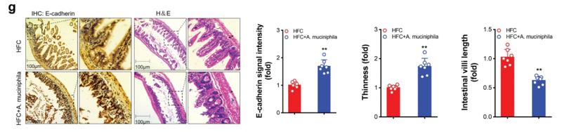
Application: IHC Species: Mice Sample: ileum
Application: WB Species: human Sample: CRC
Restrictive clause
Affinity Biosciences tests all products strictly. Citations are provided as a resource for additional applications that have not been validated by Affinity Biosciences. Please choose the appropriate format for each application and consult Materials and Methods sections for additional details about the use of any product in these publications.
For Research Use Only.
Not for use in diagnostic or therapeutic procedures. Not for resale. Not for distribution without written consent. Affinity Biosciences will not be held responsible for patent infringement or other violations that may occur with the use of our products. Affinity Biosciences, Affinity Biosciences Logo and all other trademarks are the property of Affinity Biosciences LTD.