
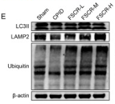
, using LAMP2 Antibody at 1/1000 dilution.
5ug/NC membrane strip.
Exposure for 30s with Affinity™ ECL Kit(#KF8001).
Bands result from membrane strip incubation.")
.
Bands result from membrane strip incubation.")


 oil red O staining and quantitative analysis of aortic sections from MiR-22-3p agomir or negative control (NC) mice; (b) Masson’s staining and quantitative analysis of aortic sections from MiR-22-3p agomir or NC mice (original magnification, × 40; bar = 500 μm); (c) sirius red staining and quantitative analysis of aortic sections from MiR-22-3p agomir or NC mice (original magnification, × 40; bar = 500 μm); (d) macrophage antigen-3 (Mac-3) immunostaining and quantitative analysis of aortic sections from MiR-22-3p agomir or NC mice (original magnification, × 40; bar = 200 μm); and (e) quantitative analysis of smooth muscle cells by α-smooth muscle actin immunostaining of aortic sections from MiR-22-3p agomir or NC mice (original magnification, × 40; bar = 500 μm). (*P")
| Product: | LAMP2 Antibody |
| Catalog: | DF6719 |
| Description: | Rabbit polyclonal antibody to LAMP2 |
| Application: | WB IHC |
| Cited expt.: | WB, IHC |
| Reactivity: | Human, Mouse, Rat |
| Prediction: | Pig, Bovine, Horse, Sheep, Rabbit, Dog |
| Mol.Wt.: | 45kDa,110kDa(Glycosylated)(Observed); 45kD(Calculated). |
| Uniprot: | P13473 |
| RRID: | AB_2838681 |
Control Products

Related Downloads
Protocols
Product Info
*The optimal dilutions should be determined by the end user. For optimal experimental results, antibody reuse is not recommended.
*Tips:
WB: For western blot detection of denatured protein samples. IHC: For immunohistochemical detection of paraffin sections (IHC-p) or frozen sections (IHC-f) of tissue samples. IF/ICC: For immunofluorescence detection of cell samples. ELISA(peptide): For ELISA detection of antigenic peptide.
Cite Format: Affinity Biosciences Cat# DF6719, RRID:AB_2838681.
Fold/Unfold
CD_antigen=CD107b;CD107 antigen like family member B;CD107 antigen-like family member B;CD107b;LAMP 2;Lamp 2b;LAMP-2;LAMP2;LAMP2_HUMAN;LAMPB;LGP110;Lysosomal associated membrane protein 2;Lysosome associated membrane glycoprotein 2;Lysosome-associated membrane glycoprotein 2;Lysosome-associated membrane protein 2;MAC3;
Immunogens
A synthesized peptide derived from human LAMP2, corresponding to a region within C-terminal amino acids.
Isoform LAMP-2A is highly expressed in placenta, lung and liver, less in kidney and pancreas, low in brain and skeletal muscle (PubMed:7488019, PubMed:26856698). Isoform LAMP-2B is detected in spleen, thymus, prostate, testis, small intestine, colon, skeletal muscle, brain, placenta, lung, kidney, ovary and pancreas and liver (PubMed:7488019, PubMed:26856698). Isoform LAMP-2C is detected in small intestine, colon, heart, brain, skeletal muscle, and at lower levels in kidney and placenta (PubMed:26856698).
- P13473 LAMP2_HUMAN:
- Protein BLAST With
- NCBI/
- ExPASy/
- Uniprot
MVCFRLFPVPGSGLVLVCLVLGAVRSYALELNLTDSENATCLYAKWQMNFTVRYETTNKTYKTVTISDHGTVTYNGSICGDDQNGPKIAVQFGPGFSWIANFTKAASTYSIDSVSFSYNTGDNTTFPDAEDKGILTVDELLAIRIPLNDLFRCNSLSTLEKNDVVQHYWDVLVQAFVQNGTVSTNEFLCDKDKTSTVAPTIHTTVPSPTTTPTPKEKPEAGTYSVNNGNDTCLLATMGLQLNITQDKVASVININPNTTHSTGSCRSHTALLRLNSSTIKYLDFVFAVKNENRFYLKEVNISMYLVNGSVFSIANNNLSYWDAPLGSSYMCNKEQTVSVSGAFQINTFDLRVQPFNVTQGKYSTAQDCSADDDNFLVPIAVGAALAGVLILVLLAYFIGLKHHHAGYEQF
Predictions
Score>80(red) has high confidence and is suggested to be used for WB detection. *The prediction model is mainly based on the alignment of immunogen sequences, the results are for reference only, not as the basis of quality assurance.
High(score>80) Medium(80>score>50) Low(score<50) No confidence
Research Backgrounds
Plays an important role in chaperone-mediated autophagy, a process that mediates lysosomal degradation of proteins in response to various stresses and as part of the normal turnover of proteins with a long biological half-live. Functions by binding target proteins, such as GAPDH and MLLT11, and targeting them for lysosomal degradation. Plays a role in lysosomal protein degradation in response to starvation (By similarity). Required for the fusion of autophagosomes with lysosomes during autophagy. Cells that lack LAMP2 express normal levels of VAMP8, but fail to accumulate STX17 on autophagosomes, which is the most likely explanation for the lack of fusion between autophagosomes and lysosomes. Required for normal degradation of the contents of autophagosomes. Required for efficient MHCII-mediated presentation of exogenous antigens via its function in lysosomal protein degradation; antigenic peptides generated by proteases in the endosomal/lysosomal compartment are captured by nascent MHCII subunits. Is not required for efficient MHCII-mediated presentation of endogenous antigens.
Modulates chaperone-mediated autophagy. Decreases presentation of endogenous antigens by MHCII. Does not play a role in the presentation of exogenous and membrane-derived antigens by MHCII.
O- and N-glycosylated; some of the 16 N-linked glycans are polylactosaminoglycans.
Cell membrane>Single-pass type I membrane protein. Endosome membrane>Single-pass type I membrane protein. Lysosome membrane>Single-pass type I membrane protein. Cytoplasmic vesicle>Autophagosome membrane.
Note: This protein shuttles between lysosomes, endosomes, and the plasma membrane.
Isoform LAMP-2A is highly expressed in placenta, lung and liver, less in kidney and pancreas, low in brain and skeletal muscle. Isoform LAMP-2B is detected in spleen, thymus, prostate, testis, small intestine, colon, skeletal muscle, brain, placenta, lung, kidney, ovary and pancreas and liver. Isoform LAMP-2C is detected in small intestine, colon, heart, brain, skeletal muscle, and at lower levels in kidney and placenta.
Belongs to the LAMP family.
Research Fields
· Cellular Processes > Transport and catabolism > Autophagy - animal. (View pathway)
· Cellular Processes > Transport and catabolism > Lysosome. (View pathway)
· Cellular Processes > Transport and catabolism > Phagosome. (View pathway)
· Human Diseases > Infectious diseases: Bacterial > Tuberculosis.
References

Application: WB Species: Mouse Sample:
Application: WB Species: Rat Sample:

Application: IF/ICC Species: Rat Sample:
Restrictive clause
Affinity Biosciences tests all products strictly. Citations are provided as a resource for additional applications that have not been validated by Affinity Biosciences. Please choose the appropriate format for each application and consult Materials and Methods sections for additional details about the use of any product in these publications.
For Research Use Only.
Not for use in diagnostic or therapeutic procedures. Not for resale. Not for distribution without written consent. Affinity Biosciences will not be held responsible for patent infringement or other violations that may occur with the use of our products. Affinity Biosciences, Affinity Biosciences Logo and all other trademarks are the property of Affinity Biosciences LTD.