
.
Bands result from membrane strip incubation.")


| Product: | GLO1 Antibody |
| Catalog: | DF6700 |
| Description: | Rabbit polyclonal antibody to GLO1 |
| Application: | WB IHC |
| Cited expt.: | WB |
| Reactivity: | Human, Mouse, Rat |
| Prediction: | Pig, Zebrafish, Bovine, Horse, Sheep, Dog |
| Mol.Wt.: | 21kDa(Observed); 21kD(Calculated). |
| Uniprot: | Q04760 |
| RRID: | AB_2838662 |
Control Products

Related Downloads
Protocols
Product Info
*The optimal dilutions should be determined by the end user. For optimal experimental results, antibody reuse is not recommended.
*Tips:
WB: For western blot detection of denatured protein samples. IHC: For immunohistochemical detection of paraffin sections (IHC-p) or frozen sections (IHC-f) of tissue samples. IF/ICC: For immunofluorescence detection of cell samples. ELISA(peptide): For ELISA detection of antigenic peptide.
Cite Format: Affinity Biosciences Cat# DF6700, RRID:AB_2838662.
Fold/Unfold
Aldoketomutase; glo1; GLOD1; Glx I; GLYI; glyoxalase domain containing 1; Glyoxalase I; Ketone aldehyde mutase; Ketone-aldehyde mutase; Lactoyl glutathione lyase; Lactoylglutathione lyase; LGUL_HUMAN; Methylglyoxalase; S D lactoylglutathione methylglyoxal lyase; S-D-lactoylglutathione methylglyoxal lyase;
Immunogens
A synthesized peptide derived from human GLO1, corresponding to a region within the internal amino acids.
- Q04760 LGUL_HUMAN:
- Protein BLAST With
- NCBI/
- ExPASy/
- Uniprot
MAEPQPPSGGLTDEAALSCCSDADPSTKDFLLQQTMLRVKDPKKSLDFYTRVLGMTLIQKCDFPIMKFSLYFLAYEDKNDIPKEKDEKIAWALSRKATLELTHNWGTEDDETQSYHNGNSDPRGFGHIGIAVPDVYSACKRFEELGVKFVKKPDDGKMKGLAFIQDPDGYWIEILNPNKMATLM
Predictions
Score>80(red) has high confidence and is suggested to be used for WB detection. *The prediction model is mainly based on the alignment of immunogen sequences, the results are for reference only, not as the basis of quality assurance.
High(score>80) Medium(80>score>50) Low(score<50) No confidence
Research Backgrounds
Catalyzes the conversion of hemimercaptal, formed from methylglyoxal and glutathione, to S-lactoylglutathione. Involved in the regulation of TNF-induced transcriptional activity of NF-kappa-B. Required for normal osteoclastogenesis.
Glutathionylation at Cys-139 inhibits enzyme activity.
Phosphorylated at Thr-107 in the presence of CaMK2. However, this is a consensus site for phosphorylation by CK2 so phosphorylation may be mediated by CK2 rather than CaMK2. Phosphorylation is induced by TNF and suppresses the TNF-induced transcriptional activity of NF-kappa-B.
Exists in a nitric oxide (NO)-modified form. The exact nature of the modification is unknown, but it suppresses the TNF-induced transcriptional activity of NF-kappa-B.
Belongs to the glyoxalase I family.
Research Fields
· Metabolism > Carbohydrate metabolism > Pyruvate metabolism.
References
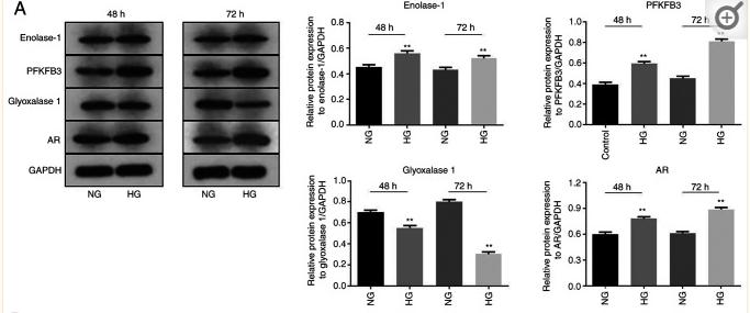
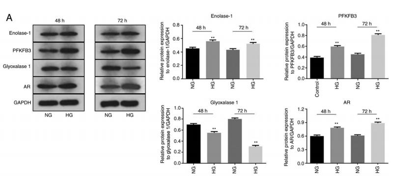
Application: WB Species: Mouse Sample: MPC5 cells
Application: WB Species: mouse Sample: MPC5 cells
Restrictive clause
Affinity Biosciences tests all products strictly. Citations are provided as a resource for additional applications that have not been validated by Affinity Biosciences. Please choose the appropriate format for each application and consult Materials and Methods sections for additional details about the use of any product in these publications.
For Research Use Only.
Not for use in diagnostic or therapeutic procedures. Not for resale. Not for distribution without written consent. Affinity Biosciences will not be held responsible for patent infringement or other violations that may occur with the use of our products. Affinity Biosciences, Affinity Biosciences Logo and all other trademarks are the property of Affinity Biosciences LTD.