Product: BUB1 Antibody
Catalog: DF6698
Description: Rabbit polyclonal antibody to BUB1
Application: WB IHC
Reactivity: Human, Mouse, Rat
Prediction: Rabbit, Dog
Mol.Wt.: 122kDa; 122kD(Calculated).
Uniprot: O43683
RRID: AB_2838660

View similar products>>

   Size Price Inventory
 100ul $280 In stock
 200ul $350 In stock

Lead Time: Same day delivery

For pricing and ordering contact:
Local distributors

Product Info

Source:
Rabbit
Application:
WB 1:500-1:2000, IHC 1:50-1:200
*The optimal dilutions should be determined by the end user.
*Tips:

WB: For western blot detection of denatured protein samples. IHC: For immunohistochemical detection of paraffin sections (IHC-p) or frozen sections (IHC-f) of tissue samples. IF/ICC: For immunofluorescence detection of cell samples. ELISA(peptide): For ELISA detection of antigenic peptide.

Reactivity:
Human,Mouse,Rat
Prediction:
Rabbit(86%), Dog(86%)
Clonality:
Polyclonal
Specificity:
BUB1 Antibody detects endogenous levels of total BUB1.
RRID:
AB_2838660
Cite Format: Affinity Biosciences Cat# DF6698, RRID:AB_2838660.
Conjugate:
Unconjugated.
Purification:
The antiserum was purified by peptide affinity chromatography using SulfoLink™ Coupling Resin (Thermo Fisher Scientific).
Storage:
Rabbit IgG in phosphate buffered saline , pH 7.4, 150mM NaCl, 0.02% sodium azide and 50% glycerol. Store at -20 °C. Stable for 12 months from date of receipt.
Alias:

Fold/Unfold

Bub1; BUB1 budding uninhibited by benzimidazoles 1 homolog; BUB1 budding uninhibited by benzimidazoles 1 homolog (yeast); BUB1 mitotic checkpoint serine/threonine kinase; BUB1, S. cerevisiae, homolog of; BUB1_HUMAN; BUB1A; BUB1L; Budding uninhibited by benzimidazoles 1 (yeast homolog); Budding uninhibited by benzimidazoles 1 homolog; Budding uninhibited by benzimidazoles 1, S. cerevisiae, homolog of; hBUB1; Homolog of mitotic checkpoint gene BUB1; Mitotic checkpoint gene BUB1; Mitotic checkpoint serine/threonine protein kinase BUB1; Mitotic checkpoint serine/threonine-protein kinase BUB1; Mitotic spindle checkpoint kinase; Putative serine/threonine protein kinase;

Immunogens

Immunogen:
Uniprot:
Gene(ID):
Expression:
O43683 BUB1_HUMAN:

High expression in testis and thymus, less in colon, spleen, lung and small intestine. Expressed in fetal thymus, bone marrow, heart, liver, spleen and thymus. Expression is associated with cells/tissues with a high mitotic index.

Description:
The Mitotic Checkpoint Complex (MCC), which contains Bub1, Bub1b, Bub3, Mad2, and Cdc20, controls chromosome segregation and monitors kinetochore-microtubule interactions (1). During mitosis, the MCC complex inhibits the ubiquitin ligase activity of the Anaphase Promoting Complex/Cyclosome (APC/C), thereby preventing cells with unaligned chromosomes from prematurely entering anaphase (2). Research studies have shown that Bub1b and Bub1 kinases are mutated in several types of human malignancies including hematopoietic, colorectal, lung, and breast cancers (3). Biallelic mutations in Bub1b have been found in mosaic variegated aneuploidy syndrome and premature chromatid separation syndrome (4). Bub1b mouse germline knockouts are embryonic lethal with heterozygous animals displaying genetic instability, early aging phenotypes, and increased cancer susceptibility (5). Bub3 binds both Bub1 and Bub1b, facilitating their recruitment to kinetochores (6), and is required for functional microtubule-kinetochore interactions (7).
Sequence:
MDTPENVLQMLEAHMQSYKGNDPLGEWERYIQWVEENFPENKEYLITLLEHLMKEFLDKKKYHNDPRFISYCLKFAEYNSDLHQFFEFLYNHGIGTLSSPLYIAWAGHLEAQGELQHASAVLQRGIQNQAEPREFLQQQYRLFQTRLTETHLPAQARTSEPLHNVQVLNQMITSKSNPGNNMACISKNQGSELSGVISSACDKESNMERRVITISKSEYSVHSSLASKVDVEQVVMYCKEKLIRGESEFSFEELRAQKYNQRRKHEQWVNEDRHYMKRKEANAFEEQLLKQKMDELHKKLHQVVETSHEDLPASQERSEVNPARMGPSVGSQQELRAPCLPVTYQQTPVNMEKNPREAPPVVPPLANAISAALVSPATSQSIAPPVPLKAQTVTDSMFAVASKDAGCVNKSTHEFKPQSGAEIKEGCETHKVANTSSFHTTPNTSLGMVQATPSKVQPSPTVHTKEALGFIMNMFQAPTLPDISDDKDEWQSLDQNEDAFEAQFQKNVRSSGAWGVNKIISSLSSAFHVFEDGNKENYGLPQPKNKPTGARTFGERSVSRLPSKPKEEVPHAEEFLDDSTVWGIRCNKTLAPSPKSPGDFTSAAQLASTPFHKLPVESVHILEDKENVVAKQCTQATLDSCEENMVVPSRDGKFSPIQEKSPKQALSSHMYSASLLRLSQPAAGGVLTCEAELGVEACRLTDTDAAIAEDPPDAIAGLQAEWMQMSSLGTVDAPNFIVGNPWDDKLIFKLLSGLSKPVSSYPNTFEWQCKLPAIKPKTEFQLGSKLVYVHHLLGEGAFAQVYEATQGDLNDAKNKQKFVLKVQKPANPWEFYIGTQLMERLKPSMQHMFMKFYSAHLFQNGSVLVGELYSYGTLLNAINLYKNTPEKVMPQGLVISFAMRMLYMIEQVHDCEIIHGDIKPDNFILGNGFLEQDDEDDLSAGLALIDLGQSIDMKLFPKGTIFTAKCETSGFQCVEMLSNKPWNYQIDYFGVAATVYCMLFGTYMKVKNEGGECKPEGLFRRLPHLDMWNEFFHVMLNIPDCHHLPSLDLLRQKLKKVFQQHYTNKIRALRNRLIVLLLECKRSRK

Predictions

Predictions:

Score>80(red) has high confidence and is suggested to be used for WB detection. *The prediction model is mainly based on the alignment of immunogen sequences, the results are for reference only, not as the basis of quality assurance.

Species
Results
Score
Rabbit
86
Dog
86
Horse
71
Bovine
57
Sheep
57
Pig
0
Xenopus
0
Zebrafish
0
Chicken
0
Model Confidence:
High(score>80) Medium(80>score>50) Low(score<50) No confidence

PTMs - O43683 As Substrate

Site PTM Type Enzyme
S17 Phosphorylation
T47 Phosphorylation
K60 Acetylation
T148 Phosphorylation O43683 (BUB1)
T173 Phosphorylation O43683 (BUB1)
K175 Ubiquitination
S176 Phosphorylation O43683 (BUB1)
K187 Ubiquitination
S194 Phosphorylation
S198 Phosphorylation O43683 (BUB1)
K203 Ubiquitination
T213 Phosphorylation O43683 (BUB1)
K216 Ubiquitination
Y219 Phosphorylation
S220 Phosphorylation
K241 Ubiquitination
S247 Phosphorylation
S250 Phosphorylation
K264 Ubiquitination
K277 Ubiquitination
K279 Ubiquitination
K290 Ubiquitination
K292 Ubiquitination
K299 Ubiquitination
T306 Phosphorylation
S307 Phosphorylation
S314 Phosphorylation Q13315 (ATM)
S331 Phosphorylation
K353 Ubiquitination
S375 Phosphorylation
T378 Phosphorylation
S402 Phosphorylation
K403 Ubiquitination
K410 Ubiquitination
S411 Phosphorylation
K416 Ubiquitination
S419 Phosphorylation O43683 (BUB1)
T435 Phosphorylation
S436 Phosphorylation
S437 Phosphorylation
T440 Phosphorylation
T441 Phosphorylation
T444 Phosphorylation
S445 Phosphorylation
T452 Phosphorylation
S454 Phosphorylation O43683 (BUB1)
K455 Ubiquitination
S459 Phosphorylation
T461 Phosphorylation
T479 Phosphorylation
K506 Ubiquitination
S510 Phosphorylation O43683 (BUB1)
S511 Phosphorylation O43683 (BUB1)
S525 Phosphorylation O43683 (BUB1)
K535 Ubiquitination
Y538 Phosphorylation
T552 Phosphorylation O43683 (BUB1)
S557 Phosphorylation
S563 Phosphorylation O43683 (BUB1)
K588 Ubiquitination
T589 Phosphorylation O43683 (BUB1)
S593 Phosphorylation P06493 (CDK1)
K595 Ubiquitination
S596 Phosphorylation
T601 Phosphorylation
S602 Phosphorylation
S608 Phosphorylation
T609 Phosphorylation P06493 (CDK1)
K613 Ubiquitination
S618 Phosphorylation O43683 (BUB1)
K625 Ubiquitination
S640 Phosphorylation
K653 Ubiquitination
S655 Phosphorylation
K660 Ubiquitination
S661 Phosphorylation
K663 Ubiquitination
S667 Phosphorylation O43683 (BUB1)
S668 Phosphorylation
Y671 Phosphorylation
S672 Phosphorylation
S679 Phosphorylation O43683 (BUB1)
S752 Phosphorylation O43683 (BUB1)
S755 Phosphorylation
K756 Ubiquitination
K770 Ubiquitination
K777 Ubiquitination
T778 Phosphorylation
K785 Ubiquitination
K813 Ubiquitination
K821 Ubiquitination
Y869 Phosphorylation
S870 Phosphorylation
K958 Ubiquitination
T960 Phosphorylation O43683 (BUB1)
S969 Phosphorylation O43683 (BUB1)
K1007 Ubiquitination
K1014 Ubiquitination
K1056 Ubiquitination
K1065 Ubiquitination
K1081 Ubiquitination

PTMs - O43683 As Enzyme

Substrate Site Source
O43683 (BUB1) T148 Uniprot
O43683 (BUB1) T173 Uniprot
O43683 (BUB1) S176 Uniprot
O43683 (BUB1) S198 Uniprot
O43683 (BUB1) T213 Uniprot
O43683 (BUB1) S419 Uniprot
O43683 (BUB1) S454 Uniprot
O43683 (BUB1) S510 Uniprot
O43683 (BUB1) S511 Uniprot
O43683 (BUB1) S525 Uniprot
O43683 (BUB1) T552 Uniprot
O43683 (BUB1) S563 Uniprot
O43683 (BUB1) T589 Uniprot
O43683 (BUB1) S618 Uniprot
O43683 (BUB1) S667 Uniprot
O43683 (BUB1) S679 Uniprot
O43683 (BUB1) S752 Uniprot
O43683 (BUB1) T960 Uniprot
O43683 (BUB1) S969 Uniprot
O43684 (BUB3) S19 Uniprot
Q12834 (CDC20) S41 Uniprot
Q12834 (CDC20) S72 Uniprot
Q12834 (CDC20) S92 Uniprot
Q12834 (CDC20) S153 Uniprot
Q12834 (CDC20) T157 Uniprot
Q12834 (CDC20) S161 Uniprot

Research Backgrounds

Function:

Serine/threonine-protein kinase that performs 2 crucial functions during mitosis: it is essential for spindle-assembly checkpoint signaling and for correct chromosome alignment. Has a key role in the assembly of checkpoint proteins at the kinetochore, being required for the subsequent localization of CENPF, BUB1B, CENPE and MAD2L1. Required for the kinetochore localization of PLK1. Required for centromeric enrichment of AUKRB in prometaphase. Plays an important role in defining SGO1 localization and thereby affects sister chromatid cohesion. Acts as a substrate for anaphase-promoting complex or cyclosome (APC/C) in complex with its activator CDH1 (APC/C-Cdh1). Necessary for ensuring proper chromosome segregation and binding to BUB3 is essential for this function. Can regulate chromosome segregation in a kinetochore-independent manner. Can phosphorylate BUB3. The BUB1-BUB3 complex plays a role in the inhibition of APC/C when spindle-assembly checkpoint is activated and inhibits the ubiquitin ligase activity of APC/C by phosphorylating its activator CDC20. This complex can also phosphorylate MAD1L1. Kinase activity is essential for inhibition of APC/CCDC20 and for chromosome alignment but does not play a major role in the spindle-assembly checkpoint activity. Mediates cell death in response to chromosome missegregation and acts to suppress spontaneous tumorigenesis.

PTMs:

Upon spindle-assembly checkpoint activation it is hyperphosphorylated and its kinase activity toward CDC20 is stimulated. Phosphorylation at Thr-609 is required for interaction with PLK1, phosphorylation at this site probably creates a binding site for the POLO-box domain of PLK1, thus enhancing the PLK1-BUB1 interaction.

Ubiquitinated and degraded during mitotic exit by APC/C-Cdh1.

Subcellular Location:

Nucleus. Chromosome>Centromere>Kinetochore.
Note: Nuclear in interphase cells. Accumulates gradually during G1 and S phase of the cell cycle, peaks at G2/M, and drops dramatically after mitosis. Localizes to the outer kinetochore. Kinetochore localization is required for normal mitotic timing and checkpoint response to spindle damage and occurs very early in prophase. AURKB, KNL1 and INCENP are required for kinetochore localization (By similarity).

Extracellular region or secreted Cytosol Plasma membrane Cytoskeleton Lysosome Endosome Peroxisome ER Golgi apparatus Nucleus Mitochondrion Manual annotation Automatic computational assertionSubcellular location
Tissue Specificity:

High expression in testis and thymus, less in colon, spleen, lung and small intestine. Expressed in fetal thymus, bone marrow, heart, liver, spleen and thymus. Expression is associated with cells/tissues with a high mitotic index.

Subunit Structure:

Interacts with BUB3 and KNL1. Interacts (when phosphorylated) with PLK1. The BUB1-BUB3 complex interacts with MAD1L1.

(Microbial infection) Interacts with SV40 Large T antigen; this interaction induces activation of a DNA damage response and promotes p53/TP53 stabilization and phosphorylation.

(Microbial infection) Interacts with herpes virus 8 protein LANA1.

Family&Domains:

The KEN box is required for its ubiquitination and degradation.

BUB1 N-terminal domain directs kinetochore localization and binding to BUB3.

Belongs to the protein kinase superfamily. Ser/Thr protein kinase family. BUB1 subfamily.

Research Fields

· Cellular Processes > Cell growth and death > Cell cycle.   (View pathway)

· Cellular Processes > Cell growth and death > Oocyte meiosis.   (View pathway)

· Organismal Systems > Endocrine system > Progesterone-mediated oocyte maturation.

References

1). BUB1 drives the occurrence and development of bladder cancer by mediating the STAT3 signaling pathway. JOURNAL OF EXPERIMENTAL & CLINICAL CANCER RESEARCH (PubMed: 34852826) [IF=11.3]

2). Inhibition of BUB1 suppresses tumorigenesis of osteosarcoma via blocking of PI3K/Akt and ERK pathways. JOURNAL OF CELLULAR AND MOLECULAR MEDICINE (PubMed: 34337852) [IF=5.3]

Application: WB    Species: Mice    Sample: OS cells

FIGURE 1 Expression of BUB1 protein in osteosarcoma (OS) tissues and cell lines. A, Representative images of different staining intensities of BUB1 protein in OS and normal bone tissues. B, Immunohistochemistry scores of OS tissues. C, Representative images showing the up- regulated expression of BUB1 protein in Saos2, U2OS and 143B cells; the ratio calculation was detected by using grey analysis. D, Significant differences in progression- free survival (PFS) (p < 0.001) and overall survival (p < 0.001) time between OS patients with high and low expression of BUB1. E, Cell viability was markedly reduced by incubating cells with BAY 1816032 for 72 h in Saos2 and U2OS cells.

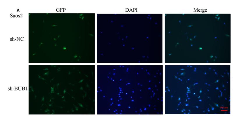
Application: IF/ICC    Species: Mice    Sample: OS cells

FIGURE 2 Effect of BUB1 knockdown on the proliferation of OS cells. A, B, Interference efficiency of stably transfected Saos2 or U2OS cells. C, D, Western blotting to verify the expression of BUB1 gene in lentivirus- transfected Saos2 or U2OS cells. E, F, Effect of BUB1 gene interference on the proliferation of Saos2 or U2OS cells detected by cell counting kit- 8 (CCK- 8). ***p < 0.001.

3). Study on the Mechanism of Sancao Tiaowei Decoction in the Treatment of MNNG-Induced Precancerous Lesions of Gastric Carcinoma Through Hedgehog Signaling Pathway. Frontiers in Oncology (PubMed: 35646631) [IF=4.7]

Application: IHC    Species: Rat    Sample: gastric tissue

Figure 7 Comparison of expression levels of Smo, IL-6, and IL-8 protein in the gastric tissue of rats in different groups. (A) The expression of Smo in the gastric tissue of rats was detected by immunohistochemistry. (B) The expression of IL-6 in the gastric tissue of rats was detected by immunohistochemistry. (C) The expression of IL-8 in the gastric tissue of rats was detected by immunohistochemistry. **P < 0.01 vs. Blank; ##P < 0.01 vs. PLGC. Scale bar: 50 µm.

4). Upregulation of ECT2 is associated with transcriptional program of cancer stem cells and predicts poor clinical outcome in gastric cancer. Oncology Letters (PubMed: 32788941) [IF=2.9]

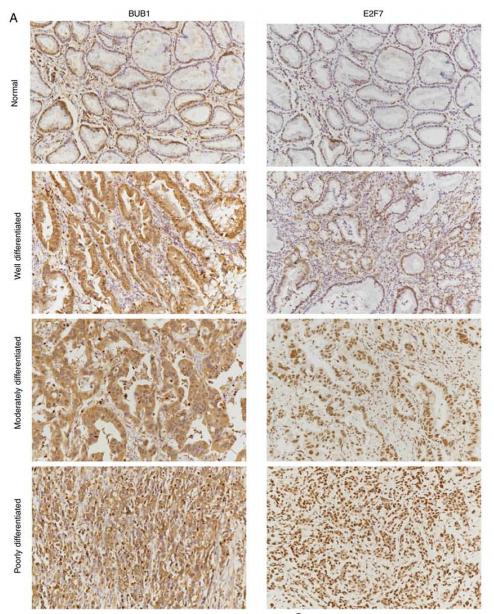
Application: IHC    Species: human    Sample: gastric cancer
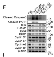
Figure 6. |BUB1 and E2F7 protein expression in human gastric cancer tissues. (A) Representative images depicting immunohistochemical staining of BUB1 (left panel) and E2F7 (right panel) in normal tissues, and well differentiated, moderately differentiated and poorly differentiated gastric cancer tissues.

Restrictive clause


Affinity Biosciences tests all products strictly. Citations are provided as a resource for additional applications that have not been validated by Affinity Biosciences. Please choose the appropriate format for each application and consult Materials and Methods sections for additional details about the use of any product in these publications.

For Research Use Only.
Not for use in diagnostic or therapeutic procedures. Not for resale. Not for distribution without written consent. Affinity Biosciences will not be held responsible for patent infringement or other violations that may occur with the use of our products. Affinity Biosciences, Affinity Biosciences Logo and all other trademarks are the property of Affinity Biosciences LTD.