.
Bands result from membrane strip incubation.")


 and mouse anti-beta tubulin Ab(#T0023) for 1 hour at 37°C. An AlexaFluor594 conjugated goat anti-rabbit IgG Ab(Red) and an AlexaFluor488 conjugated goat anti-mouse IgG Ab(Green) were used as the secondary antibody.
The nuclear counter stain is DAPI (blue).")
 of the patella and trochlea induced by patellar ligament shortening (PLS) surgery in the rat model of patellofemoral joint osteoarthritis (PFJOA). (A) Representative immunohistochemical images of both cathepsin K and osteoprotegerin (OPG) in the subchondral bone.")
 Quantitative real-time PCR analyzed the expression level of Cathepsin K mRNA in MDA-MB231 and SK-BR-3 cells treated with different compounds. (B) Western blot was performed to analyze the expression level of Cathepsin K protein in MDA-MB-231 and SK-BR-3 cells treated with different compounds.")
 The Alizarin Red S (yellow arrow head) and TRAP staining (red arrow head) of bone tissues in groups. (B) Immunohistochemical analysis of bone tissue among groups for MMP9 and RUNX2 (x 400). (C, D) Western blotting results of MMP, RUNX2, Cath-K, OPG and RANKL expression in bone marrow from rat femurs. The data are expressed as the means ± SD (n = 6 in each group). ***P<0.001; ****P<0.0001 vs. SHAM, and #P<0.05; ####P<0.0001 vs. OVX by one-way ANOVA and Tukey’s post hoc test")
 and RANKL (30 ng/ml) and then treated with or without rSj‑Cys (0.3 µM) for different time periods (24, 48 or 72 h). The expression levels of genes and proteins associated with osteoclast phenotype markers (CTSK, TRAP, ITGB3) and cell surface receptors of osteoclast precursors (RANK, OSCAR, TREM‑2)were determined by (A) reverse transcription‑quantitative PCR and (B) western blot analysis.")
 The RT‐qPCR assay detected the relative mRNA expression of osteoclastogenesis-associated marker genes, including NFATc1, c-fos, TRAP, Rank, cathepsin K and MMP9. (B) Western-blot analysis for OB’s effects on protein levels of NFATc1 and c-fos. (C) Western-blot analysis for OB’s effects on protein levels of MMP9 and cathepsin K. (D) Quantitative analysis of NFATc1 and c-fos. (E) Quantitative analysis of MMP9 and cathepsin K. (F) Immunofluorescence images for OB’s effects on protein expression of NFATc1. ns, no statistical significance")
 TRAP activity in the serum. (b) Expression levels of CTSK and ACP5 in the femoral heads. ∗∗p < 0.01; ns, no significant. TRAP, tartrate-resistant acid phosphatase; CTSK, cathepsin K; ACP5, acid phosphatase 5.")
 inhibited the differentiation of osteoclasts. A) RANK-positive rate of RAW264.7 cells was detected by flow cytometry. B) The protein expressions of OCs markers Cathepsin K and CTR were detected by Western blot. C) The area of F-actin ring was evaluated by immunofluorescence staining.")
 and Siha (B) were measured by Western blotting. The full length of blots was provided in Supplemental materials. *P < 0.05, **P < 0.01.")
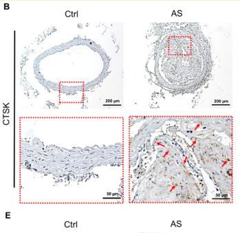
 and CTSK (B) was analyzed by immunohistochemistry. Scale = 50 μm *P < 0.05, **P < 0.01.")
 Light microscopy images of TRAP staining after osteoclast induction (100x). The blue arrows in the figure indicate macrophages, while the white arrows represent osteoclasts. (B) Western blot analysis (n = 3). (C) Statistical graph of macrophage counts, osteoclast counts and the proportion of osteoclasts in the total cell count (n = 3). Ns, p > 0.05. *, p < 0.05. **, p < 0.01. (D) Statistical graph of the relative expression of Cathepsin K, TRAP and RANK (n = 3). Ns > 0.05, ****p < 0.0001. (E) Relative mRNA expression of osteoclast-related genes (CTSK and ACP5) and macrophage-related gene (RANK). Ns > 0.05, ***p < 0.001.")
, MMP-13 (B), and sclerostin (C) within the osteocyte LCS of db/db mice compared to WT mice. Linear regression analysis (D) reveals a significant positive correlation between sclerostin expression and CTSK and MMP-13 using combined data from both groups. Data are presented as mean ± SD. Statistical analysis was performed using an unpaired Student’s t-test for (A–C) (n = 5 mice per group) and Pearson correlation for (D).")
 suppresses bone resorption activity of osteoclasts. a) Toluidine blue staining images of bovine bone slices in each group (100×, scale bar = 200 μm). b) and c) Expression and quantification of bone resorption-related proteins in osteoclasts of each group. d) Fluorescence expression of bone resorption-related proteins in each group (200×, scale bar = 100 μm). CTSK, cathepsin K; DAPI, 4′,6-diamidino-2-phenylindole; MMP-9, matrix metalloproteinase 9; TRAP, tartrate-resistant acid phosphatase.")
 RT-qPCR analysis of osteoclastogenic marker genes in BMMs treated with various concentrations of INS (0, 15, and 30 μM) for 5 days in the presence of RANKL and M-CSF. Relative mRNA expression levels of Acp5, c-Fos, Ctsk, and Nfatc1 were normalized to β-actin. (E, F) Representative Western blot images and quantification showing that INS dose-dependently upregulates the expression of antioxidant-related proteins (Nrf2, HO-1, SOD1, and catalase). (G, H) Representative Western blot images and quantification of osteoclastogenic marker proteins (CTSK, NFATc1, MMP9, and c-Fos) in RANKL-stimulated BMMs treated with INS (0, 15, and 30 μM) at days 1, 3, and 5 of differentiation. Data are presented as mean ± SD from three independent experiments performed in triplicate. *P < 0.05, **P < 0.01, ***P < 0.001 compared to the RANKL-stimulated control group (0 μM INS).")
| Product: | CTSK Antibody |
| Catalog: | DF6614 |
| Description: | Rabbit polyclonal antibody to CTSK |
| Application: | WB IHC IF/ICC |
| Cited expt.: | WB, IHC |
| Reactivity: | Human, Mouse, Rat |
| Prediction: | Pig, Horse, Sheep, Dog |
| Mol.Wt.: | 39kDa(Observed); 37kD(Calculated). |
| Uniprot: | P43235 |
| RRID: | AB_2838576 |
Control Products

Related Downloads
Protocols
Product Info
*The optimal dilutions should be determined by the end user. For optimal experimental results, antibody reuse is not recommended.
*Tips:
WB: For western blot detection of denatured protein samples. IHC: For immunohistochemical detection of paraffin sections (IHC-p) or frozen sections (IHC-f) of tissue samples. IF/ICC: For immunofluorescence detection of cell samples. ELISA(peptide): For ELISA detection of antigenic peptide.
Cite Format: Affinity Biosciences Cat# DF6614, RRID:AB_2838576.
Fold/Unfold
Cathepsin K; Cathepsin O; Cathepsin O1; Cathepsin O2; Cathepsin X; CATK_HUMAN; CTS02; Ctsk; CTSO; CTSO1; CTSO2; MGC23107; PKND; PYCD;
Immunogens
A synthesized peptide derived from human CTSK, corresponding to a region within N-terminal amino acids.
Predominantly expressed in osteoclasts (bones) (PubMed:7805878). Expressed in thyroid epithelial cells (PubMed:11082042).
- P43235 CATK_HUMAN:
- Protein BLAST With
- NCBI/
- ExPASy/
- Uniprot
MWGLKVLLLPVVSFALYPEEILDTHWELWKKTHRKQYNNKVDEISRRLIWEKNLKYISIHNLEASLGVHTYELAMNHLGDMTSEEVVQKMTGLKVPLSHSRSNDTLYIPEWEGRAPDSVDYRKKGYVTPVKNQGQCGSCWAFSSVGALEGQLKKKTGKLLNLSPQNLVDCVSENDGCGGGYMTNAFQYVQKNRGIDSEDAYPYVGQEESCMYNPTGKAAKCRGYREIPEGNEKALKRAVARVGPVSVAIDASLTSFQFYSKGVYYDESCNSDNLNHAVLAVGYGIQKGNKHWIIKNSWGENWGNKGYILMARNKNNACGIANLASFPKM
Predictions
Score>80(red) has high confidence and is suggested to be used for WB detection. *The prediction model is mainly based on the alignment of immunogen sequences, the results are for reference only, not as the basis of quality assurance.
High(score>80) Medium(80>score>50) Low(score<50) No confidence
Research Backgrounds
Thiol protease involved in osteoclastic bone resorption and may participate partially in the disorder of bone remodeling. Displays potent endoprotease activity against fibrinogen at acid pH. May play an important role in extracellular matrix degradation. Involved in the release of thyroid hormone thyroxine (T4) by limited proteolysis of TG/thyroglobulin in the thyroid follicle lumen.
Lysosome. Secreted. Apical cell membrane>Peripheral membrane protein>Extracellular side.
Note: Localizes to the lumen of thyroid follicles and to the apical membrane of thyroid epithelial cells.
Predominantly expressed in osteoclasts (bones). Expressed in thyroid epithelial cells.
Belongs to the peptidase C1 family.
Research Fields
· Cellular Processes > Transport and catabolism > Lysosome. (View pathway)
· Cellular Processes > Cell growth and death > Apoptosis. (View pathway)
· Human Diseases > Immune diseases > Rheumatoid arthritis.
· Organismal Systems > Development > Osteoclast differentiation. (View pathway)
· Organismal Systems > Immune system > Toll-like receptor signaling pathway. (View pathway)
References
Application: IHC Species: Mouse Sample:

Application: WB Species: Mouse Sample:
Restrictive clause
Affinity Biosciences tests all products strictly. Citations are provided as a resource for additional applications that have not been validated by Affinity Biosciences. Please choose the appropriate format for each application and consult Materials and Methods sections for additional details about the use of any product in these publications.
For Research Use Only.
Not for use in diagnostic or therapeutic procedures. Not for resale. Not for distribution without written consent. Affinity Biosciences will not be held responsible for patent infringement or other violations that may occur with the use of our products. Affinity Biosciences, Affinity Biosciences Logo and all other trademarks are the property of Affinity Biosciences LTD.