Product: SERPINF1 Antibody
Catalog: DF6547
Description: Rabbit polyclonal antibody to SERPINF1
Application: WB IHC IF/ICC
Reactivity: Human, Mouse, Rat
Prediction: Pig, Bovine, Horse, Sheep, Rabbit, Dog
Mol.Wt.: 46kDa; 46kD(Calculated).
Uniprot: P36955
RRID: AB_2838509

View similar products>>

   Size Price Inventory
 100ul $280 In stock
 200ul $350 In stock

Lead Time: Same day delivery

For pricing and ordering contact:
Local distributors

Product Info

Source:
Rabbit
Application:
WB 1:500-1:2000, IHC 1:50-1:200, IF/ICC 1:100-1:500
*The optimal dilutions should be determined by the end user.
*Tips:

WB: For western blot detection of denatured protein samples. IHC: For immunohistochemical detection of paraffin sections (IHC-p) or frozen sections (IHC-f) of tissue samples. IF/ICC: For immunofluorescence detection of cell samples. ELISA(peptide): For ELISA detection of antigenic peptide.

Reactivity:
Human,Mouse,Rat
Prediction:
Pig(100%), Bovine(100%), Horse(92%), Sheep(100%), Rabbit(92%), Dog(100%)
Clonality:
Polyclonal
Specificity:
SERPINF1 Antibody detects endogenous levels of total SERPINF1.
RRID:
AB_2838509
Cite Format: Affinity Biosciences Cat# DF6547, RRID:AB_2838509.
Conjugate:
Unconjugated.
Purification:
The antiserum was purified by peptide affinity chromatography using SulfoLink™ Coupling Resin (Thermo Fisher Scientific).
Storage:
Rabbit IgG in phosphate buffered saline , pH 7.4, 150mM NaCl, 0.02% sodium azide and 50% glycerol. Store at -20 °C. Stable for 12 months from date of receipt.
Alias:

Fold/Unfold

Cell proliferation-inducing gene 35 protein; EPC 1; EPC-1; EPC1; OI12; OI6; PEDF; PEDF_HUMAN; PIG 35; PIG35; Pigment epithelium derived factor; Pigment epithelium-derived factor; Proliferation inducing protein 35; serine (or cysteine) proteinase inhibitor, clade F (alpha-2 antiplasmin, pigment epithelium derived factor), member 1; Serpin F1; Serpin family F member 1; Serpin peptidase inhibitor clade F member 1; serpin peptidase inhibitor, clade F (alpha-2 antiplasmin, pigment epithelium derived factor), member 1; SERPINF 1; Serpinf1;

Immunogens

Immunogen:
Uniprot:
Gene(ID):
Expression:
P36955 PEDF_HUMAN:

Retinal pigment epithelial cells and blood plasma.

Description:
Pigment epithelium-derived growth factor (PEDF), also known as EPC-1 (early population doubling level cDNA-1), is a glycoprotein found naturally in the normal eye. PEDF has reported neuroprotective and differentiation properties and is secreted in abundance by retinal pigment epithelium cells. It belongs to the serine protease inhibitor (Serpin) superfamily and has been reported to inhibit angiogenesis and proliferation of several cell types. The “pooling” of PEDF within the interphotoreceptor matrix places this molecule in a prime physical location to affect the underlying neural retina. Additionally, PEDF induces neuronal differentiation and promotes survival of neurons of the central nervous system from degeneration caused by serum withdrawal or glutamate cytotoxicity.
Sequence:
MQALVLLLCIGALLGHSSCQNPASPPEEGSPDPDSTGALVEEEDPFFKVPVNKLAAAVSNFGYDLYRVRSSTSPTTNVLLSPLSVATALSALSLGAEQRTESIIHRALYYDLISSPDIHGTYKELLDTVTAPQKNLKSASRIVFEKKLRIKSSFVAPLEKSYGTRPRVLTGNPRLDLQEINNWVQAQMKGKLARSTKEIPDEISILLLGVAHFKGQWVTKFDSRKTSLEDFYLDEERTVRVPMMSDPKAVLRYGLDSDLSCKIAQLPLTGSMSIIFFLPLKVTQNLTLIEESLTSEFIHDIDRELKTVQAVLTVPKLKLSYEGEVTKSLQEMKLQSLFDSPDFSKITGKPIKLTQVEHRAGFEWNEDGAGTTPSPGLQPAHLTFPLDYHLNQPFIFVLRDTDTGALLFIGKILDPRGP

Predictions

Predictions:

Score>80(red) has high confidence and is suggested to be used for WB detection. *The prediction model is mainly based on the alignment of immunogen sequences, the results are for reference only, not as the basis of quality assurance.

Species
Results
Score
Pig
100
Bovine
100
Sheep
100
Dog
100
Horse
92
Rabbit
92
Chicken
58
Xenopus
55
Zebrafish
0
Model Confidence:
High(score>80) Medium(80>score>50) Low(score<50) No confidence

PTMs - P36955 As Substrate

Site PTM Type Enzyme
S24 Phosphorylation P68400 (CSNK2A1)
S114 Phosphorylation P68400 (CSNK2A1)
S152 Phosphorylation
S153 Phosphorylation
S227 Phosphorylation P17612 (PRKACA)
T283 Phosphorylation
N285 N-Glycosylation
T287 Phosphorylation
T294 Phosphorylation
S295 Phosphorylation
K316 Methylation

Research Backgrounds

Function:

Neurotrophic protein; induces extensive neuronal differentiation in retinoblastoma cells. Potent inhibitor of angiogenesis. As it does not undergo the S (stressed) to R (relaxed) conformational transition characteristic of active serpins, it exhibits no serine protease inhibitory activity.

PTMs:

The N-terminus is blocked. Extracellular phosphorylation enhances antiangiogenic activity.

N- and O-glycosylated. O-glycosylated with a core 1 or possibly core 8 glycan.

Subcellular Location:

Secreted. Melanosome.
Note: Enriched in stage I melanosomes.

Extracellular region or secreted Cytosol Plasma membrane Cytoskeleton Lysosome Endosome Peroxisome ER Golgi apparatus Nucleus Mitochondrion Manual annotation Automatic computational assertionSubcellular location
Tissue Specificity:

Retinal pigment epithelial cells and blood plasma.

Family&Domains:

The N-terminal (AA 44-121) exhibits neurite outgrowth-inducing activity. The C-terminal exposed loop (AA 382-418) is essential for serpin activity.

Belongs to the serpin family.

Research Fields

· Environmental Information Processing > Signal transduction > Wnt signaling pathway.   (View pathway)

References

1). PEDF is an antifibrosis factor that inhibits the activation of fibroblasts in a bleomycin-induced pulmonary fibrosis rat model. Respiratory Research (PubMed: 35459189) [IF=5.8]

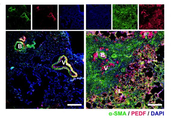
Application: IF/ICC    Species: rat    Sample: lung

Fig. 2 |Angiogenesis and PEDF expression and distribution.F Immunofuorescent staining showed the localization and expression of PEDF in the lung from normal and BLM-exposed (28 days) rats. Bar = 200 μm

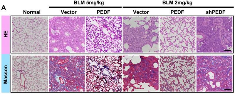
Application: IHC    Species: rat    Sample: lung

Fig. 3| PEDF could inhibit fbrosis induced by BLM. A Representative images of the H&E and Masson-stained cross-section of the lung from the experimental group (BLM 5 mg/kg or 2 mg/kg) and PEDF-AAV (denoted as PEDF) treatment group or PEDF-shRNA-AAV (denoted as shPEDF)treatment group. BLM (2 mg/kg or 5 mg/kg) represents lung tissue from rats 28 days after BLM instillation. Bar=200 μm.

Application: WB    Species: rat    Sample: fibroblasts

Fig. 4| PEDF inhibited fbroblast activation by inhibiting the TGF-β1/smad2/3 pathway. D and E Western blot to detect the efect of diferent concentrations of TGF-β1 on PEDF expression.

2). PEDF Protects Endothelial Barrier Integrity during Acute Myocardial Infarction via 67LR. International Journal of Molecular Sciences (PubMed: 36769107) [IF=5.6]

3). Preparation and Characterization of Protein-loaded PFC Nanoemulsions for the Treatment of Heart Diseases by Pulmonary Administration. European Journal of Pharmaceutical Sciences (PubMed: 33359617) [IF=4.6]

4). A "Hibernating-like" viable state induced by Lentiviral Vector-Mediated PEDF overexpression in rat acute ischemic myocardium. Human Gene Therapy (PubMed: 30734585) [IF=4.2]

Application: WB    Species: rat    Sample: cardiomyocytes

Figure 6. |Cardioprotective effect of PEDF in neonatal cardiomyocytes and contractile recoverability of anoxic cardiomyocytes. (A) Representative western blots of PEDF and RIP3 protein in neonatal cardiomyocytes transduced by PEDF-LVs or not and subjected to anoxic insult at 0-12 hours. 0 hour represents normal control. The labels 0 + P, 1 + P, 2 + P, 4 + P, 8 + P, and 12 + P represent relative anoxic period plus PEDF-LVs transduced.

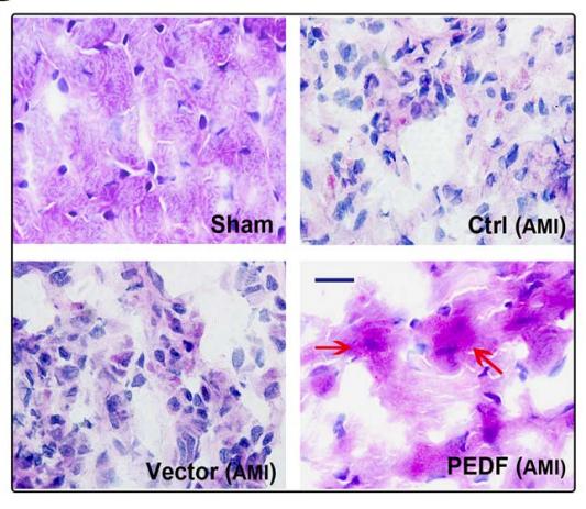
Application: IHC    Species: rat    Sample: myocardium

Figure 5. |Ultrastructure change observed by electron microscopy and glycogen deposition demonstrated by PAS staining. (A) Representative ultrastructure images of myocardium observed by electron microscopy. The black arrow indicates the area of glycogen. The blue arrow indicates the area of mitochondria. The yellow arrow indicates the reserved contractile material (bar = 2 μm). (B) PAS staining images of myocardium observed by light microscopy. The red arrow indicates the area of glycogen (bar = 10 μm). All Biopsy sections were taken from the LAD distribution of the animals sacrificed after PET and echocardiographic studies 3 days after AMI.

5). PEDF inhibits non‑small cell lung cancer proliferation by suppressing autophagy through downregulation of AMPK‑ULK1 signaling. Oncology Reports (PubMed: 36281945) [IF=4.2]

Restrictive clause


Affinity Biosciences tests all products strictly. Citations are provided as a resource for additional applications that have not been validated by Affinity Biosciences. Please choose the appropriate format for each application and consult Materials and Methods sections for additional details about the use of any product in these publications.

For Research Use Only.
Not for use in diagnostic or therapeutic procedures. Not for resale. Not for distribution without written consent. Affinity Biosciences will not be held responsible for patent infringement or other violations that may occur with the use of our products. Affinity Biosciences, Affinity Biosciences Logo and all other trademarks are the property of Affinity Biosciences LTD.