


 and mouse anti-beta tubulin Ab(T0023 1:200) for 1 hour at 37°C. An AlexaFluor594 conjugated goat anti-rabbit IgG(H+L) Ab(Red) and an AlexaFluor488 conjugated goat anti-mouse IgG(H+L) Ab(Green) were used as the secondary antibody.
The nuclear counter stain is DAPI(blue).")
| Product: | Syndecan 4 Antibody |
| Catalog: | AF0831 |
| Description: | Rabbit polyclonal antibody to Syndecan 4 |
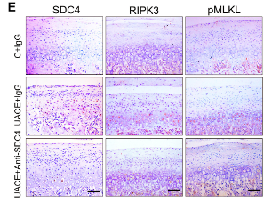
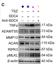
| Application: | WB IHC IF/ICC |
| Cited expt.: | WB, IHC |
| Reactivity: | Human, Mouse, Rat |
| Prediction: | Pig, Zebrafish, Bovine, Horse, Sheep, Rabbit, Dog, Chicken |
| Mol.Wt.: | 22kDa(Observed); 22kD(Calculated). |
| Uniprot: | P31431 |
| RRID: | AB_2833791 |
Control Products

Related Downloads
Protocols
Product Info
*The optimal dilutions should be determined by the end user. For optimal experimental results, antibody reuse is not recommended.
*Tips:
WB: For western blot detection of denatured protein samples. IHC: For immunohistochemical detection of paraffin sections (IHC-p) or frozen sections (IHC-f) of tissue samples. IF/ICC: For immunofluorescence detection of cell samples. ELISA(peptide): For ELISA detection of antigenic peptide.
Cite Format: Affinity Biosciences Cat# AF0831, RRID:AB_2833791.
Fold/Unfold
Amphiglycan; MGC22217; OTTHUMP00000031788; ryudocan amphiglycan; Ryudocan; Ryudocan core protein; SDC 4; Sdc4; SDC4_HUMAN; SYND 4; SYND4; syndecan 4; syndecan 4 (amphiglycan, ryudocan); syndecan proteoglycan 4; Syndecan-4; Syndecan4;
Immunogens
A synthesized peptide derived from human Syndecan 4, corresponding to a region within C-terminal amino acids.
- P31431 SDC4_HUMAN:
- Protein BLAST With
- NCBI/
- ExPASy/
- Uniprot
MAPARLFALLLFFVGGVAESIRETEVIDPQDLLEGRYFSGALPDDEDVVGPGQESDDFELSGSGDLDDLEDSMIGPEVVHPLVPLDNHIPERAGSGSQVPTEPKKLEENEVIPKRISPVEESEDVSNKVSMSSTVQGSNIFERTEVLAALIVGGIVGILFAVFLILLLMYRMKKKDEGSYDLGKKPIYKKAPTNEFYA
Predictions
Score>80(red) has high confidence and is suggested to be used for WB detection. *The prediction model is mainly based on the alignment of immunogen sequences, the results are for reference only, not as the basis of quality assurance.
High(score>80) Medium(80>score>50) Low(score<50) No confidence
Research Backgrounds
Cell surface proteoglycan that bears heparan sulfate. Regulates exosome biogenesis in concert with SDCBP and PDCD6IP.
Shedding is enhanced by a number of factors such as heparanase, thrombin or EGF. Also by stress and wound healing. PMA-mediated shedding is inhibited by TIMP3.
Membrane>Single-pass type I membrane protein. Secreted.
Note: Shedding of the ectodomain produces a soluble form.
Secreted.
Expressed in epithelial and fibroblastic cells.
Belongs to the syndecan proteoglycan family.
Research Fields
· Environmental Information Processing > Signaling molecules and interaction > ECM-receptor interaction. (View pathway)
· Environmental Information Processing > Signaling molecules and interaction > Cell adhesion molecules (CAMs). (View pathway)
· Human Diseases > Cancers: Overview > Proteoglycans in cancer.
References
Application: IHC Species: Rat Sample:
Application: WB Species: Rat Sample:

Application: IHC Species: Rat Sample:

Application: WB Species: human Sample: AGS and MKN-74 cell
Restrictive clause
Affinity Biosciences tests all products strictly. Citations are provided as a resource for additional applications that have not been validated by Affinity Biosciences. Please choose the appropriate format for each application and consult Materials and Methods sections for additional details about the use of any product in these publications.
For Research Use Only.
Not for use in diagnostic or therapeutic procedures. Not for resale. Not for distribution without written consent. Affinity Biosciences will not be held responsible for patent infringement or other violations that may occur with the use of our products. Affinity Biosciences, Affinity Biosciences Logo and all other trademarks are the property of Affinity Biosciences LTD.