


 antibody(Red), diluted at 1/600 was used as secondary antibody.")
| Product: | SCARB1 Antibody |
| Catalog: | DF6479 |
| Description: | Rabbit polyclonal antibody to SCARB1 |
| Application: | WB IHC IF/ICC |
| Cited expt.: | WB |
| Reactivity: | Human, Mouse, Rat |
| Prediction: | Pig, Horse, Sheep, Rabbit, Dog |
| Mol.Wt.: | 70~80kDa(Observed); 61kD(Calculated). |
| Uniprot: | Q8WTV0 |
| RRID: | AB_2838441 |
Control Products

Related Downloads
Protocols
Product Info
*The optimal dilutions should be determined by the end user. For optimal experimental results, antibody reuse is not recommended.
*Tips:
WB: For western blot detection of denatured protein samples. IHC: For immunohistochemical detection of paraffin sections (IHC-p) or frozen sections (IHC-f) of tissue samples. IF/ICC: For immunofluorescence detection of cell samples. ELISA(peptide): For ELISA detection of antigenic peptide.
Cite Format: Affinity Biosciences Cat# DF6479, RRID:AB_2838441.
Fold/Unfold
CD36 and LIMPII analogous 1; CD36; CD36 Antigen like 1; CD36 antigen-like 1; CD36L1; CLA 1; CLA-1; CLA1; Collagen type I receptor; HDLQTL6; MGC138242; SCARB1; Scavebger Receptor Class B Member 1; Scavenger receptor class B member 1; Scavenger Receptor Class B Type 1; SCRB1_HUMAN; SR BI; SR-BI; SRB1; SRBI; Thrombospondin receptor like 1; thrombospondin receptor-like 1;
Immunogens
A synthesized peptide derived from human SCARB1, corresponding to a region within the internal amino acids.
- Q8WTV0 SCRB1_HUMAN:
- Protein BLAST With
- NCBI/
- ExPASy/
- Uniprot
MGCSAKARWAAGALGVAGLLCAVLGAVMIVMVPSLIKQQVLKNVRIDPSSLSFNMWKEIPIPFYLSVYFFDVMNPSEILKGEKPQVRERGPYVYREFRHKSNITFNNNDTVSFLEYRTFQFQPSKSHGSESDYIVMPNILVLGAAVMMENKPMTLKLIMTLAFTTLGERAFMNRTVGEIMWGYKDPLVNLINKYFPGMFPFKDKFGLFAELNNSDSGLFTVFTGVQNISRIHLVDKWNGLSKVDFWHSDQCNMINGTSGQMWPPFMTPESSLEFYSPEACRSMKLMYKESGVFEGIPTYRFVAPKTLFANGSIYPPNEGFCPCLESGIQNVSTCRFSAPLFLSHPHFLNADPVLAEAVTGLHPNQEAHSLFLDIHPVTGIPMNCSVKLQLSLYMKSVAGIGQTGKIEPVVLPLLWFAESGAMEGETLHTFYTQLVLMPKVMHYAQYVLLALGCVLLLVPVICQIRSQVGAGQRAARADSHSLACWGKGASDRTLWPTAAWSPPPAAVLRLCRSGSGHCWGLRSTLASFACRVATTLPVLEGLGPSLGGGTGS
Predictions
Score>80(red) has high confidence and is suggested to be used for WB detection. *The prediction model is mainly based on the alignment of immunogen sequences, the results are for reference only, not as the basis of quality assurance.
High(score>80) Medium(80>score>50) Low(score<50) No confidence
Research Backgrounds
Receptor for different ligands such as phospholipids, cholesterol ester, lipoproteins, phosphatidylserine and apoptotic cells. Receptor for HDL, mediating selective uptake of cholesteryl ether and HDL-dependent cholesterol efflux. Also facilitates the flux of free and esterified cholesterol between the cell surface and apoB-containing lipoproteins and modified lipoproteins, although less efficiently than HDL. May be involved in the phagocytosis of apoptotic cells, via its phosphatidylserine binding activity.
(Microbial infection) Acts as a receptor for hepatitis C virus in hepatocytes and appears to facilitate its cell entry. Binding between SCARB1 and the hepatitis C virus glycoprotein E2 is independent of the genotype of the viral isolate. Mediates uptake of M.fortuitum, E.coli and S.aureus.
N-glycosylated.
The six cysteines of the extracellular domain are all involved in intramolecular disulfide bonds.
Cell membrane>Multi-pass membrane protein. Membrane>Caveola>Multi-pass membrane protein.
Note: Predominantly localized to cholesterol and sphingomyelin-enriched domains within the plasma membrane, called caveolae.
Widely expressed.
Belongs to the CD36 family.
Research Fields
· Cellular Processes > Transport and catabolism > Phagosome. (View pathway)
· Human Diseases > Infectious diseases: Viral > Hepatitis C.
· Organismal Systems > Endocrine system > Ovarian steroidogenesis.
· Organismal Systems > Endocrine system > Aldosterone synthesis and secretion.
· Organismal Systems > Digestive system > Fat digestion and absorption.
· Organismal Systems > Digestive system > Vitamin digestion and absorption.
· Organismal Systems > Digestive system > Cholesterol metabolism.
References
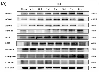
Application: WB Species: Mouse Sample: brain
Application: WB Species: rat Sample: EPCs

Application: WB Species: Mouse Sample:
Restrictive clause
Affinity Biosciences tests all products strictly. Citations are provided as a resource for additional applications that have not been validated by Affinity Biosciences. Please choose the appropriate format for each application and consult Materials and Methods sections for additional details about the use of any product in these publications.
For Research Use Only.
Not for use in diagnostic or therapeutic procedures. Not for resale. Not for distribution without written consent. Affinity Biosciences will not be held responsible for patent infringement or other violations that may occur with the use of our products. Affinity Biosciences, Affinity Biosciences Logo and all other trademarks are the property of Affinity Biosciences LTD.