Product: G6PD Antibody
Catalog: DF6444
Description: Rabbit polyclonal antibody to G6PD
Application: WB IHC IF/ICC
Reactivity: Human, Mouse, Rat
Prediction: Pig, Zebrafish, Bovine, Horse, Rabbit, Dog, Xenopus
Mol.Wt.: 59kDa; 59kD(Calculated).
Uniprot: P11413
RRID: AB_2838407

View similar products>>

   Size Price Inventory
 100ul $280 In stock
 200ul $350 In stock

Lead Time: Same day delivery

For pricing and ordering contact:
Local distributors

Product Info

Source:
Rabbit
Application:
IF/ICC 1:100-1:500, WB 1:500-1:2000, IHC 1:50-1:200
*The optimal dilutions should be determined by the end user.
*Tips:

WB: For western blot detection of denatured protein samples. IHC: For immunohistochemical detection of paraffin sections (IHC-p) or frozen sections (IHC-f) of tissue samples. IF/ICC: For immunofluorescence detection of cell samples. ELISA(peptide): For ELISA detection of antigenic peptide.

Reactivity:
Human,Mouse,Rat
Prediction:
Pig(90%), Zebrafish(90%), Bovine(90%), Horse(90%), Rabbit(90%), Dog(90%), Xenopus(100%)
Clonality:
Polyclonal
Specificity:
G6PD Antibody detects endogenous levels of total G6PD.
RRID:
AB_2838407
Cite Format: Affinity Biosciences Cat# DF6444, RRID:AB_2838407.
Conjugate:
Unconjugated.
Purification:
The antiserum was purified by peptide affinity chromatography using SulfoLink™ Coupling Resin (Thermo Fisher Scientific).
Storage:
Rabbit IgG in phosphate buffered saline , pH 7.4, 150mM NaCl, 0.02% sodium azide and 50% glycerol. Store at -20 °C. Stable for 12 months from date of receipt.
Alias:

Fold/Unfold

G6PD; G6PD_HUMAN; G6PD1; G6pdx; Glucose 6 phosphate 1 dehydrogenase; Glucose 6 phosphate dehydrogenase; Glucose 6 phosphate dehydrogenase, G6PD; Glucose-6-phosphate 1-dehydrogenase; MET19; POS10; Zwf1p;

Immunogens

Immunogen:
Uniprot:
Gene(ID):
Expression:
P11413 G6PD_HUMAN:

Isoform Long is found in lymphoblasts, granulocytes and sperm.

Description:
Glucose-6-phosphate dehydrogenase (G6PD) catalyses the first and rate-limiting step of the pentose phosphate pathway (1). The NADPH generated from this reaction is essential to protect cells from oxidative stress (1). Recent studies have shown that p53 interacts with G6PD and inhibits its activity, therefore suppressing glucose consumption through the pentose phosphate pathway (2). In cancer cells with p53 mutations, the increased glucose consumption is directed towards increased biosynthesis, which is critical for cancer cell proliferation (2).
Sequence:
MAEQVALSRTQVCGILREELFQGDAFHQSDTHIFIIMGASGDLAKKKIYPTIWWLFRDGLLPENTFIVGYARSRLTVADIRKQSEPFFKATPEEKLKLEDFFARNSYVAGQYDDAASYQRLNSHMNALHLGSQANRLFYLALPPTVYEAVTKNIHESCMSQIGWNRIIVEKPFGRDLQSSDRLSNHISSLFREDQIYRIDHYLGKEMVQNLMVLRFANRIFGPIWNRDNIACVILTFKEPFGTEGRGGYFDEFGIIRDVMQNHLLQMLCLVAMEKPASTNSDDVRDEKVKVLKCISEVQANNVVLGQYVGNPDGEGEATKGYLDDPTVPRGSTTATFAAVVLYVENERWDGVPFILRCGKALNERKAEVRLQFHDVAGDIFHQQCKRNELVIRVQPNEAVYTKMMTKKPGMFFNPEESELDLTYGNRYKNVKLPDAYERLILDVFCGSQMHFVRSDELREAWRIFTPLLHQIELEKPKPIPYIYGSRGPTEADELMKRVGFQYEGTYKWVNPHKL

Predictions

Predictions:

Score>80(red) has high confidence and is suggested to be used for WB detection. *The prediction model is mainly based on the alignment of immunogen sequences, the results are for reference only, not as the basis of quality assurance.

Species
Results
Score
Xenopus
100
Pig
90
Horse
90
Bovine
90
Dog
90
Zebrafish
90
Rabbit
90
Sheep
0
Chicken
0
Model Confidence:
High(score>80) Medium(80>score>50) Low(score<50) No confidence

PTMs - P11413 As Substrate

Site PTM Type Enzyme
A2 Acetylation
S8 Phosphorylation
R9 Methylation
T10 Phosphorylation
T76 Phosphorylation
K82 Ubiquitination
K89 Acetylation
K89 Ubiquitination
K95 Acetylation
K95 Ubiquitination
K97 Acetylation
K97 Ubiquitination
S106 Phosphorylation
Y107 Phosphorylation
Y112 Phosphorylation
S123 Phosphorylation
S132 Phosphorylation
Y139 Phosphorylation
Y147 Phosphorylation
K171 Acetylation
K171 Ubiquitination
S180 Phosphorylation Q05655 (PRKCD)
R182 Methylation
Y202 Phosphorylation
K205 Ubiquitination
T236 Phosphorylation Q05655 (PRKCD)
Y249 Phosphorylation
K293 Ubiquitination
S296 Phosphorylation
K320 Ubiquitination
Y322 Phosphorylation
K386 Acetylation
K386 Ubiquitination
Y401 Phosphorylation
T402 Phosphorylation
K403 Acetylation
K403 Ubiquitination
K408 Acetylation
K408 Ubiquitination
S418 Phosphorylation
T423 Phosphorylation
Y424 Phosphorylation
Y428 Phosphorylation P12931 (SRC)
K432 Acetylation
K432 Ubiquitination
S448 Phosphorylation
T490 Phosphorylation
K497 Acetylation
K497 Ubiquitination
Y503 Phosphorylation
T506 Phosphorylation
Y507 Phosphorylation P12931 (SRC)
K514 Acetylation
K514 Ubiquitination

Research Backgrounds

Function:

Cytosolic glucose-6-phosphate dehydrogenase that catalyzes the first and rate-limiting step of the oxidative branch within the pentose phosphate pathway/shunt, an alternative route to glycolysis for the dissimilation of carbohydrates and a major source of reducing power and metabolic intermediates for fatty acid and nucleic acid biosynthetic processes.

PTMs:

Acetylated by ELP3 at Lys-403; acetylation inhibits its homodimerization and enzyme activity. Deacetylated by SIRT2 at Lys-403; deacetylation stimulates its enzyme activity.

Subcellular Location:

Cytoplasm>Cytosol. Membrane>Peripheral membrane protein.

Extracellular region or secreted Cytosol Plasma membrane Cytoskeleton Lysosome Endosome Peroxisome ER Golgi apparatus Nucleus Mitochondrion Manual annotation Automatic computational assertionSubcellular location
Tissue Specificity:

Isoform Long is found in lymphoblasts, granulocytes and sperm.

Subunit Structure:

Homotetramer; dimer of dimers. Interacts with SIRT2; the interaction is enhanced by H(2)O(2) treatment.

Family&Domains:

Belongs to the glucose-6-phosphate dehydrogenase family.

Research Fields

· Human Diseases > Cancers: Overview > Central carbon metabolism in cancer.   (View pathway)

· Metabolism > Carbohydrate metabolism > Pentose phosphate pathway.

· Metabolism > Metabolism of other amino acids > Glutathione metabolism.

· Metabolism > Global and overview maps > Metabolic pathways.

· Metabolism > Global and overview maps > Carbon metabolism.

References

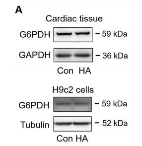
1). Hypoxic acclimation improves cardiac redox homeostasis and protects heart against ischemia-reperfusion injury through upregulation of O-GlcNAcylation. Redox Biology, 2021 (PubMed: 33964586) [IF=11.4]

Application: WB    Species: rat    Sample: cardiac tissues and H9c2 cells

Fig. 5. |PPP was enhanced via O-GlcNAcylation-mediated activation of G6PDH. A, Representative blots of G6PDH in cardiac tissues and H9c2 cells (n = 4 per group). B–C, Representative blots of O-GlcNAc modified G6PDH (B) and G6PDH activity (C) in H9c2 cells treated with IL-1β for 4 or 24 h (n = 6 per group).

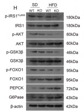
2). Nr2e1 ablation impairs liver glucolipid metabolism and induces inflammation, high-fat diets amplify the damage. Biomedicine & Pharmacotherapy, 2019 (PubMed: 31590127) [IF=4.8]

Application: WB    Species: mouse    Sample: liver

Fig. 3. |Nr2e1 deficiency impaired glucose tolerance and insulin sensitivity, the damages were exacerbated by HFD.Western blots measured the expression of phosphorylated protein of IRS1, AKT, GSK3β and FOXO1 in the liver samples after 12 weeks of HFD or SD feeding (3 H). Phosphorylated protein levels were normalized to the respective total protein levels (3I).

3). Oxidative stress accompanies HIF1-dependent impairment of glucose metabolism in the hippocampus of adult rats survived prenatal severe hypoxia. Developmental neuroscience, 2023 (PubMed: 37980886) [IF=2.9]

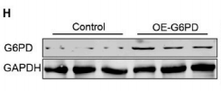
4). Exploring Senescence-Related Transcriptional Signatures to Uncover Molecular Subtypes and Key Genes in Hepatocellular Carcinoma. , 2023

Application: WB    Species: Human    Sample: HepG2 cells

Figure 7. Differential Gene Screening Among Three Subtypes and Risk Model Construction. Volcano plot for differential analysis of cluster1 vs. no_cluster1 (A), cluster2 vs. no_cluster2 (B), and cluster3 vs. no_cluster3 (C). D. Identification of 729 candidates among differentially expressed genes. E. Trajectory of each independent variable as lambda changes. F. Confidence interval under lambda. G. Coefficients of the seven genes in the model. H. Western blot assay to evaluate G6PD protein expression in HepG2 cells. I. Colony formation assay to evaluate HepG2 cell proliferation. J-K. Transwell assay assesses HepG2 cell migration (J) and invasion (K).

Restrictive clause


Affinity Biosciences tests all products strictly. Citations are provided as a resource for additional applications that have not been validated by Affinity Biosciences. Please choose the appropriate format for each application and consult Materials and Methods sections for additional details about the use of any product in these publications.

For Research Use Only.
Not for use in diagnostic or therapeutic procedures. Not for resale. Not for distribution without written consent. Affinity Biosciences will not be held responsible for patent infringement or other violations that may occur with the use of our products. Affinity Biosciences, Affinity Biosciences Logo and all other trademarks are the property of Affinity Biosciences LTD.