
 Expression of BDNF in different groups (B) Quantitative analysis of expression of BDNF in different groups (mean ± SD, ∗P < 0.05, ∗∗∗P < 0.01, one-way ANOVA).")


brain-derived neurotrophic factor (BDNF) and tropomyosin-related
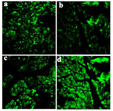
kinase B (TrkB) in morphine-induced conditioned place preference (CPP). a The mRNA expression of BDNF. F(2,6)=6.716. b The
mRNA expression of TrkB. F(2,6)=7.553. c The representative immunoblots. d Graphic representation of relative expression of BDNF to
β-actin. F(2,6)=9.132. e Graphic representation of relative expression of TrkB to β-actin. F(2,6)=12.13. f Immunohistochemical staining
for BDNF and TrkB expression. Figures were magnifed by ×200. g
Comparison of the percentage of BDNF-positive cells. F(2,6)=8.633.
h Comparison of the percentage of TrkB-positive cells. F(2,6)=9.624.
Data are presented as the mean±SEM, n=3. One-way ANOVA with
a post hoc Newman–Keuls test. *:vs the con group, P<0.05; **:vs
the con group, P<0.001; #:vs the mod group, P<0.05")

brain-derived neurotrophic factor (BDNF) and tropomyosin-related
kinase B (TrkB) in morphine-induced conditioned place preference (CPP). a The mRNA expression of BDNF. F(2,6)=6.716. b The
mRNA expression of TrkB. F(2,6)=7.553. c The representative immunoblots. d Graphic representation of relative expression of BDNF to
β-actin. F(2,6)=9.132. e Graphic representation of relative expression of TrkB to β-actin. F(2,6)=12.13. f Immunohistochemical staining
for BDNF and TrkB expression. Figures were magnifed by ×200. g
Comparison of the percentage of BDNF-positive cells. F(2,6)=8.633.
h Comparison of the percentage of TrkB-positive cells. F(2,6)=9.624.
Data are presented as the mean±SEM, n=3. One-way ANOVA with
a post hoc Newman–Keuls test. *:vs the con group, P<0.05; **:vs
the con group, P<0.001; #:vs the mod group, P<0.05")
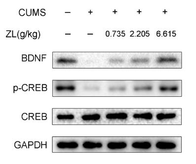
 mRNA expression; (b) protein expression. Compared with the control group, ∗P < 0.05; compared with the model group, #P < 0.05 (N = 5 in each group).")
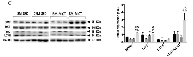
 Mitochondrial morphology by Mitotracker Red staining. (B) Cell viability quantified using a Cell Counting Kit-8 (CCK-8) assay. (C) Brain-derived neurotrophic factor (BDNF) level in cell supernatant of
the cultured astrocytes. (D) Glial cell-derived neurotrophic factor (GDNF) level in cell supernatant of the cultured astrocytes. (E) Levels of BDNF and GDNF in
cultured astrocytes. (F–G) Relative protein levels of BDNF and GDNF to β-actin, as determined using ImageJ. ##P < 0.01 vs. high glucose (HG), & P < 0.05 vs.
recurrent low glucose (0.1 mM) (RLG) under conditions of high glucose (HG + RLG), && P < 0.01 vs. HG + RLG.")
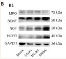
 Representative HE-stained images of prostate tissues. (b) Inflammation grades for prostate samples were scored on the basis of HE sections (×400 magnification). The columns represent five random spots of one rat, while the rows denote six animals involved in each group. (c) The mRNA expression of proinflammatory cytokines (TNF-α, IL-1β, and IFN-γ) and chemokines (CCL2, CCL3) relative to β-actin was performed by the RT-PCR method. (d) The protein levels of immunological mediators (COX-2 and IL-2) and cytokines (IL-6, VCAM-1, and BDNF) were performed using the western blot method, and the relative quantitative results are shown. Data was presented as mean ± SEM. #P < 0.05 versus the control group; ##P < 0.01 versus the control group; ###P < 0.001 versus the control group; ∗P < 0.05 versus the EAP group; ∗∗P < 0.01 versus the EAP group; ∗∗∗P < 0.001 versus the EAP group.")
–(c) Western blot analysis of BDNF and TrkB in the hippocampus 4 weeks after RCIR. n = 6 in each group. (d) Representative micrographs for TUNEL staining in CA1 subfields of the hippocampus 4 weeks after RCIR. 400× magnification was shown. Scale bar = 50 μm. (e) The apoptotic index was calculated as the number of TUNEL (+) cells divided by the total number of cells. n = 4 in each group. (f)–(h) Western blot analysis demonstrated Bax and Bcl-2 expression at 4 weeks after RCIR injury. n = 6 in each group. β-Actin was used as an internal control. ∗∗∗P < 0.001, the RCIR group vs. the sham group; #P < 0.05, ##P < 0.01, ###P < 0.001, the NBP80 group or NBP120 group vs. the RCIR group; $P < 0.05, the NBP80 group vs. the NBP120 group. Values are expressed as the mean ± SD.")
 cAMP level in hippocampus was analyzed by Elisa. (b–e) Western blot was used to analyze the protein expression of PKA, p-CREB, and BDNF in hippocampus. The above data are presented in the form of mean ± SEM, Independent Samples t Test, * p < 0.05 vs. D-Gal group, ** p < 0.01 vs. D-Gal group. Control: young group treated with saline, D-Gal Group: aging group induced by D-galactose, D-Gal+E Group: aging group induced by D-galactose that performed treadmill exercise.")
 Western blot assays of protein expression levels of the ERK, p-CREB and BDNF within the prefrontal cortex (N = 6 rats per group). Q-PCR showing mRNA levels of BDNF (B), cleaved caspase-3 (C), caspase-9 (D), Bax (E) and Bcl2 (F). Band intensities were normalized to GAPDH (N = 6 rats per group). (G) The ratio of mRNA levels of Bax and Bcl2 (N = 6 rats per group). (H) Immunofluorescent staining shows the TUNEL (green) and NeuN (red) double labeling of the prefrontal cortex. Nuclei (blue) are stained with DAPI. Scale bar is 50 μm. (I) Quantifications of the proportion of TUNEL-positive neuron in prefrontal cortex (N = 6 rats per group) (J) Representative images of Hoechst-33258 staining to observe morphological changes in nuclei. Scale bar is 50 µm. (K) Quantifications of chromatin-condensing cell numbers in prefrontal cortex (N = 6 rats per group). *p < 0.05, **p < 0.01, ***p < 0.001 and ****p < 0.0001, LPS vs Control group; #p < 0.05, ##p < 0.01 and ###p < 0.001, LPS vs LPS + LVM (LVM, Levomilnacipran). The data are presented as the means ± SEMs.")
 with different high frequencies on BDNF protein in the substantia nigra and striatum. Representative BDNF western blot images in the substantia nigra (a) and striata (c) in the groups were shown. The averaged data of BDNF proteins in the substantia nigra (b) and striata (d) were displayed. Values are expressed as the mean ± SEM. *p < .05, **p < .01, ***p < .001, ****p < .0001. I: Control group; II: MPTP/p group; III: MPTP/p + 5 Hz group; IV: MPTP/p + 10 Hz group; V: MPTP/p + 15 Hz group; VI: MPTP/p + 20 Hz group; VII: MPTP/p + iTBS group. B: F(6,14) = 11.760, D: F(6,14) = 9.717.")
 Representative immunofluorescence images of BDNF and pTrkB in the in LC. Scale bar = 50 μm. (B, D) Quantitative analysis of the average fluorescence intensity in the left and right LC regions of BDNF and pTrkB. Data are presented as mean ± SD, n = 3, *P")
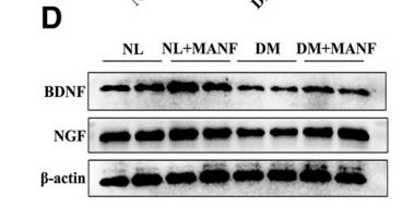
; all bar charts were created using GraphPad Prism software, and error bars (SEM) were added. A WB analysis of BDNF and NGF expression. B (a) TUNEL staining to observe apoptosis in brain. B (b–d) IHC for BDNF, NGF, and TH in brain. B (e) Nissl staining to observe neuronal cells. (f–j) Bar charts corresponding to (a–e)")
| Product: | BDNF Antibody |
| Catalog: | DF6387 |
| Description: | Rabbit polyclonal antibody to BDNF |
| Application: | WB IHC IF/ICC |
| Cited expt.: | WB, IHC, IF/ICC |
| Reactivity: | Human, Mouse, Rat |
| Prediction: | Pig, Zebrafish, Bovine, Horse, Sheep, Rabbit, Dog, Chicken, Xenopus |
| Mol.Wt.: | 15kD(mature), 28kDa(precursor), 45kD(unprocessed)(Observed); 28kD(Calculated). |
| Uniprot: | P23560 |
| RRID: | AB_2838350 |
Control Products

Related Downloads
Protocols
Product Info
*The optimal dilutions should be determined by the end user. For optimal experimental results, antibody reuse is not recommended.
*Tips:
WB: For western blot detection of denatured protein samples. IHC: For immunohistochemical detection of paraffin sections (IHC-p) or frozen sections (IHC-f) of tissue samples. IF/ICC: For immunofluorescence detection of cell samples. ELISA(peptide): For ELISA detection of antigenic peptide.
Cite Format: Affinity Biosciences Cat# DF6387, RRID:AB_2838350.
Fold/Unfold
Abrineurin; ANON2; BDNF; BDNF_HUMAN; Brain Derived Neurotrophic Factor; Brain-derived neurotrophic factor; BULN2; MGC34632; Neurotrophin;
Immunogens
A synthesized peptide derived from human BDNF, corresponding to a region within the internal amino acids.
Detected in blood plasma and in saliva (at protein level) (PubMed:11152678, PubMed:19467646). Brain. Highly expressed in hippocampus, amygdala, cerebral cortex and cerebellum. Also expressed in heart, lung, skeletal muscle, testis, prostate and placenta.
- P23560 BDNF_HUMAN:
- Protein BLAST With
- NCBI/
- ExPASy/
- Uniprot
MTILFLTMVISYFGCMKAAPMKEANIRGQGGLAYPGVRTHGTLESVNGPKAGSRGLTSLADTFEHVIEELLDEDQKVRPNEENNKDADLYTSRVMLSSQVPLEPPLLFLLEEYKNYLDAANMSMRVRRHSDPARRGELSVCDSISEWVTAADKKTAVDMSGGTVTVLEKVPVSKGQLKQYFYETKCNPMGYTKEGCRGIDKRHWNSQCRTTQSYVRALTMDSKKRIGWRFIRIDTSCVCTLTIKRGR
Predictions
Score>80(red) has high confidence and is suggested to be used for WB detection. *The prediction model is mainly based on the alignment of immunogen sequences, the results are for reference only, not as the basis of quality assurance.
High(score>80) Medium(80>score>50) Low(score<50) No confidence
Research Backgrounds
Important signaling molecule that activates signaling cascades downstream of NTRK2. During development, promotes the survival and differentiation of selected neuronal populations of the peripheral and central nervous systems. Participates in axonal growth, pathfinding and in the modulation of dendritic growth and morphology. Major regulator of synaptic transmission and plasticity at adult synapses in many regions of the CNS. The versatility of BDNF is emphasized by its contribution to a range of adaptive neuronal responses including long-term potentiation (LTP), long-term depression (LTD), certain forms of short-term synaptic plasticity, as well as homeostatic regulation of intrinsic neuronal excitability.
Important signaling molecule that activates signaling cascades downstream of NTRK2. Activates signaling cascades via the heterodimeric receptor formed by NGFR and SORCS2. Signaling via NGFR and SORCS2 plays a role in synaptic plasticity and long-term depression (LTD). Binding to NGFR and SORCS2 promotes neuronal apoptosis. Promotes neuronal growth cone collapse (By similarity).
N-glycosylated and glycosulfated, contrary to mature BDNF.
Mature BDNF is produced by proteolytic removal of the propeptide, catalyzed by a FURIN family member. In addition, the precursor form is proteolytically cleaved within the propeptide, but this is not an obligatory intermediate for the production of mature BDNF. Can be converted into mature BDNF by plasmin (PLG).
Secreted.
Secreted.
Note: A proportion of BDNF is secreted as immature precursor (proBDNF).
Detected in blood plasma and in saliva (at protein level). Brain. Highly expressed in hippocampus, amygdala, cerebral cortex and cerebellum. Also expressed in heart, lung, skeletal muscle, testis, prostate and placenta.
Belongs to the NGF-beta family.
Research Fields
· Environmental Information Processing > Signal transduction > MAPK signaling pathway. (View pathway)
· Environmental Information Processing > Signal transduction > Ras signaling pathway. (View pathway)
· Environmental Information Processing > Signal transduction > cAMP signaling pathway. (View pathway)
· Environmental Information Processing > Signal transduction > PI3K-Akt signaling pathway. (View pathway)
· Human Diseases > Neurodegenerative diseases > Huntington's disease.
· Human Diseases > Substance dependence > Cocaine addiction.
· Human Diseases > Substance dependence > Alcoholism.
· Organismal Systems > Nervous system > Neurotrophin signaling pathway. (View pathway)
References

Application: WB Species: Mouse Sample:
Application: WB Species: mice Sample: corneal epithelial cells
Application: WB Species: Rat Sample:
Application: WB Species: rat Sample: hippocampus
Application: WB Species: rat Sample: gastrocnemius muscles
Application: IF/ICC Species: rat Sample: gastrocnemius muscles
Restrictive clause
Affinity Biosciences tests all products strictly. Citations are provided as a resource for additional applications that have not been validated by Affinity Biosciences. Please choose the appropriate format for each application and consult Materials and Methods sections for additional details about the use of any product in these publications.
For Research Use Only.
Not for use in diagnostic or therapeutic procedures. Not for resale. Not for distribution without written consent. Affinity Biosciences will not be held responsible for patent infringement or other violations that may occur with the use of our products. Affinity Biosciences, Affinity Biosciences Logo and all other trademarks are the property of Affinity Biosciences LTD.