Product: DR5 Antibody
Catalog: DF6368
Description: Rabbit polyclonal antibody to DR5
Application: WB IHC
Reactivity: Human
Mol.Wt.: 48kDa; 48kD(Calculated).
Uniprot: O14763
RRID: AB_2838332

View similar products>>

   Size Price Inventory
 100ul $280 In stock
 200ul $350 In stock

Lead Time: Same day delivery

For pricing and ordering contact:
Local distributors

Product Info

Source:
Rabbit
Application:
WB 1:500-1:2000, IHC 1:50-1:200
*The optimal dilutions should be determined by the end user.
*Tips:

WB: For western blot detection of denatured protein samples. IHC: For immunohistochemical detection of paraffin sections (IHC-p) or frozen sections (IHC-f) of tissue samples. IF/ICC: For immunofluorescence detection of cell samples. ELISA(peptide): For ELISA detection of antigenic peptide.

Reactivity:
Human
Clonality:
Polyclonal
Specificity:
DR5 Antibody detects endogenous levels of total DR5.
RRID:
AB_2838332
Cite Format: Affinity Biosciences Cat# DF6368, RRID:AB_2838332.
Conjugate:
Unconjugated.
Purification:
The antiserum was purified by peptide affinity chromatography using SulfoLink™ Coupling Resin (Thermo Fisher Scientific).
Storage:
Rabbit IgG in phosphate buffered saline , pH 7.4, 150mM NaCl, 0.02% sodium azide and 50% glycerol. Store at -20 °C. Stable for 12 months from date of receipt.
Alias:

Fold/Unfold

Fas like protein; Apoptosis inducing protein TRICK2A/2B; Apoptosis inducing receptor TRAIL R2; CD262; CD262 antigen; Cytotoxic TRAIL receptor 2; Death domain containing receptor for TRAIL/Apo 2L; Death receptor 5; DR5; KILLER; KILLER/DR5; OTTHUMP00000123492; OTTHUMP00000123493; p53 regulated DNA damage inducible cell death receptor(killer); TNF related apoptosis inducing ligand receptor 2; TNF-related apoptosis-inducing ligand receptor 2; TNFRSF10B; TR10B_HUMAN; TRAIL R2; TRAIL receptor 2; TRAIL-R2; TRAILR2; TRICK2; TRICK2A; TRICK2B; TRICKB; Tumor necrosis factor receptor like protein ZTNFR9; Tumor necrosis factor receptor superfamily member 10B; Tumor necrosis factor receptor superfamily, member 10b; ZTNFR9;

Immunogens

Immunogen:
Uniprot:
Gene(ID):
Expression:
O14763 TR10B_HUMAN:

Widely expressed in adult and fetal tissues; very highly expressed in tumor cell lines such as HeLaS3, K-562, HL-60, SW480, A-549 and G-361; highly expressed in heart, peripheral blood lymphocytes, liver, pancreas, spleen, thymus, prostate, ovary, uterus, placenta, testis, esophagus, stomach and throughout the intestinal tract; not detectable in brain.

Description:
The tumor necrosis factor receptor family, which includes TNF-RI, Fas, DR3, DR4, DR5, and DR6, plays an important role in the regulation of apoptosis in various physiological systems (1,2). The receptors are activated by a family of cytokines that include TNF, FasL, and TRAIL. They are characterized by a highly conserved extracellular region containing cysteine-rich repeats and a conserved intracellular region of about 80 amino acids termed the death domain (DD). The DD is important for transducing the death signal by recruiting other DD containing adaptor proteins (FADD, TRADD, RIP) to the death-inducing signaling complex (DISC), resulting in activation of caspases. DR5 is a receptor for TNF-related apoptosis inducing ligand (TRAIL), which has been been shown to induce apoptosis in variety of cell types and has been targeted for cancer therapy (1-5). Structurally, DR5 contains an amino-terminal leader cleavage site followed by an extracellular region containing two cysteine-rich repeats, then a central transmembrane domain and a carboxy-terminal death domain. DR5 is expressed in a wide variety of tissues and is transcriptional target for p53 (6-8). It induces apoptosis through a FADD-dependent pathway. Deletion of DR5 leads to resistance in TRAIL-mediated apoptosis as well as an abrogated response to DNA-damaging stimuli (9).
Sequence:
MEQRGQNAPAASGARKRHGPGPREARGARPGPRVPKTLVLVVAAVLLLVSAESALITQQDLAPQQRAAPQQKRSSPSEGLCPPGHHISEDGRDCISCKYGQDYSTHWNDLLFCLRCTRCDSGEVELSPCTTTRNTVCQCEEGTFREEDSPEMCRKCRTGCPRGMVKVGDCTPWSDIECVHKESGTKHSGEVPAVEETVTSSPGTPASPCSLSGIIIGVTVAAVVLIVAVFVCKSLLWKKVLPYLKGICSGGGGDPERVDRSSQRPGAEDNVLNEIVSILQPTQVPEQEMEVQEPAEPTGVNMLSPGESEHLLEPAEAERSQRRRLLVPANEGDPTETLRQCFDDFADLVPFDSWEPLMRKLGLMDNEIKVAKAEAAGHRDTLYTMLIKWVNKTGRDASVHTLLDALETLGERLAKQKIEDHLLSSGKFMYLEGNADSAMS

PTMs - O14763 As Substrate

Site PTM Type Enzyme
K245 Ubiquitination
K360 Ubiquitination
K369 Ubiquitination
T381 Phosphorylation
Y383 Phosphorylation
K417 Ubiquitination

Research Backgrounds

Function:

Receptor for the cytotoxic ligand TNFSF10/TRAIL. The adapter molecule FADD recruits caspase-8 to the activated receptor. The resulting death-inducing signaling complex (DISC) performs caspase-8 proteolytic activation which initiates the subsequent cascade of caspases (aspartate-specific cysteine proteases) mediating apoptosis. Promotes the activation of NF-kappa-B. Essential for ER stress-induced apoptosis.

Subcellular Location:

Membrane>Single-pass type I membrane protein.

Extracellular region or secreted Cytosol Plasma membrane Cytoskeleton Lysosome Endosome Peroxisome ER Golgi apparatus Nucleus Mitochondrion Manual annotation Automatic computational assertionSubcellular location
Tissue Specificity:

Widely expressed in adult and fetal tissues; very highly expressed in tumor cell lines such as HeLaS3, K-562, HL-60, SW480, A-549 and G-361; highly expressed in heart, peripheral blood lymphocytes, liver, pancreas, spleen, thymus, prostate, ovary, uterus, placenta, testis, esophagus, stomach and throughout the intestinal tract; not detectable in brain.

Subunit Structure:

Monomer. Can interact with TRADD and RIPK1. Interacts with HCMV protein UL141; this interaction prevents TNFRSF10B cell surface expression. Two TNFRSF10B monomers interact with a UL141 homodimer. Three TNFRSF10B molecules interact with TNFSF10 homotrimer. In the absence of stimulation, interacts with BIRC2, DDX3X and GSK3B. The interaction with BIRC2 and DDX3X is further enhanced upon receptor stimulation and accompanied by DDX3X and BIRC2 cleavage.

Research Fields

· Cellular Processes > Cell growth and death > p53 signaling pathway.   (View pathway)

· Cellular Processes > Cell growth and death > Apoptosis.   (View pathway)

· Cellular Processes > Cell growth and death > Necroptosis.   (View pathway)

· Environmental Information Processing > Signaling molecules and interaction > Cytokine-cytokine receptor interaction.   (View pathway)

· Human Diseases > Infectious diseases: Viral > Measles.

· Human Diseases > Infectious diseases: Viral > Influenza A.

· Organismal Systems > Immune system > Natural killer cell mediated cytotoxicity.   (View pathway)

References

1). Akkermansia muciniphila Aspartic Protease Amuc_1434* Inhibits Human Colorectal Cancer LS174T Cell Viability via TRAIL-Mediated Apoptosis Pathway. INTERNATIONAL JOURNAL OF MOLECULAR SCIENCES (PubMed: 32403433) [IF=5.6]

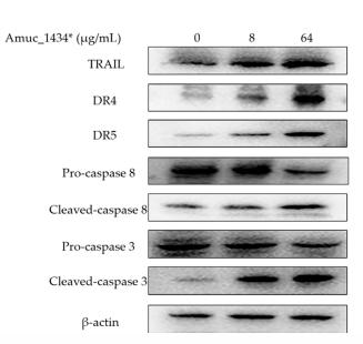
Application: WB    Species: human    Sample: LS174T cells

Figure 5. | Amuc_1434* mediated the activation of the apoptosis pathway in LS174T cells. (A) The expression of death receptor 4 (DR4), death receptor 5 (DR5), cysteinyl aspartate specific proteinase 8(caspase 8) and cysteinyl aspartate specific proteinase 3 (caspase 3) induced by Amuc_1434* in LS174T cells was dependent on tumor necrosis factor-related apoptosis-inducing ligand (TRAIL). (a) LS174T cells were treated with 8 and 64 µg/mL Amuc_1434* for 24 h, respectively. The cell lysates were analyzed by Western blot.

2). Bisimidazolium Salt Glycosyltransferase Inhibitors Suppress Hepatocellular Carcinoma Progression In Vitro and In Vivo. Pharmaceuticals (PubMed: 35745636) [IF=4.6]

Application: WB    Species: Human    Sample: HepG2 cells

Figure 5 C20/C22 increased the sensitivity of HepG2 cells to TRAIL-induced apoptosis. (A,B) WB analysis of the expression of DR4 and 5 in HepG2 cells that were treated with 2 μM C20/C22 for 0, 6, 12, and 24 h. ImageJ software was used to calculate the gray value (n = 3). (C,D) The immunofluorescence assays used to determine the intracellular and cell surface distribution of DR4/5. (E,F) Annexin V/PI double staining was used to detect the apoptotic cells after the indicated treatments (n = 3). (G,H) WB analysis of the expression of Fas in HepG2 cells treated with C20/C22 for 0, 6, 12, and 24 h. (I) The immunofluorescence assays used to determine the intracellular and cell surface distribution of Fas. (J,K) Flow cytometry analysis of the apoptotic cells after the indicated treatments (n = 3). Scale bars, 10 µm. Data shown correspond to the mean ± SD of three independent experiments. The p-value for all datasets was analyzed by one-way ANOVA followed by Tukey’s test using GraphPad Prism version 8.00, except the data in (B) whose p-value was analyzed by nonparametric Dunnett’s test using GraphPad Prism version 8.00. * p < 0.05, ** p < 0.01, *** p < 0.001, **** p < 0.0001, and ns nonsignificant vs. 0 h/control group.

Application: IF/ICC    Species: Human    Sample: HepG2 cells

Figure 5 C20/C22 increased the sensitivity of HepG2 cells to TRAIL-induced apoptosis. (A,B) WB analysis of the expression of DR4 and 5 in HepG2 cells that were treated with 2 μM C20/C22 for 0, 6, 12, and 24 h. ImageJ software was used to calculate the gray value (n = 3). (C,D) The immunofluorescence assays used to determine the intracellular and cell surface distribution of DR4/5. (E,F) Annexin V/PI double staining was used to detect the apoptotic cells after the indicated treatments (n = 3). (G,H) WB analysis of the expression of Fas in HepG2 cells treated with C20/C22 for 0, 6, 12, and 24 h. (I) The immunofluorescence assays used to determine the intracellular and cell surface distribution of Fas. (J,K) Flow cytometry analysis of the apoptotic cells after the indicated treatments (n = 3). Scale bars, 10 µm. Data shown correspond to the mean ± SD of three independent experiments. The p-value for all datasets was analyzed by one-way ANOVA followed by Tukey’s test using GraphPad Prism version 8.00, except the data in (B) whose p-value was analyzed by nonparametric Dunnett’s test using GraphPad Prism version 8.00. * p < 0.05, ** p < 0.01, *** p < 0.001, **** p < 0.0001, and ns nonsignificant vs. 0 h/control group.

3). Sphingomyelin synthase 2 overexpression promotes cisplatin-induced apoptosis of HepG2 cells. Oncology Letters (PubMed: 29375716) [IF=2.9]

Application: WB    Species: human    Sample: HepG2 cells

Figure 2.| Expression of DR4, DR5, cleaved caspase‑3 and c‑Myc in HepG2 cells. (A) The protein expression of DR4, DR5, cleaved caspase‑3 and c‑Myc was investigated using western blotting. Quantification and statistical analysis was performed on (B) DR4, (C) DR5, (D) cleaved caspase‑3 and (E) c‑Myc. n=3, *P<0.05, **P<0.001 vs. control group; #P<0.05, ##P<0.001 vs. control + DDP group. DR. death receptor; DDP, cisplatin; SMS2, sphyngomyelin synthase 2; c‑Myc, avian myelocytomatosis viral oncogene homolog.

Restrictive clause


Affinity Biosciences tests all products strictly. Citations are provided as a resource for additional applications that have not been validated by Affinity Biosciences. Please choose the appropriate format for each application and consult Materials and Methods sections for additional details about the use of any product in these publications.

For Research Use Only.
Not for use in diagnostic or therapeutic procedures. Not for resale. Not for distribution without written consent. Affinity Biosciences will not be held responsible for patent infringement or other violations that may occur with the use of our products. Affinity Biosciences, Affinity Biosciences Logo and all other trademarks are the property of Affinity Biosciences LTD.