Product: AMPK alpha Antibody
Catalog: DF6361
Description: Rabbit polyclonal antibody to AMPK alpha
Application: WB IHC IF/ICC
Reactivity: Human, Mouse, Rat
Prediction: Zebrafish, Bovine, Sheep, Rabbit, Dog, Chicken
Mol.Wt.: 63kDa; 64kD,62kD(Calculated).
Uniprot: Q13131 | P54646
RRID: AB_2838325

View similar products>>

   Size Price Inventory
 100ul $280 In stock
 200ul $350 In stock

Lead Time: Same day delivery

For pricing and ordering contact:
Local distributors

Product Info

Source:
Rabbit
Application:
WB 1:500-1:2000, IHC 1:50-1:200, IF/ICC 1:100
*The optimal dilutions should be determined by the end user.
*Tips:

WB: For western blot detection of denatured protein samples. IHC: For immunohistochemical detection of paraffin sections (IHC-p) or frozen sections (IHC-f) of tissue samples. IF/ICC: For immunofluorescence detection of cell samples. ELISA(peptide): For ELISA detection of antigenic peptide.

Reactivity:
Human,Mouse,Rat
Prediction:
Zebrafish(88%), Bovine(100%), Sheep(100%), Rabbit(88%), Dog(100%), Chicken(100%)
Clonality:
Polyclonal
Specificity:
AMPK alpha Antibody detects endogenous levels of total AMPK alpha.
RRID:
AB_2838325
Cite Format: Affinity Biosciences Cat# DF6361, RRID:AB_2838325.
Conjugate:
Unconjugated.
Purification:
The antiserum was purified by peptide affinity chromatography using SulfoLink™ Coupling Resin (Thermo Fisher Scientific).
Storage:
Rabbit IgG in phosphate buffered saline , pH 7.4, 150mM NaCl, 0.02% sodium azide and 50% glycerol. Store at -20 °C. Stable for 12 months from date of receipt.
Alias:

Fold/Unfold

5 AMP activated protein kinase alpha 1catalytic subunit; 5 AMP activated protein kinase catalytic alpha 1 chain; 5' AMP activated protein kinase catalytic subunit alpha 1; 5'-AMP-activated protein kinase catalytic subunit alpha-1; AAPK1; AAPK1_HUMAN; ACACA kinase; acetyl CoA carboxylase kinase; AI194361; AI450832; AL024255; AMP -activate kinase alpha 1 subunit; AMP-activated protein kinase, catalytic, alpha -1; AMPK 1; AMPK alpha 1; AMPK alpha 1 chain; AMPK; AMPK subunit alpha-1; AMPK1; AMPKa1; AMPKalpha1; C130083N04Rik; cb116; EC 2.7.11.1; HMG CoA reductase kinase; HMGCR kinase; hormone sensitive lipase kinase; Hydroxymethylglutaryl CoA reductase kinase; im:7154392; kinase AMPK alpha1; MGC33776; MGC57364; OTTHUMP00000161795; OTTHUMP00000161796; PRKAA 1; PRKAA1; Protein kinase AMP activated alpha 1 catalytic subunit; SNF1-like protein AMPK; SNF1A; Tau protein kinase PRKAA1; wu:fa94c10; 5'-AMP-activated protein kinase catalytic subunit alpha-2; AAPK2_HUMAN; ACACA kinase; Acetyl-CoA carboxylase kinase; AMPK alpha 2 chain; AMPK subunit alpha-2; AMPK2; AMPKa2; AMPKalpha2; HMGCR kinase; Hydroxymethylglutaryl-CoA reductase kinase; PRKAA; PRKAA2; Protein kinase AMP activated alpha 2 catalytic subunit; Protein kinase AMP activated catalytic subunit alpha 2;

Immunogens

Immunogen:
Uniprot:
Gene(ID):
Description:
AMP-activated protein kinase (AMPK) is highly conserved from yeast to plants and animals and plays a key role in the regulation of energy homeostasis (1). AMPK is a heterotrimeric complex composed of a catalytic α subunit and regulatory β and γ subunits, each of which is encoded by two or three distinct genes (α1, 2; β1, 2; γ1, 2, 3) (2). The kinase is activated by an elevated AMP/ATP ratio due to cellular and environmental stress, such as heat shock, hypoxia, and ischemia (1). The tumor suppressor LKB1, in association with accessory proteins STRAD and MO25, phosphorylates AMPKα at Thr172 in the activation loop, and this phosphorylation is required for AMPK activation (3-5). AMPKα is also phosphorylated at Thr258 and Ser485 (for α1; Ser491 for α2). The upstream kinase and the biological significance of these phosphorylation events have yet to be elucidated (6). The β1 subunit is post-translationally modified by myristoylation and multi-site phosphorylation including Ser24/25, Ser96, Ser101, Ser108, and Ser182 (6,7). Phosphorylation at Ser108 of the β1 subunit seems to be required for the activation of AMPK enzyme, while phosphorylation at Ser24/25 and Ser182 affects AMPK localization (7). Several mutations in AMPKγ subunits have been identified, most of which are located in the putative AMP/ATP binding sites (CBS or Bateman domains). Mutations at these sites lead to reduction of AMPK activity and cause glycogen accumulation in heart or skeletal muscle (1,2). Accumulating evidence indicates that AMPK not only regulates the metabolism of fatty acids and glycogen, but also modulates protein synthesis and cell growth through EF2 and TSC2/mTOR pathways, as well as blood flow via eNOS/nNOS (1).
Sequence:
MRRLSSWRKMATAEKQKHDGRVKIGHYILGDTLGVGTFGKVKVGKHELTGHKVAVKILNRQKIRSLDVVGKIRREIQNLKLFRHPHIIKLYQVISTPSDIFMVMEYVSGGELFDYICKNGRLDEKESRRLFQQILSGVDYCHRHMVVHRDLKPENVLLDAHMNAKIADFGLSNMMSDGEFLRTSCGSPNYAAPEVISGRLYAGPEVDIWSSGVILYALLCGTLPFDDDHVPTLFKKICDGIFYTPQYLNPSVISLLKHMLQVDPMKRATIKDIREHEWFKQDLPKYLFPEDPSYSSTMIDDEALKEVCEKFECSEEEVLSCLYNRNHQDPLAVAYHLIIDNRRIMNEAKDFYLATSPPDSFLDDHHLTRPHPERVPFLVAETPRARHTLDELNPQKSKHQGVRKAKWHLGIRSQSRPNDIMAEVCRAIKQLDYEWKVVNPYYLRVRRKNPVTSTYSKMSLQLYQVDSRTYLLDFRSIDDEITEAKSGTATPQRSGSVSNYRSCQRSDSDAEAQGKSSEVSLTSSVTSLDSSPVDLTPRPGSHTIEFFEMCANLIKILAQ

MAEKQKHDGRVKIGHYVLGDTLGVGTFGKVKIGEHQLTGHKVAVKILNRQKIRSLDVVGKIKREIQNLKLFRHPHIIKLYQVISTPTDFFMVMEYVSGGELFDYICKHGRVEEMEARRLFQQILSAVDYCHRHMVVHRDLKPENVLLDAHMNAKIADFGLSNMMSDGEFLRTSCGSPNYAAPEVISGRLYAGPEVDIWSCGVILYALLCGTLPFDDEHVPTLFKKIRGGVFYIPEYLNRSVATLLMHMLQVDPLKRATIKDIREHEWFKQDLPSYLFPEDPSYDANVIDDEAVKEVCEKFECTESEVMNSLYSGDPQDQLAVAYHLIIDNRRIMNQASEFYLASSPPSGSFMDDSAMHIPPGLKPHPERMPPLIADSPKARCPLDALNTTKPKSLAVKKAKWHLGIRSQSKPYDIMAEVYRAMKQLDFEWKVVNAYHLRVRRKNPVTGNYVKMSLQLYLVDNRSYLLDFKSIDDEVVEQRSGSSTPQRSCSAAGLHRPRSSFDSTTAESHSLSGSLTGSLTGSTLSSVSPRLGSHTMDFFEMCASLITTLAR

Predictions

Predictions:

Score>80(red) has high confidence and is suggested to be used for WB detection. *The prediction model is mainly based on the alignment of immunogen sequences, the results are for reference only, not as the basis of quality assurance.

Species
Results
Score
Bovine
100
Sheep
100
Dog
100
Chicken
100
Zebrafish
88
Rabbit
88
Xenopus
75
Pig
0
Horse
0
Model Confidence:
High(score>80) Medium(80>score>50) Low(score<50) No confidence

PTMs - Q13131/P54646 As Substrate

Site PTM Type Enzyme
S6 Phosphorylation
T32 Phosphorylation
K40 Ubiquitination
K45 Ubiquitination
K52 Ubiquitination
K56 Ubiquitination
K71 Ubiquitination
K80 Acetylation
K80 Ubiquitination
K152 Ubiquitination
S172 Phosphorylation
S176 Phosphorylation
T183 Phosphorylation Q8TDC3-2 (BRSK1) , Q16584 (MAP3K11) , Q15831 (STK11) , Q13554 (CAMK2B) , Q8N5S9 (CAMKK1) , Q13131 (PRKAA1) , Q96RR4 (CAMKK2) , Q8IWQ3 (BRSK2)
S184 Phosphorylation
C185 S-Nitrosylation
S187 Phosphorylation
Y190 Phosphorylation
Y247 Phosphorylation
K266 Ubiquitination
K271 Ubiquitination
K280 Ubiquitination
K285 Ubiquitination
S293 Phosphorylation
Y294 Phosphorylation
T355 Phosphorylation
S356 Phosphorylation
S360 Phosphorylation Q13131 (PRKAA1) , O75385 (ULK1)
T368 Phosphorylation O75385 (ULK1)
T382 Phosphorylation
T388 Phosphorylation Q13131 (PRKAA1)
K396 Ubiquitination
S397 Phosphorylation O75385 (ULK1)
K406 Sumoylation
K406 Ubiquitination
K429 Ubiquitination
Y441 Phosphorylation
Y442 Phosphorylation
K448 Ubiquitination
Y463 Phosphorylation
S467 Phosphorylation
T482 Phosphorylation
K485 Ubiquitination
S486 Phosphorylation P49841 (GSK3B) , O75385 (ULK1) , Q13131 (PRKAA1) , P17612 (PRKACA)
T488 Phosphorylation O75385 (ULK1)
T490 Phosphorylation P49841 (GSK3B)
S494 Phosphorylation Q13131 (PRKAA1)
S496 Phosphorylation Q13131 (PRKAA1) , P31749 (AKT1)
S498 Phosphorylation
Y500 Phosphorylation
S506 Phosphorylation
S508 Phosphorylation
K515 Ubiquitination
S520 Phosphorylation
T522 Phosphorylation
S523 Phosphorylation
S524 Phosphorylation
T526 Phosphorylation
S527 Phosphorylation
S531 Phosphorylation
Site PTM Type Enzyme
K41 Ubiquitination
K45 Ubiquitination
K69 Acetylation
K69 Ubiquitination
K141 Ubiquitination
S161 Phosphorylation
S165 Phosphorylation
T172 Phosphorylation Q96RR4 (CAMKK2) , O43318 (MAP3K7) , Q15831 (STK11)
S173 Phosphorylation P17612 (PRKACA)
C174 S-Nitrosylation
S176 Phosphorylation
Y179 Phosphorylation
R227 Methylation
Y232 Phosphorylation
K260 Ubiquitination
K269 Ubiquitination
Y324 Phosphorylation
S377 Phosphorylation
K391 Ubiquitination
K401 Sumoylation
K401 Ubiquitination
Y436 Phosphorylation
S481 Phosphorylation
S483 Phosphorylation
T485 Phosphorylation Q15831 (STK11)
S489 Phosphorylation
S491 Phosphorylation P31749 (AKT1)
S500 Phosphorylation
S501 Phosphorylation
S509 Phosphorylation
S511 Phosphorylation
S515 Phosphorylation
T521 Phosphorylation
T524 Phosphorylation
S527 Phosphorylation
S534 Phosphorylation

PTMs - Q13131/P54646 As Enzyme

Substrate Site Source
O00418 (EEF2K) S78 Uniprot
O00418 (EEF2K) S366 Uniprot
O00418 (EEF2K) S398 Uniprot
O00429 (DNM1L) S637 Uniprot
O00763 (ACACB) S222 Uniprot
O15350 (TP73) S426 Uniprot
O15360 (FANCA) S347 Uniprot
O43524 (FOXO3) T179 Uniprot
O43524 (FOXO3) S399 Uniprot
O43524 (FOXO3) S413 Uniprot
O43524 (FOXO3) S555 Uniprot
O43524 (FOXO3) S588 Uniprot
O43524 (FOXO3) S626 Uniprot
O60825 (PFKFB2) S466 Uniprot
O75385 (ULK1) S317 Uniprot
O75385 (ULK1) S638 Uniprot
O95278 (EPM2A) S25 Uniprot
O95863 (SNAI1) S11 Uniprot
O95863 (SNAI1) S92 Uniprot
P00533 (EGFR) T892 Uniprot
P04049 (RAF1) S259 Uniprot
P04049 (RAF1) S621 Uniprot
P04406 (GAPDH) S122 Uniprot
P04626 (ERBB2) T900 Uniprot
P04637 (TP53) S15 Uniprot
P04637 (TP53) T18 Uniprot
P04637 (TP53) S20 Uniprot
P06400 (RB1) S811 Uniprot
P10636-8 (MAPT) S214 Uniprot
P10636-8 (MAPT) T231 Uniprot
P10636 (MAPT) S255 Uniprot
P10636-8 (MAPT) S262 Uniprot
P10636 (MAPT) S355 Uniprot
P10636-8 (MAPT) S356 Uniprot
P10636 (MAPT) S396 Uniprot
P10636-8 (MAPT) S422 Uniprot
P13569 (CFTR) S737 Uniprot
P13569 (CFTR) S768 Uniprot
P13569 (CFTR) S813 Uniprot
P14859 (POU2F1) S335 Uniprot
P14859 (POU2F1) S385 Uniprot
P15056 (BRAF) S729 Uniprot
P15531 (NME1) S122 Uniprot
P15531 (NME1) S144 Uniprot
P17600 (SYN1) S9 Uniprot
P19429 (TNNI3) S23 Uniprot
P19429 (TNNI3) S24 Uniprot
P19429 (TNNI3) S150 Uniprot
P29474 (NOS3) T495 Uniprot
P29474 (NOS3) S633 Uniprot
P29474 (NOS3) S1177 Uniprot
P36956-3 (SREBF1) S372 Uniprot
P41235 (HNF4A) S303 Uniprot
P41235 (HNF4A) S313 Uniprot
P42345 (MTOR) T2446 Uniprot
P43405 (SYK) S178 Uniprot
P46527 (CDKN1B) T198 Uniprot
P46937 (YAP1) S61 Uniprot
P46937 (YAP1) S94 Uniprot
P49116 (NR2C2) S351 Uniprot
P49674 (CSNK1E) S389 Uniprot
P50552 (VASP) T278 Uniprot
P52292 (KPNA2) S105 Uniprot
P54840 (GYS2) S8 Uniprot
P55011 (SLC12A2) S77 Uniprot
P63244 (RACK1) T50 Uniprot
Q04759 (PRKCQ) T538 Uniprot
Q05469 (LIPE) S855 Uniprot
Q06210 (GFPT1) S242 Uniprot
Q06210-2 (GFPT1) S243 Uniprot
Q06210 (GFPT1) S261 Uniprot
Q07866 (KLC1) S521 Uniprot
Q09472 (EP300) S89 Uniprot
Q12778 (FOXO1) T649 Uniprot
Q13002-1 (GRIK2) S715 Uniprot
Q13085-2 (ACACA) S22 Uniprot
Q13085 (ACACA) S80 Uniprot
Q13085-4 (ACACA) S117 Uniprot
Q13085-1 (ACACA) S1201 Uniprot
Q13085-4 (ACACA) S1238 Uniprot
Q13131 (PRKAA1) T183 Uniprot
Q13131 (PRKAA1) S360 Uniprot
Q13131 (PRKAA1) T388 Uniprot
Q13131 (PRKAA1) S486 Uniprot
Q13131 (PRKAA1) S494 Uniprot
Q13131 (PRKAA1) S496 Uniprot
Q13362 (PPP2R5C) S298 Uniprot
Q13362 (PPP2R5C) S336 Uniprot
Q13363 (CTBP1) S158 Uniprot
Q13621 (SLC12A1) S122 Uniprot
Q13621 (SLC12A1) S130 Uniprot
Q15036 (SNX17) S437 Uniprot
Q16526 (CRY1) S71 Uniprot
Q16875 (PFKFB3) S461 Uniprot
Q53ET0 (CRTC2) S170 Uniprot
Q6N021 (TET2) S99 Uniprot
Q7Z3C6 (ATG9A) S761 Uniprot
Q7Z628 (NET1) S100 Uniprot
Q86TI0 (TBC1D1) S237 Uniprot
Q86TI0 (TBC1D1) T596 Uniprot
Q8IXJ6 (SIRT2) T101 Uniprot
Q8N122 (RPTOR) S722 Uniprot
Q8N122 (RPTOR) S792 Uniprot
Q8WUI4 (HDAC7) S155 Uniprot
Q8WUI4 (HDAC7) S358 Uniprot
Q92538 (GBF1) T1337 Uniprot
Q9BU19 (ZNF692) S470 Uniprot
Q9BZL4 (PPP1R12C) S452 Uniprot
Q9GZY8 (MFF) S155 Uniprot
Q9GZY8 (MFF) S172 Uniprot
Q9H0B6 (KLC2) S539 Uniprot
Q9H0B6 (KLC2) S545 Uniprot
Q9H0B6 (KLC2) S581 Uniprot
Q9H0B6 (KLC2) S582 Uniprot
Q9NYV6 (RRN3) S635 Uniprot
Q9P2M7 (CGN) S131 Uniprot
Q9UBK2 (PPARGC1A) T178 Uniprot
Q9UQK1 (PPP1R3C) S33 Uniprot
Q9UQK1 (PPP1R3C) S293 Uniprot
Q9UQL6 (HDAC5) S259 Uniprot
Q9UQL6 (HDAC5) S498 Uniprot
Q9Y478 (PRKAB1) S24 Uniprot
Q9Y478 (PRKAB1) T80 Uniprot
Q9Y478 (PRKAB1) S108 Uniprot
Q9Y478 (PRKAB1) T148 Uniprot
Q9Y478 (PRKAB1) T158 Uniprot
Q9Y478 (PRKAB1) S174 Uniprot
Q9Y478 (PRKAB1) S177 Uniprot
Substrate Site Source
F1D8S2 (NR2A1) S313 Uniprot
O00418 (EEF2K) S78 Uniprot
O00418 (EEF2K) S366 Uniprot
O00418 (EEF2K) S398 Uniprot
O00763-1 (ACACB) S222 Uniprot
O14920 (IKBKB) S177 Uniprot
O14920 (IKBKB) S181 Uniprot
O15151 (MDM4) S342 Uniprot
O43524 (FOXO3) T179 Uniprot
O43524 (FOXO3) S399 Uniprot
O43524 (FOXO3) S413 Uniprot
O43524 (FOXO3) S555 Uniprot
O43524 (FOXO3) S588 Uniprot
O43524 (FOXO3) S626 Uniprot
O60825 (PFKFB2) S466 Uniprot
O75385 (ULK1) S317 Uniprot
O75385 (ULK1) S556 Uniprot
O75385 (ULK1) S638 Uniprot
P05549 (TFAP2A) S219 Uniprot
P06241 (FYN) T12 Uniprot
P08151 (GLI1) S102 Uniprot
P08151 (GLI1) S408 Uniprot
P08151 (GLI1) T1074 Uniprot
P09874 (PARP1) S177 Uniprot
P28562 (DUSP1) S334 Uniprot
P29474 (NOS3) S633 Uniprot
P29474 (NOS3) S1177 Uniprot
P30260 (CDC27) S379 Uniprot
P35222 (CTNNB1) S552 Uniprot
P36956 (SREBF1) S396 Uniprot
P41235-3 (HNF4A) S313 Uniprot
P49815 (TSC2) S1387 Uniprot
P50552 (VASP) T278 Uniprot
P50552 (VASP) S322 Uniprot
P55011 (SLC12A2) S77 Uniprot
P55011 (SLC12A2) S242 Uniprot
Q13085-2 (ACACA) S22 Uniprot
Q13085 (ACACA) S78 Uniprot
Q13085 (ACACA) S80 Uniprot
Q13085-4 (ACACA) S117 Uniprot
Q13177 (PAK2) S20 Uniprot
Q13393 (PLD1) S505 Uniprot
Q15121 (PEA15) S116 Uniprot
Q53ET0 (CRTC2) S171 Uniprot
Q86TI0 (TBC1D1) S237 Uniprot
Q8IY63 (AMOTL1) S793 Uniprot
Q8N122 (RPTOR) S863 Uniprot
Q8NFG4 (FLCN) S62 Uniprot
Q92819 (HAS2) T110 Uniprot
Q96EB6 (SIRT1) T344 Uniprot
Q9BZL4 (PPP1R12C) S452 Uniprot
Q9UQB8 (BAIAP2) S366 Uniprot
Q9UQL6 (HDAC5) S259 Uniprot
Q9UQL6 (HDAC5) S498 Uniprot
Q9Y2I7 (PIKFYVE) S307 Uniprot

Research Backgrounds

Function:

Catalytic subunit of AMP-activated protein kinase (AMPK), an energy sensor protein kinase that plays a key role in regulating cellular energy metabolism. In response to reduction of intracellular ATP levels, AMPK activates energy-producing pathways and inhibits energy-consuming processes: inhibits protein, carbohydrate and lipid biosynthesis, as well as cell growth and proliferation. AMPK acts via direct phosphorylation of metabolic enzymes, and by longer-term effects via phosphorylation of transcription regulators. Also acts as a regulator of cellular polarity by remodeling the actin cytoskeleton; probably by indirectly activating myosin. Regulates lipid synthesis by phosphorylating and inactivating lipid metabolic enzymes such as ACACA, ACACB, GYS1, HMGCR and LIPE; regulates fatty acid and cholesterol synthesis by phosphorylating acetyl-CoA carboxylase (ACACA and ACACB) and hormone-sensitive lipase (LIPE) enzymes, respectively. Regulates insulin-signaling and glycolysis by phosphorylating IRS1, PFKFB2 and PFKFB3. AMPK stimulates glucose uptake in muscle by increasing the translocation of the glucose transporter SLC2A4/GLUT4 to the plasma membrane, possibly by mediating phosphorylation of TBC1D4/AS160. Regulates transcription and chromatin structure by phosphorylating transcription regulators involved in energy metabolism such as CRTC2/TORC2, FOXO3, histone H2B, HDAC5, MEF2C, MLXIPL/ChREBP, EP300, HNF4A, p53/TP53, SREBF1, SREBF2 and PPARGC1A. Acts as a key regulator of glucose homeostasis in liver by phosphorylating CRTC2/TORC2, leading to CRTC2/TORC2 sequestration in the cytoplasm. In response to stress, phosphorylates 'Ser-36' of histone H2B (H2BS36ph), leading to promote transcription. Acts as a key regulator of cell growth and proliferation by phosphorylating TSC2, RPTOR and ATG1/ULK1: in response to nutrient limitation, negatively regulates the mTORC1 complex by phosphorylating RPTOR component of the mTORC1 complex and by phosphorylating and activating TSC2. In response to nutrient limitation, promotes autophagy by phosphorylating and activating ATG1/ULK1. In that process also activates WDR45. In response to nutrient limitation, phosphorylates transcription factor FOXO3 promoting FOXO3 mitochondrial import (By similarity). AMPK also acts as a regulator of circadian rhythm by mediating phosphorylation of CRY1, leading to destabilize it. May regulate the Wnt signaling pathway by phosphorylating CTNNB1, leading to stabilize it. Also has tau-protein kinase activity: in response to amyloid beta A4 protein (APP) exposure, activated by CAMKK2, leading to phosphorylation of MAPT/TAU; however the relevance of such data remains unclear in vivo. Also phosphorylates CFTR, EEF2K, KLC1, NOS3 and SLC12A1.

PTMs:

Ubiquitinated.

Phosphorylated at Thr-183 by STK11/LKB1 in complex with STE20-related adapter-alpha (STRADA) pseudo kinase and CAB39. Also phosphorylated at Thr-183 by CAMKK2; triggered by a rise in intracellular calcium ions, without detectable changes in the AMP/ATP ratio. CAMKK1 can also phosphorylate Thr-183, but at a much lower level. Dephosphorylated by protein phosphatase 2A and 2C (PP2A and PP2C). Phosphorylated by ULK1 and ULK2; leading to negatively regulate AMPK activity and suggesting the existence of a regulatory feedback loop between ULK1, ULK2 and AMPK. Dephosphorylated by PPM1A and PPM1B.

Subcellular Location:

Cytoplasm. Nucleus.
Note: In response to stress, recruited by p53/TP53 to specific promoters.

Extracellular region or secreted Cytosol Plasma membrane Cytoskeleton Lysosome Endosome Peroxisome ER Golgi apparatus Nucleus Mitochondrion Manual annotation Automatic computational assertionSubcellular location
Subunit Structure:

AMPK is a heterotrimer of an alpha catalytic subunit (PRKAA1 or PRKAA2), a beta (PRKAB1 or PRKAB2) and a gamma non-catalytic subunits (PRKAG1, PRKAG2 or PRKAG3). Interacts with FNIP1 and FNIP2.

Family&Domains:

The AIS (autoinhibitory sequence) region shows some sequence similarity with the ubiquitin-associated domains and represses kinase activity.

Belongs to the protein kinase superfamily. CAMK Ser/Thr protein kinase family. SNF1 subfamily.

Function:

Catalytic subunit of AMP-activated protein kinase (AMPK), an energy sensor protein kinase that plays a key role in regulating cellular energy metabolism. In response to reduction of intracellular ATP levels, AMPK activates energy-producing pathways and inhibits energy-consuming processes: inhibits protein, carbohydrate and lipid biosynthesis, as well as cell growth and proliferation. AMPK acts via direct phosphorylation of metabolic enzymes, and by longer-term effects via phosphorylation of transcription regulators. Also acts as a regulator of cellular polarity by remodeling the actin cytoskeleton; probably by indirectly activating myosin. Regulates lipid synthesis by phosphorylating and inactivating lipid metabolic enzymes such as ACACA, ACACB, GYS1, HMGCR and LIPE; regulates fatty acid and cholesterol synthesis by phosphorylating acetyl-CoA carboxylase (ACACA and ACACB) and hormone-sensitive lipase (LIPE) enzymes, respectively. Regulates insulin-signaling and glycolysis by phosphorylating IRS1, PFKFB2 and PFKFB3. Involved in insulin receptor/INSR internalization. AMPK stimulates glucose uptake in muscle by increasing the translocation of the glucose transporter SLC2A4/GLUT4 to the plasma membrane, possibly by mediating phosphorylation of TBC1D4/AS160. Regulates transcription and chromatin structure by phosphorylating transcription regulators involved in energy metabolism such as CRTC2/TORC2, FOXO3, histone H2B, HDAC5, MEF2C, MLXIPL/ChREBP, EP300, HNF4A, p53/TP53, SREBF1, SREBF2 and PPARGC1A. Acts as a key regulator of glucose homeostasis in liver by phosphorylating CRTC2/TORC2, leading to CRTC2/TORC2 sequestration in the cytoplasm. In response to stress, phosphorylates 'Ser-36' of histone H2B (H2BS36ph), leading to promote transcription. Acts as a key regulator of cell growth and proliferation by phosphorylating TSC2, RPTOR and ATG1/ULK1: in response to nutrient limitation, negatively regulates the mTORC1 complex by phosphorylating RPTOR component of the mTORC1 complex and by phosphorylating and activating TSC2. In response to nutrient limitation, promotes autophagy by phosphorylating and activating ATG1/ULK1. In that process also activates WDR45. AMPK also acts as a regulator of circadian rhythm by mediating phosphorylation of CRY1, leading to destabilize it. May regulate the Wnt signaling pathway by phosphorylating CTNNB1, leading to stabilize it. Also phosphorylates CFTR, EEF2K, KLC1, NOS3 and SLC12A1. Plays an important role in the differential regulation of pro-autophagy (composed of PIK3C3, BECN1, PIK3R4 and UVRAG or ATG14) and non-autophagy (composed of PIK3C3, BECN1 and PIK3R4) complexes, in response to glucose starvation. Can inhibit the non-autophagy complex by phosphorylating PIK3C3 and can activate the pro-autophagy complex by phosphorylating BECN1 (By similarity).

PTMs:

Ubiquitinated.

Phosphorylated at Thr-172 by STK11/LKB1 in complex with STE20-related adapter-alpha (STRADA) pseudo kinase and CAB39. Also phosphorylated at Thr-172 by CAMKK2; triggered by a rise in intracellular calcium ions, without detectable changes in the AMP/ATP ratio. CAMKK1 can also phosphorylate Thr-172, but at much lower level. Dephosphorylated by protein phosphatase 2A and 2C (PP2A and PP2C). Phosphorylated by ULK1; leading to negatively regulate AMPK activity and suggesting the existence of a regulatory feedback loop between ULK1 and AMPK. Dephosphorylated by PPM1A and PPM1B at Thr-172 (mediated by STK11/LKB1).

Subcellular Location:

Cytoplasm. Nucleus.
Note: In response to stress, recruited by p53/TP53 to specific promoters.

Extracellular region or secreted Cytosol Plasma membrane Cytoskeleton Lysosome Endosome Peroxisome ER Golgi apparatus Nucleus Mitochondrion Manual annotation Automatic computational assertionSubcellular location
Subunit Structure:

AMPK is a heterotrimer of an alpha catalytic subunit (PRKAA1 or PRKAA2), a beta (PRKAB1 or PRKAB2) and a gamma non-catalytic subunits (PRKAG1, PRKAG2 or PRKAG3). Interacts with FNIP1 and FNIP2. Associates with internalized insulin receptor/INSR complexes on Golgi/endosomal membranes; PRKAA2/AMPK2 together with ATIC and HACD3/PTPLAD1 is proposed to be part of a signaling network regulating INSR autophosphorylation and endocytosis.

Family&Domains:

The AIS (autoinhibitory sequence) region shows some sequence similarity with the ubiquitin-associated domains and represses kinase activity.

Belongs to the protein kinase superfamily. CAMK Ser/Thr protein kinase family. SNF1 subfamily.

Research Fields

· Cellular Processes > Transport and catabolism > Autophagy - animal.   (View pathway)

· Cellular Processes > Cellular community - eukaryotes > Tight junction.   (View pathway)

· Environmental Information Processing > Signal transduction > FoxO signaling pathway.   (View pathway)

· Environmental Information Processing > Signal transduction > mTOR signaling pathway.   (View pathway)

· Environmental Information Processing > Signal transduction > PI3K-Akt signaling pathway.   (View pathway)

· Environmental Information Processing > Signal transduction > AMPK signaling pathway.   (View pathway)

· Environmental Information Processing > Signal transduction > Apelin signaling pathway.   (View pathway)

· Human Diseases > Endocrine and metabolic diseases > Insulin resistance.

· Human Diseases > Endocrine and metabolic diseases > Non-alcoholic fatty liver disease (NAFLD).

· Human Diseases > Cardiovascular diseases > Hypertrophic cardiomyopathy (HCM).

· Organismal Systems > Aging > Longevity regulating pathway.   (View pathway)

· Organismal Systems > Aging > Longevity regulating pathway - multiple species.   (View pathway)

· Organismal Systems > Environmental adaptation > Circadian rhythm.   (View pathway)

· Organismal Systems > Endocrine system > Insulin signaling pathway.   (View pathway)

· Organismal Systems > Endocrine system > Adipocytokine signaling pathway.

· Organismal Systems > Endocrine system > Oxytocin signaling pathway.

· Organismal Systems > Endocrine system > Glucagon signaling pathway.

References

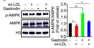
1). Gastrodin induces lysosomal biogenesis and autophagy to prevent the formation of foam cells via AMPK‐FoxO1‐TFEB signalling axis. Journal of Cellular and Molecular Medicine, 2021 (PubMed: 33973365) [IF=5.3]

Application: WB    Species: Mice    Sample: macrophages

FIGURE 6 AMPK is a critical upstream regulator of FoxO1 and TFEB. A and B, Gastrodin activated AMPK in the foam cells. A, Representative blots of AMPK and p‐AMPK in macrophages. B, Immunofluorescence analysis of p‐AMPK in macrophages. C and D, The inhibition of AMPK activity decreased the phosphorylation of FoxO1 and nuclear translocation of TFEB. Macrophages were treated with CC (10μM) for 1 h. The phosphorylation level of FoxO1 was analysed by Western blotting C, and nuclear translocation of TFEB was determined by immunofluorescence D. *P < .05; **P < .01. Results are presented as mean ± SD of three independent experiments. The value represents fold of vehicle. CC, Dorsomorphin dihydrochloride

2). Asiatic acid attenuates hypertrophic and fibrotic differentiation of articular chondrocytes via AMPK/PI3K/AKT signaling pathway. ARTHRITIS RESEARCH & THERAPY, 2020 (PubMed: 32398124) [IF=4.9]

Application: WB    Species: human    Sample: human OA chondrocytes

Fig. 5| AA treatment activated the AMPK and inhibited PI3K/AKT signaling pathway. The cells were treated with or without AA (5 μM) for 3 days. aRepresentative western blot of p-AMPK, AMPK, p-PI3K, PI3K, p-AKT, and AKT. GAPDH was served as a loading control.

Application: WB    Species: Human    Sample: OA chondrocytes

Fig. 5 AA treatment activated the AMPK and inhibited PI3K/AKT signaling pathway. The cells were treated with or without AA (5 μM) for 3 days. a Representative western blot of p-AMPK, AMPK, p-PI3K, PI3K, p-AKT, and AKT. GAPDH was served as a loading control. b Relative protein expression of p-AMPK/AMPK, p-PI3K/PI3K, and p-AKT/AKT. Unpaired t test, *P < 0.05, ***P < 0.001. Each experiment was repeated three times (p-AMPK, phosphorylated AMP-activated protein kinase; AMPK, AMP-activated protein kinase; p-PI3K, phosphorylated phosphoinositide-3 kinase; PI3K, phosphoinositide-3 kinase; p-AKT, phosphorylated protein kinase B; AKT, protein kinase B)

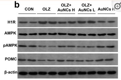
3). Gold nanoclusters eliminate obesity induced by antipsychotics. Scientific Reports, 2022 (PubMed: 35365730) [IF=4.6]

Application: WB    Species: Rat    Sample: hypothalamus

Figure 3 Effects of olanzapine and AuNCs co-treatment on H1R-AMPK signaling, POMC protein expression and POMC immunofluorescence staining in the hypothalamus. (a) TEM image of a hypothalamic slice at 6th h after IP injection of 20 mg/kg AuNCs in rats. The presence of AuNCs was marked by red arrows. (b) Representative western blot figures of H1R, AMPK, pAMPK and POMC in the hypothalamus after co-treatment of olanzapine and AuNCs. (c–e) Densitometry analysis of H1R expression (c), pAMPK/AMPK (d) and POMC expression (e). (f–k) POMC immunofluorescence staining in the hypothalamic Arc of rats in CON (f), OLZ (g), O + AuNCs H (h), O + AuNCs L (i), AuNCs H (j) group and the corresponding quantification of POMC fluorescence intensity (k). n = 4/group. All data were presented as mean ± SEM. *p < 0.05, **p < 0.01, ***p < 0.0001, OLZ vs. CON; #p < 0.05, ##p < 0.01, ###p < 0.0001, OLZ + AuNCs H vs. OLZ; $p < 0.05, OLZ + AuNCs L vs. OLZ. Original figures were shown in Fig. S8.

4). High expressions of LDHA and AMPK as prognostic biomarkers for breast cancer. BREAST, 2016 (PubMed: 27598996) [IF=3.9]

Application: WB    Species: human    Sample:

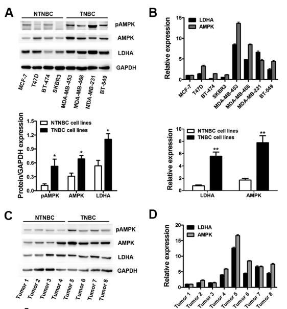
Fig. 1. LDHA and AMPK were up-regulated synchronously in breast cancer. A. Expression levels of LDHA, AMPK and pAMPK were assessed by Western blot (above) and gray image scanning (below) in eight different breast cancer cell lines, including four NTNBC cell lines and four TNBC cell lines. GAPDH was used as a loading control. B. Expression levels of LDHA and AMPK were determined by qRT-PCR (above). The differences between TNBC and NTNBC cell lines were analyzed (below). GAPDH was used as an internal control. C. Expression levels of LDHA, AMPK and pAMPK were assessed by Western blot (above) and gray image scanning (below) in eight different breast cancer tissues, including four NTNBC tissues and four TNBC tissues. GAPDH was used as a loading control. D. Expression levels of LDHA and AMPK were determined by qRT-PCR (above). The diffe

Application: IHC    Species: human    Sample:

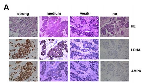
Fig. 2. The expression of LDHA and AMPK showed a positive correlation in breast cancer. A. The expression of LDHA and AMPK were detected by IHC using breast cancer TMAs of 112 patients. Representative IHC images of four staining degrees (no-weak-medium-strong) of LDHA and AMPK expression under a microscope were showed (400). B. The correlation between LDHA and AMPK expression scores of 112 breast cancer patients was analyzed and a positive correlation between them was showed.

5). FGF1 reduces cartilage injury in osteoarthritis via regulating AMPK/Nrf2 pathway. Journal of Molecular Histology, 2023 (PubMed: 37659992) [IF=3.2]

6). Myostatin inhibits eEF2K-eEF2 by regulating AMPK to suppress protein synthesis. BIOCHEMICAL AND BIOPHYSICAL RESEARCH COMMUNICATIONS, 2017 (PubMed: 29024627) [IF=3.1]

Application: WB    Species: mouse    Sample:

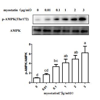
Fig. 4. Myostatin regulated translation elongation through AMP. C2C12 myotubes were treated with various concentration recombinant myostatin (0, 0.01,0.1, 1, 2, 3 µg/ml) for 48 h andthen lysed and cellular extracts were analyzed by Western blot with anti-AMPK(A).

Application: WB    Species: mouse    Sample: C2C12 myotubes

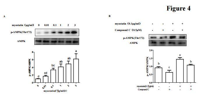
Fig. 4. |Myostatin regulated translation elongation through AMPK.C2C12 myotubes were treated with various concentration recombinant myostatin (0, 0.01,429 0.1, 1, 2, 3 µg/ml) for 48 h andthen lysed and cellular extracts were analyzed by Western blot with 430 anti-AMPK(A).

Restrictive clause


Affinity Biosciences tests all products strictly. Citations are provided as a resource for additional applications that have not been validated by Affinity Biosciences. Please choose the appropriate format for each application and consult Materials and Methods sections for additional details about the use of any product in these publications.

For Research Use Only.
Not for use in diagnostic or therapeutic procedures. Not for resale. Not for distribution without written consent. Affinity Biosciences will not be held responsible for patent infringement or other violations that may occur with the use of our products. Affinity Biosciences, Affinity Biosciences Logo and all other trademarks are the property of Affinity Biosciences LTD.