TPM1 Antibody - #DF6291
| Product: | TPM1 Antibody |
| Catalog: | DF6291 |
| Description: | Rabbit polyclonal antibody to TPM1 |
| Application: | WB IHC |
| Cited expt.: | WB |
| Reactivity: | Human, Mouse, Rat |
| Prediction: | Pig, Zebrafish, Bovine, Sheep, Rabbit, Dog, Chicken, Xenopus |
| Mol.Wt.: | 33kDa; 33kD(Calculated). |
| Uniprot: | P09493 |
| RRID: | AB_2838257 |
Related Downloads
Protocols
Product Info
*The optimal dilutions should be determined by the end user. For optimal experimental results, antibody reuse is not recommended.
*Tips:
WB: For western blot detection of denatured protein samples. IHC: For immunohistochemical detection of paraffin sections (IHC-p) or frozen sections (IHC-f) of tissue samples. IF/ICC: For immunofluorescence detection of cell samples. ELISA(peptide): For ELISA detection of antigenic peptide.
Cite Format: Affinity Biosciences Cat# DF6291, RRID:AB_2838257.
Fold/Unfold
AA986836; AI854628; Alpha tropomyosin; alpha-TM; Alpha-tropomyosin; C15orf13; cardiomyopathy, hypertrophic 3; CMD1Y; CMH3; HTM alpha; HTM-alpha; OTTHUMP00000163688; sarcomeric tropomyosin kappa; TM2; Tmpa; TMSA; Tpm-1; TPM1; TPM1_HUMAN; tropomyosin 1 (alpha); tropomyosin 1 (alpha) isoform 1; tropomyosin 1 (alpha) isoform 2; tropomyosin 1 (alpha) isoform 3; tropomyosin 1 (alpha) isoform 4; tropomyosin 1 (alpha) isoform 5; tropomyosin 1 (alpha) isoform 6; tropomyosin 1 (alpha) isoform 7; Tropomyosin 1; Tropomyosin alpha 1 chain; Tropomyosin alpha-1 chain; Tropomyosin, skeletal muscle alpha; Tropomyosin-1;
Immunogens
A synthesized peptide derived from human TPM1, corresponding to a region within N-terminal amino acids.
Detected in primary breast cancer tissues but undetectable in normal breast tissues in Sudanese patients. Isoform 1 is expressed in adult and fetal skeletal muscle and cardiac tissues, with higher expression levels in the cardiac tissues. Isoform 10 is expressed in adult and fetal cardiac tissues, but not in skeletal muscle.
- P09493 TPM1_HUMAN:
- Protein BLAST With
- NCBI/
- ExPASy/
- Uniprot
MDAIKKKMQMLKLDKENALDRAEQAEADKKAAEDRSKQLEDELVSLQKKLKGTEDELDKYSEALKDAQEKLELAEKKATDAEADVASLNRRIQLVEEELDRAQERLATALQKLEEAEKAADESERGMKVIESRAQKDEEKMEIQEIQLKEAKHIAEDADRKYEEVARKLVIIESDLERAEERAELSEGKCAELEEELKTVTNNLKSLEAQAEKYSQKEDRYEEEIKVLSDKLKEAETRAEFAERSVTKLEKSIDDLEDELYAQKLKYKAISEELDHALNDMTSI
Predictions
Score>80(red) has high confidence and is suggested to be used for WB detection. *The prediction model is mainly based on the alignment of immunogen sequences, the results are for reference only, not as the basis of quality assurance.
High(score>80) Medium(80>score>50) Low(score<50) No confidence
Research Backgrounds
Binds to actin filaments in muscle and non-muscle cells. Plays a central role, in association with the troponin complex, in the calcium dependent regulation of vertebrate striated muscle contraction. Smooth muscle contraction is regulated by interaction with caldesmon. In non-muscle cells is implicated in stabilizing cytoskeleton actin filaments.
Phosphorylated at Ser-283 by DAPK1 in response to oxidative stress and this phosphorylation enhances stress fiber formation in endothelial cells.
Cytoplasm>Cytoskeleton.
Note: Associates with F-actin stress fibers.
Detected in primary breast cancer tissues but undetectable in normal breast tissues in Sudanese patients. Isoform 1 is expressed in adult and fetal skeletal muscle and cardiac tissues, with higher expression levels in the cardiac tissues. Isoform 10 is expressed in adult and fetal cardiac tissues, but not in skeletal muscle.
The molecule is in a coiled coil structure that is formed by 2 polypeptide chains. The sequence exhibits a prominent seven-residues periodicity.
Belongs to the tropomyosin family.
Research Fields
· Human Diseases > Cancers: Overview > MicroRNAs in cancer.
· Human Diseases > Cardiovascular diseases > Hypertrophic cardiomyopathy (HCM).
· Human Diseases > Cardiovascular diseases > Dilated cardiomyopathy (DCM).
· Organismal Systems > Circulatory system > Cardiac muscle contraction. (View pathway)
· Organismal Systems > Circulatory system > Adrenergic signaling in cardiomyocytes. (View pathway)
References
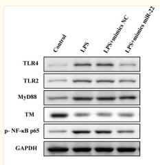
Application: WB Species: Rat Sample: HBZY-1 cells
Restrictive clause
Affinity Biosciences tests all products strictly. Citations are provided as a resource for additional applications that have not been validated by Affinity Biosciences. Please choose the appropriate format for each application and consult Materials and Methods sections for additional details about the use of any product in these publications.
For Research Use Only.
Not for use in diagnostic or therapeutic procedures. Not for resale. Not for distribution without written consent. Affinity Biosciences will not be held responsible for patent infringement or other violations that may occur with the use of our products. Affinity Biosciences, Affinity Biosciences Logo and all other trademarks are the property of Affinity Biosciences LTD.


