

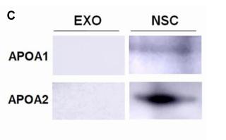
, using APOA1 Antibody at 1/1000 dilution.
5ug/NC membrane strip.
Exposure for 5s with Affinity™ ECL Kit(#KF8001).
Bands result from membrane strip incubation.")


| Product: | APOA1 Antibody |
| Catalog: | DF6264 |
| Description: | Rabbit polyclonal antibody to APOA1 |
| Application: | WB IHC |
| Cited expt.: | WB |
| Reactivity: | Human, Mouse, Rat |
| Prediction: | Pig, Bovine, Horse, Sheep, Rabbit, Dog |
| Mol.Wt.: | 31kDa(Observed); 31kD(Calculated). |
| Uniprot: | P02647 |
| RRID: | AB_2838230 |
Control Products

Related Downloads
Protocols
Product Info
*The optimal dilutions should be determined by the end user. For optimal experimental results, antibody reuse is not recommended.
*Tips:
WB: For western blot detection of denatured protein samples. IHC: For immunohistochemical detection of paraffin sections (IHC-p) or frozen sections (IHC-f) of tissue samples. IF/ICC: For immunofluorescence detection of cell samples. ELISA(peptide): For ELISA detection of antigenic peptide.
Cite Format: Affinity Biosciences Cat# DF6264, RRID:AB_2838230.
Fold/Unfold
Apo-AI; ApoA I; ApoA-I; APOA1; APOA1_HUMAN; Apolipoprotein A-I(1-242); Apolipoprotein A1; Apolipoprotein AI; Brp14; Ltw1; Lvtw1; Sep1; Sep2;
Immunogens
A synthesized peptide derived from human APOA1, corresponding to a region within N-terminal amino acids.
Major protein of plasma HDL, also found in chylomicrons. Synthesized in the liver and small intestine. The oxidized form at Met-110 and Met-136 is increased in individuals with increased risk for coronary artery disease, such as in carrier of the eNOSa/b genotype and exposure to cigarette smoking. It is also present in increased levels in aortic lesions relative to native ApoA-I and increased levels are seen with increasing severity of disease.
- P02647 APOA1_HUMAN:
- Protein BLAST With
- NCBI/
- ExPASy/
- Uniprot
MKAAVLTLAVLFLTGSQARHFWQQDEPPQSPWDRVKDLATVYVDVLKDSGRDYVSQFEGSALGKQLNLKLLDNWDSVTSTFSKLREQLGPVTQEFWDNLEKETEGLRQEMSKDLEEVKAKVQPYLDDFQKKWQEEMELYRQKVEPLRAELQEGARQKLHELQEKLSPLGEEMRDRARAHVDALRTHLAPYSDELRQRLAARLEALKENGGARLAEYHAKATEHLSTLSEKAKPALEDLRQGLLPVLESFKVSFLSALEEYTKKLNTQ
Predictions
Score>80(red) has high confidence and is suggested to be used for WB detection. *The prediction model is mainly based on the alignment of immunogen sequences, the results are for reference only, not as the basis of quality assurance.
High(score>80) Medium(80>score>50) Low(score<50) No confidence
Research Backgrounds
Participates in the reverse transport of cholesterol from tissues to the liver for excretion by promoting cholesterol efflux from tissues and by acting as a cofactor for the lecithin cholesterol acyltransferase (LCAT). As part of the SPAP complex, activates spermatozoa motility.
Glycosylated.
Palmitoylated.
Phosphorylation sites are present in the extracellular medium.
Secreted.
Major protein of plasma HDL, also found in chylomicrons. Synthesized in the liver and small intestine. The oxidized form at Met-110 and Met-136 is increased in individuals with increased risk for coronary artery disease, such as in carrier of the eNOSa/b genotype and exposure to cigarette smoking. It is also present in increased levels in aortic lesions relative to native ApoA-I and increased levels are seen with increasing severity of disease.
Belongs to the apolipoprotein A1/A4/E family.
Research Fields
· Human Diseases > Infectious diseases: Parasitic > African trypanosomiasis.
· Organismal Systems > Endocrine system > PPAR signaling pathway.
· Organismal Systems > Digestive system > Fat digestion and absorption.
· Organismal Systems > Digestive system > Vitamin digestion and absorption.
· Organismal Systems > Digestive system > Cholesterol metabolism.
References

Application: WB Species: Mouse Sample: neural stem cells (NSC)
Application: WB Species: mouse Sample: NSCs

Application: WB Species: porcine Sample:
Restrictive clause
Affinity Biosciences tests all products strictly. Citations are provided as a resource for additional applications that have not been validated by Affinity Biosciences. Please choose the appropriate format for each application and consult Materials and Methods sections for additional details about the use of any product in these publications.
For Research Use Only.
Not for use in diagnostic or therapeutic procedures. Not for resale. Not for distribution without written consent. Affinity Biosciences will not be held responsible for patent infringement or other violations that may occur with the use of our products. Affinity Biosciences, Affinity Biosciences Logo and all other trademarks are the property of Affinity Biosciences LTD.