


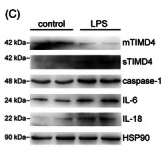
, using IL18 Antibody at 1/1000 dilution.
5ug/NC membrane strip.
Exposure for 5s with Affinity™ ECL Kit(#KF8001).
Bands result from membrane strip incubation.")
 and mouse anti-beta tubulin Ab(T0023) for 1 hour at 37°C. An AlexaFluor594 conjugated goat anti-rabbit IgG(H+L) Ab(Red) and an AlexaFluor488 conjugated goat anti-mouse IgG(H+L) Ab(Green) were used as the secondary antibody.
The nuclear counter stain is DAPI (blue).")
 and mouse anti-beta tubulin Ab(T0023) for 1 hour at 37°C. An AlexaFluor594 conjugated goat anti-rabbit IgG(H+L) Ab(Red) and an AlexaFluor488 conjugated goat anti-mouse IgG(H+L) Ab(Green) were used as the secondary antibody.
The nuclear counter stain is DAPI (blue).")

, cleaved caspase-1 (c), and IL-18 e) were shown.")
, the NLRP3 inflammasome was activated in diabetic mice kidney (DN) and high-dose AB-38b (DN + AB38b) inhibited its activation. Densitometry analyses of NLRP3")

 for 24 h, the expression of OCTN1 (62 kDa), OCTN2 (63 kDa), IL6 (24 kDa), IL18 (26 kDa), IL1β (31 kDa),and TNF-α (26 kDa) was significantly regulated in human respiratory epithelial cells;β-actin (42 kDa) was used as a loading control. Bands were analysed using ImageJ 1.45s (National Institutes of Health, USA). NC indicates the normal control groups. Data are shown as the mean ± SD (n = 3).")
 The effects of oroxylin A on the levels of IL-1β, IL-6,IL-18 and TNF-α in skin tumor and blood were determined respectively (n = 3). (E) Protein expression of IL-1β, NLRP3, IL-6, IL-18 and TNF-α were measured by Western blot assays (n = 3). GAPDH was used as an invariant control for equal loading. Representative blots are shown with densitometry.")
 Representative blots and histograms showing expression of (B) SIRT1; (C) active caspase‑1; (D) active caspase‑11; and (E) IL‑1β. (F) Representative blots. Histograms showing expression of (G) NLRP3 and (H) IL‑18.")
; db/db + QH: Quercetin (70 mg/kg/d). Data represent mean ± SEM (n = 8 per group). # P < .05, ## P < .01, ### P < .001 vs db/m; *P < .05, **P < .01, ***P < .001 vs db/db")
 The expressions of NLRP3, ASC, caspase-1, IL-18, and IL-1β. Carnosine-L: carnosine (100 mg kg−1 d−1
); carnosine-H: carnosine (200 mg kg−1 d−1
). Data represent mean ± SEM (n = 10 per group). #p < 0.05, ##p < 0.01, ###p <
0.001 vs. SAMR1db m−1
; *p < 0.05, **p < 0.01, ***p < 0.001 vs. SAMP8.")
 Detection of cell viability (CCK-8 assay) after (1) 72 h exposure to different concentrations of VbP, and (2) exposure to 15 μM VbP for various times. (B) ELISA results showing secretion of IL-18 (1) and IL-1β (2) by macrophages treated for 72 h with different concentrations of VbP. (C) Dose-dependent expression of pyroptosis-related proteins in untreated (control) and VbP-treated macrophages (72 h exposure). (D) Time-dependent expression of pyroptosis-related proteins in untreated (control) and VbP-treated (15 μM) macrophages. (E) Dose-response analysis of NLRP1 inflammasome expression in VbP-treated macrophages (72 h exposure). (F) Time-response analysis of NLRP1 expression in macrophages exposed to 15 μM VbP. n = 3; *P<0.05, **P<0.01, and ***P<0.001 vs. control cells.")
 Rats livers from each group were subjected to protein extraction and western blotting for p-mTOR, mTOR and GAPDH proteins. (B) Rat livers in each group were
subjected to protein extraction and western blotting for p-P70S6K, P70S6K, p-4EBP1 and 4EBP1. (C–D) Rats livers in each group were subjected to protein extraction
and western blotting for NLRP3, ASC2, IL-1β, IL-18 and cleaved caspase-1. NC, normal chow; HF, high-fat diet; HF + L, 100 μg/kg liraglutide group; All data are
presented as the mean ± SD, n = 3,a
P < 0.05, aaP < 0.01 vs NC; bbP < 0.01 vs HF.")
 The activation level of NLRP3
inflammasome and inflammatory factors in the brain of rats. After FMT from healthy rats, the expression of IL-1β, IL-18, NLRP3, and TLR4 in brain was significantly
downregulated, which were significantly different from that in Mn-treated group. *P < 0.05, **P < 0.01, ***P < 0.001.")
; and aging + QH: quercetin (70 mg kg−1 d−1
). Data are presented as mean ± SEM (n = 10 per group). #P <
0.05, ##P < 0.01 vs. control group; *P < 0.05, **P < 0.01, ***P < 0.001 vs. aging group.")
 and ASC
(B). (C, D) The representative photographs and grayscale analysis of NLRP3, Pro-casp-1, casp-1, Pro-IL-1β, and IL-1β in the liver by Western blotting. Data was shown
as the mean ± SEM, n = 4, **p < 0.01, *p < 0.05, compared with control group")
 The mRNA expression level of Bmal1 and Reverba in BMDMs derived from
Bmal1f/f and Bmal1f/f, LysMcre/+ mice. (B-E) The transcriptional level of Tnf (B), Il6 (C), Il1b (D), and Il18 (E) at ZT6 or ZT18 in LPS-treated BMDMs with or without
SR9009 treatment. (F) Representative blots showing the IL-1β protein expression in LPS-stimulated BMDMs at ZT6 or ZT18. (G-H) Quantification of pro-IL-1β (G) and
mature IL-1β (H) in (F). (I) Representative image showing the IL-18 protein expression in LPS-treated BMDMs. (J-K) Quantification of pro-IL-18 (J) and mature IL-18
(K) in (I). The data represent means ± SEM from at least 3 independent experiments. * indicates p < 0.05, ** indicates p < 0.01, *** indicates p < 0.001, ****
indicates p < 0.0001, and n.s indicates no statistical significance.")
 Correlation between TN-C and NLRP3 in heart from the GEPIA database. (B) (C) qPCR and immunohistochemistry staining analysis of NLRP3, IL-18, and IL-1β in
cardiac tissues of WT Sham, TN-C KO Sham, WT MI, and TN-C KO MI groups. (D) Representative western blot analyses of NLRP3, ASC, IL-18, and IL-1β in cardiac
tissues. Data are mean ± SD (n = 6 per group), scale bar = 20 μm. *P < 0.05, **P < 0.01, ***P < 0.001. NLRP indicates NOD-like receptor protein; IL, interleukin;
ASC, apoptosis-associated speck-like protein containing CARD; TN-C, tenascin-C; qPCR, quantitative real-time polymerase chain reaction; WT, wild type; TN-C KO,
TN-C knockout mice; MI, myocardial infarction.")
 NLRP3, (D) caspase-1, (E) IL-1β and (F) IL-18 mRNA levels on LPS-induced DPFs detected by qPCR and (G) their protein levels tested by western blotting (normalized to that of β-tubulin).")
 Immunohistochemistry results of NLRP3 and caspase-1 in rats. Relative percentages of (B) NLRP3 and (C) caspase-1 positive area to total area. (D) Western blot and (E) ELISA results of IL-1β in kidney of rats. (F) Western blot and (G) ELISA results of IL-18 in kidney and serum of rats. Data are presented as mean ± SEM (n = 3–8 for each group). ††P < 0.01 vs. CON, ###P < 0.001 vs. CON, *P < 0.05, **P < 0.01, ***P < 0.001 vs. DKD.")

 Western blotting of Calpain 1, Calpain 2, Klotho, AIM2, NLRP3, pro-Caspase 1, cleaved-Caspase 1, IL-1β and IL-18 in the kidney of all mice. (B) Quantitative determination of Calpain 1, Calpain 2, Klotho, AIM2, NLRP3, cleaved-Caspase 1 and IL-18. (C) The mRNA levels of Calpain 2, Klotho, AIM2, ASC and GSDMD in the kidney among different groups. (D) The calpain activity of renal issues was measured by the relative fluorescence units (400/505 nm). (E) The cathepsin B activity of kidney issues was tested by the relative fluorescence units (400/505 nm). (F) Representative immunohistochemical micrographs from the kidney issues of different groups stained with Calpain 1, Calpain 2 and AIM2. ×400, bar = 50 μm. All data were presented as mean ± SEM (n = 3). NS, no significance; *p < 0.05 vs. sham group; **p < 0.01 vs. sham group; ***p < 0.001 vs. sham group; #p < 0.05 vs. IR group; ##p < 0.01 vs. IR group; ###p < 0.001 vs. IR group. CP, calpeptin; IR, ischemia/reperfusion; RFU, relative fluorescence units.")
 (Mean ± SD, n = 3). The results of western blot are shown in Figure 10A (1: unstressed group, 2: CUMS group, 3: FH group, 4: TTWC-L group, 5: TTWC-M group, 6: TTWC-H group). Values are expressed as (Mean ± SD). * p < 0.05, ** p < 0.01, *** p < 0.001 vs. vehicle treated CUMS group. ### p < 0.001 vs. unstressed group.")
 Western blot analysis of caspase-1, cleaved caspase-1, IL-1β, IL-18, and GSDMD expression six weeks after HIE. (b) Quantitative analyses of these pyroptosis-related molecules. Pyroptosis-related molecules were partly downregulated after Tuina treatment. Data are presented as the means ± SD; ∗p < 0.05 compared with the sham group; #p < 0.05 compared with the model group.")
 Western blot analysis of NLRP3, caspase1, cleaved-caspase1, GSDMD-N, ASC, IL-18, and IL-1β protein abundance. (f) The mRNA expression of IL-1β, IL-18, caspase1, and GSDMD genes. (g) Representative immunofluorescent images co-stained with PGC-1α (red), DAPI (blue) and both channels merged (400x magnification). The graphs (down panel) show the fluorescence intensity profiles in two fluorescence channels along the arrow and the white arrows represent nucleus PGC-1α; the yellow arrowheads show cytoplasm PGC-1α, whereas a clear nuclear translocation (white arrow) and shrinkage of PGC-1α (red) is seen in quercetin treatment and inhibited by ethanol. Data are expressed as mean ± SD (n = 6). Different subscript letters indicate significant differences among the groups (p < 0.05).")
 Western blot analysis of IL-1β, IL-18 and TNF-α in mice liver. (B) Real-time PCR of IL-1β, IL-18 and TNF-α in mice liver. (C) Immunohistochemistry of IL-1β, IL-18, TNF-α and F4/80 in mice liver. (D) ELISA analysis of serum IL-1β, IL-18 and TNF-α. Results represent means ± SEM for 6–8 mice. *p < 0.05, **p < 0.01, ***p < 0.001 vs db/m. #p < 0.05, ##p < 0.01, ###p < 0.001 vs db/db.")
 Western blot analysis of IL-1β, IL-18 and TNF-α in mice liver. (B) Real-time PCR of IL-1β, IL-18 and TNF-α in mice liver. (C) Immunohistochemistry of IL-1β, IL-18, TNF-α and F4/80 in mice liver. (D) ELISA analysis of serum IL-1β, IL-18 and TNF-α. Results represent means ± SEM for 6–8 mice. *p < 0.05, **p < 0.01, ***p < 0.001 vs db/m. #p < 0.05, ##p < 0.01, ###p < 0.001 vs db/db.")
 The mRNA expression levels of NLRP3, ASC, GSDMD, and caspase 1 in CPB-induced lung injury rats treated with XFZYD, Ac-YVAD-CMK, and Bay-11-7082 were measured via RT-qPCR. (b and c) western blots were performed to determine NLRP3, ASC, Caspase-1 p20, Pro-GSDMD, GSDMD p30, IL-18, IL-1β p-P65, P65, p-IKBα, and IKBα levels in lung tissues of rats with CPB-induced acute lung injury. β-actin was used as a loading control for the blots. All the data were presented as the means ± SD. from independent experiments performed in triplicate. ∗P < 0.05; ∗∗P < 0.01, vs. CPB group.")
 Immunofluorescence staining for F4/80 (macrophages) and chemerin of placenta tissue in normal and GDM pregnant women. (B) Content of IL-18 and IL-1β in placenta tissue of normal and GDM pregnant women was measured by ELISA (n = 3). (C) Content of IL-18 and IL-1β in placenta tissue of Ctrl, CHM+sh-NC, and CHM+sh-chemR23 mice was measured by ELISA (n = 3). (D) Protein levels of NOD-like receptor family pyrin domain containing 3 (NRLP3), apoptosis-associated speck-like protein containing CARD (Asc), pro caspase 3, cleaved caspase 3, pro caspase 9, cleaved caspase 9, pro caspase 1, cleaved caspase 1, pro-IL-1β, and pro-IL-18 in placental macrophages were measured by Western blotting. Protein levels of cleaved caspase 1, IL-1β, and IL-18 in the culture supernatants of placental macrophages were measured by Western blotting as well. Glyceraldehyde-3-phosphate dehydrogenase (GAPDH) served as the internal control (n = 3). (E) Trophoblast cells were co-incubated with control (Ctrl), the supernatant from macrophage, macrophage+CHM+sh-NC, and macrophage+CHM+sh-chemR23 for 24 h, then relative exosomal miR-140-3p and miR-574-3p expression was measured by qRT-PCR (n = 3). (F) Trophoblast cells were treated with Control, recombinant protein IL-18, recombinant protein IL-1β, and recombinant protein IL-18+IL-1β. Relative exosomal miR-140-3p and miR-574-3p expression was measured by qRT-PCR (n = 3). (G) Changed protein levels of IL-18 and IL-1β in macrophage after treatment with chemerin and siRNA NC (CHM+si-NC), IL-1β siRNA (CHM+si-IL-1β), IL-18 siRNA (CHM+si-IL-18) and IL-1β siRNA and IL-18 siRNA (CHM+si-IL-1β+si-IL-18) were measured by Western blotting (n = 3). (H) Trophoblast cells were co-incubated with the supernatant of macrophage+CHM+si-NC, macrophage+CHM+si-IL-1β, macrophage+CHM+si-IL-18, and macrophage+CHM+si-IL-1β+si-IL-18, then relative exosomal miR-140-3p and miR-574-3p expression was measured by qRT-PCR (n = 3).")
 Skin tissue sections were stained with anti-GSDMD-N (green) or anti-caspase 1 (CASP1; red) immunofluorescence antibodies (DAPI staining of the nuclei). Scale bar: 10 μm. The number of positive cells is shown on the right of representative images (n = 6 mice per group). (e,f) CHBP treatment reduced the expression of NLRP3, ASC, caspase 1 (CASP1), GSDMD-N, IL-1β and IL-18 in the context of ischaemia–reperfusion injury. Densitometric quantification is shown on the right (n = 6 mice per group). (g–j) CHBP treatment attenuated the activation of GSDMD, IL-1β, IL-18 and caspase 1 in the skin tissues detected by ELISA kits (n = 6 mice per group). Data shown are means ± SEM. *P")

 Immunofluorescence staining of Caspase 1 (pyroptosis-related marker, red) and NeuN (indicating neurons, green) (original magnification 30×) in spinal cord sections. The optical densities of Caspase 1 were markedly decreased in the SCI + Bex group and significantly increased in the SCI group. Scale bars: 25 μm. (B) Quantification of Caspase-1 in neurons of spinal cords in A. (C) Immunofluorescence staining of GSDMD-N (pyroptosis-related protein, green) and NeuN (indicating neurons, red) (original magnification 30×). Scale bars: 25 μm. (D) Quantification of GSDMD in neurons of spinal cords in C. (E–H) Evaluation of CASP1, GSDMD, IL-18, and IL-1β in the spinal cord by ELISA. (I) Western blot assay of GSDMD-N, NLRP3, Caspase-1, IL-1β, IL-18, and ASC. (J) Quantitative analyses of data from I; data were normalized to GAPDH. Data are expressed as the mean ± SEM (n = 6 mice per group). **P < 0.01, vs. sham group; ##P < 0.01, vs. SCI group (one-way analysis of variance with the least significance difference post hoc test). ASC: Apoptosis-associated speck-like protein containing a CARD; Bex: bexarotene; C-CASP-1: cleaved Caspase 1; DAPI: 4′,6-diamidino-2-phenylindole; ELISA: enzyme-linked immunosorbent assay; GAPDH: glyceraldehyde-3- phosphate dehydrogenase; GSDMD-N: gasdermin D-N; IL: interleukin; IOD: integrated optical density; NLRP3: NOD-like receptor thermal protein domain associated protein 3; SCI: spinal cord injury.")
 Immunofluorescence staining of Caspase 1 (pyroptosis-related marker, red) and NeuN (indicating neurons, green) (original magnification 30×) in spinal cord sections. The optical densities of Caspase 1 were markedly decreased in the SCI + Bex group and significantly increased in the SCI group. Scale bars: 25 μm. (B) Quantification of Caspase-1 in neurons of spinal cords in A. (C) Immunofluorescence staining of GSDMD-N (pyroptosis-related protein, green) and NeuN (indicating neurons, red) (original magnification 30×). Scale bars: 25 μm. (D) Quantification of GSDMD in neurons of spinal cords in C. (E–H) Evaluation of CASP1, GSDMD, IL-18, and IL-1β in the spinal cord by ELISA. (I) Western blot assay of GSDMD-N, NLRP3, Caspase-1, IL-1β, IL-18, and ASC. (J) Quantitative analyses of data from I; data were normalized to GAPDH. Data are expressed as the mean ± SEM (n = 6 mice per group). **P < 0.01, vs. sham group; ##P < 0.01, vs. SCI group (one-way analysis of variance with the least significance difference post hoc test). ASC: Apoptosis-associated speck-like protein containing a CARD; Bex: bexarotene; C-CASP-1: cleaved Caspase 1; DAPI: 4′,6-diamidino-2-phenylindole; ELISA: enzyme-linked immunosorbent assay; GAPDH: glyceraldehyde-3- phosphate dehydrogenase; GSDMD-N: gasdermin D-N; IL: interleukin; IOD: integrated optical density; NLRP3: NOD-like receptor thermal protein domain associated protein 3; SCI: spinal cord injury.")

. BBR, berberine (20 μM); UA, uric acid (20 mg/dL); cl-Caspase1, cleaved-Caspase1.")
. B. Morphology images of HCT116 and HT29 cells treated with 100 μM of Que for 48 h. Black arrowheads indicate the large bubbles emerging from the plasma membrane and cytoplasmic swelling (n=3, where “n” represents the number of independent repeated experiments. Scale bar: 100 μm, 200×). C-F. Protein expression of GSDMD, N-GSDMD, IL-1β, and IL-18 (pro and mature) in Que-treated HCT116 and HT29 cells was measured using western blotting (n=3, where “n” represents the number of independent repeated experiments). G. Release of IL-1β and IL-18 from Que-treated HCT116 and HT29 cells was measured using ELISA kits, with three technical replicates per group, and the mean value utilized for quantitative analysis (n=5, where “n” represents the number of independent repeated experiments).")
 NLRP3, ASC, caspase-1, IL-18, and IL-1β protein expression levels were evaluated by Western blotting in THP-1 cells. (B) The concentrations of LDH released by THP-1-derived MΦs were determined using an LDH assay kit to evaluate cell integrity. n = 3 in each group. *p < 0.05 between the indicated groups. (C) Mice were treated with LAA for 1 h before S. aureus infection. The protein expression of ASC, caspase-1, IL-18, and IL-1β in the lung tissue was measured by Western blotting. THP-1-derived MΦs were primed with LPS and then stimulated with LAA for 6 h with or without the NLRP3 inflammasome activator nigericin or ATP for 0.5 h before the end of the experiment. (D) NLRP3, caspase-1 and IL-1β protein expression levels were evaluated by Western blotting. (E) The levels of IL-1β were tested by ELISA. n = 4 in each group. **p < 0.01 between the indicated groups.")
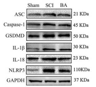
 NLRP3 (A), ASC (B), GSDMD (C), and Caspase-1 (D) mRNA expression levels were measured by quantitative reverse transcription-polymerase chain reaction. (E) Western blot analysis of inflammation- and pyroptosis-associated protein expression levels. β-Actin was used as an internal reference for NLRP3, ASC, GADMD, IL-18, and IL-1β. GAPDH was used as an internal reference for TLR4, P65, p-P65, IκBα, and p-IκBα. (F–M) NLRP3 (F), ASC (G), GSDMD (H), IL-18 (I), IL-1β (J), TLR4 (K), p-P65/P65 (L), and p-IκBα/IκBα (M) protein expression levels were measured by Western blot assay. All western blot data were normalized to the sham group. Data are expressed as mean ± SD (n = 3). *P < 0.05, **P < 0.01, vs. sham group; #P < 0.05, ##P < 0.01, vs. model group (one-way analysis of variance followed by Tukey's post hoc test). ASC: Apoptosis-associated speck-like protein containing CARD; BA: Biochanin A; GAPDH: glyceraldehyde-3-phosphate dehydrogenase; GSDMD: gasdermin D; IL-18: interleukin-18; IL-1β: interleukin-1β; NLRP3: NOD-like receptor thermal protein domain associated protein 3; PC: positive control; p-P65: phospho-NF-κB P65; p-IκBα: phospho-NF-kappa-B inhibitor alpha; SCI: spinal cord injury; TLR4: Toll-like receptor 4.")
 Expression of the protein SIRT2 associated with pyroptosis in the thoracic aorta of diabetic mice was assessed using western blotting. (C, D) Effect of 1,8-cineole on the levels of IL-18 and IL-1β in murine serum. (E) Effect of 1,8-cineole on the concentration of lactate dehydrogenase (LDH) in mouse serum. Results are presented as the mean ± SD (n = 6), *p < 0.05, **p < 0.01 versus control group; #p < 0.05, ##p < 0.01 versus DM group.")
 The expressions of NLRP3, ASC, caspase-1, and IL-1β mRNA were tested by RT-PCR. (E–J) The expressions of NLRP3, ASC, caspase-1, IL-1β, and IL-18 protein were examined by western blotting. Data are showed as means ± SEM. **P < 0.01 vs control group, #P < 0.05, ##P < 0.01 vs DSS group.")
.*P")
. Note: Panel A depicts a representative western blot, while Panels B, C, D, and E present the protein levels of NLRP3, caspase-1-2/caspase-1-1, IL-1β-2/IL-1β-1, and IL-18. Notably, NLRP3 and IL-18 are depicted as ratios relative to GAPDH protein levels; caspase-1 and IL-1β exhibit both total and activated forms, with the comparison method being the ratio of activated form to total protein. *P")
 The expressions of NLRP3, ASC, caspase-1, and IL-1β mRNA were tested by RT-PCR. (E–J) The expressions of NLRP3, ASC, caspase-1, IL-1β, and IL-18 protein were examined by western blotting. Data are showed as means ± SEM. **P < 0.01 vs control group, #P < 0.05, ##P < 0.01 vs DSS group.")
 TUNEL staining was used to assess the levels of apoptosis in the prostate tissue of mice in the control, EAP, EAP + Low JUG, and EAP + High JUG groups. (B-D) The expression levels of NLRP3, ASC, and caspase-1 in prostate tissue from control, EAP, EAP + Low JUG, and EAP + High JUG mice were assessed by IHC. (E) Immunofluorescence was used to assess the expression of GSDMD in the macrophages of prostate tissue from the control, EAP, EAP + Low JUG, and EAP + High JUG groups. (F-l) The activation of the NLRP3 in prostate tissue from control, EAP, EAP + Low JUG, and EAP + High JUG mice was assessed using WB analysis (NLRP3, ASC, and procaspase-1 and cleaved-caspase-1, GSDMD, GSDMD-N, cleaved-IL-1β/18). (M-N) The levels of the serum proinflammatory cytokines IL-1β/18 in the control, EAP, EAP + Low JUG, and EAP + High JUG groups were quantified by ELISA. (O-Q) The oxidative stress levels of the control, EAP, EAP + Low JUG, and EAP + High JUG groups were assessed according to the MDA, SOD, and GPx levels. *p < 0.05; **p < 0.01; ***p < 0.001; ****p < 0.0001; TUNEL, terminal-deoxynucleotidyl transferase-mediated nick end labelling; IHC, immunohistochemical; WB, Western blot; ELISA, enzyme-linked immunosorbent assay; MDA, malonaldehyde; SOD, superoxide dismutase; GPx, glutathione peroxidase.")
 Protein expressions of NLRP3, C-Caspase-1, GSDMD, IL-18, ASC, IL-1β, and GAPDH were evaluated by Western blot. GAPDH was used as an internal control for sample loading. (B-G) Quantitative analyses of the expression levels of NLRP3, C-Caspase-1, GSDMD, IL-18, ASC, and IL-1β. (n = 3 per group). Values are presented as the mean ± SD. *p")
. a) The expression of melatonin receptor type 1A (MT- 1A- R) and cleaved caspase- 1 (CASP- 1) of NPCs was detected by Western blot. b) The influence of hydrogen peroxide with different concentrations on NPC viability was detected by the cell counting kit- 8 test. c) The intracellular reactive oxygen species (ROS) levels of NPCs were detected by flow cytometry through DCFH- DA staining. The P1 value represents the fluorescence intensity of each 1×10⁴ NPCs. d) The comparison of the fluorescence intensity of NPCs treated with and without hydrogen peroxide. e) The expression of pyroptosis- related protein NLRP3, cleaved CASP- 1, N- terminal fragment of gasdermin D (GSDMD- N), interleukin (IL)- 18, and IL- 1β expressed in NPCs with and without treatment of hydrogen peroxide (200 μM for three hours) was detected by Western blot. f) The panel showed the gray histogram of the Western blot band in Figure 2e. g) The influence of ML385 with different concentrations on cell viability of NPCs. *p<0.05, **p < 0.01. GAPDH, glyceraldehyde 3- phosphate dehydrogenase.")
. C Quantitative results of TNF-α, IL-1β and IL-18 assays in serum by ELISA (TNF-α: t6 = 3.757, p = 0.0094; IL-1β: t6 = 3.507, p = 0.0127; IL-18: t6 = 2.663, p = 0.0374; n = 4 mice per group). D Representative immunoblotting bands of Atg7, p62, Atg5 and Beclin1. E Quantitative results of Atg7, p62, Atg5 and Beclin1 immunoblotting assays (Atg7: t10 = 2.460, p = 0.0337; p62: t10 = 7.415, p")
 Immunofluorescent staining of NLRP3 and quantitative analysis. (C) Expression of TXNIP, NLRP3, GSDMD and IL-1β and IL-18. (D-H) Quantitative analysis of C.*P")

 Representative western blot showing a downregulated NLRP3 inflammatory pathway after LIPUS irradiation in DSS-induced colitis mice. (b) Representative immunofluorescence images of Iba-1. (c) Quantitative analysis of Iba-1. PCR results demonstrating that levels of IL-1β (d), IL-18 (e), and TNF-α (f) in the brain were significantly reduced after LIPUS irradiation.")
 IL-1β and (B) IL-18 levels in rat hearts. (C, D) NLRP3 protein localization in cardiac tissue sections by immunohistochemistry (scale bar = 50 μm). (E) Representative immunoblots of SOX9 and NLRP3 inflammasome components in rat hearts. (F) Densitometric quantification of protein expression. N = 10 animals per group. Data are presented as the mean ± standard deviation.")
. qRT-PCR analysis of NLRP3, IL-1β, ASC, IL-18, and caspase-1 mRNA expression in LPS-induced ALI in vivo (G–K). Western blot analysis of NLRP3, IL-1β, and ASC protein expression in LPS-induced ALI in vitro (L–O). (R–S) Immunofluorescence staining of NLRP3 and ASC in LPS-induced ALI in vitro. Fluorescence intensity analysis of ASC and cleaved-caspase-1 in LPS-induced ALI in vitro (P–Q).")
 Images of GSDMD and TUNEL fluorescence double co-localization. (b) IHC images of IL-1β and IL-18 (magnification = 630 × , scale bar = 50 μm). (c–e) Statistical results of GSDMD and TUNEL fluorescence double-stained pyroptotic cells, pyroptosis-related inflammatory factor IL-1β and IL-18 in nasal mucosa (n = 3). (compared with Control group: **P < 0.01; compared with Model group: #P < 0.05, ##P < 0.01; compared with 10.92 g/kg XQLD group: ☆P < 0.05).")
Control Products

Related Downloads
Protocols
Product Info
*The optimal dilutions should be determined by the end user. For optimal experimental results, antibody reuse is not recommended.
*Tips:
WB: For western blot detection of denatured protein samples. IHC: For immunohistochemical detection of paraffin sections (IHC-p) or frozen sections (IHC-f) of tissue samples. IF/ICC: For immunofluorescence detection of cell samples. ELISA(peptide): For ELISA detection of antigenic peptide.
Cite Format: Affinity Biosciences Cat# DF6252, RRID:AB_2838218.
Fold/Unfold
Iboctadekin; IFN gamma inducing factor; IFN-gamma-inducing factor; IGIF; IL 1 gamma; IL 18; IL 1g; IL-1 gamma; IL-18; IL1 gamma; IL18; IL18 protein; IL18_HUMAN; IL1F4; IL1g; IL1gamma; ILIF4; Interferon gamma inducing factor; Interferon gamma-inducing factor; Interleukin 1 gamma; Interleukin 18 (interferon-gamma-inducing factor); Interleukin 18; Interleukin-1 gamma; Interleukin-18; Interleukin18; MGC12320;
Immunogens
A synthesized peptide derived from human IL18, corresponding to a region within the internal amino acids.
- Q14116 IL18_HUMAN:
- Protein BLAST With
- NCBI/
- ExPASy/
- Uniprot
MAAEPVEDNCINFVAMKFIDNTLYFIAEDDENLESDYFGKLESKLSVIRNLNDQVLFIDQGNRPLFEDMTDSDCRDNAPRTIFIISMYKDSQPRGMAVTISVKCEKISTLSCENKIISFKEMNPPDNIKDTKSDIIFFQRSVPGHDNKMQFESSSYEGYFLACEKERDLFKLILKKEDELGDRSIMFTVQNED
Research Backgrounds
A proinflammatory cytokine primarily involved in polarized T-helper 1 (Th1) cell and natural killer (NK) cell immune responses (Probable). Upon binding to IL18R1 and IL18RAP, forms a signaling ternary complex which activates NF-kappa-B, triggering synthesis of inflammatory mediators. Synergizes with IL12/interleukin-12 to induce IFNG synthesis from T-helper 1 (Th1) cells and natural killer (NK) cells (Probable).
The pro-IL-18 precursor is processed by CASP1 or CASP4 to yield the active form.
Secreted.
Belongs to the IL-1 family.
Research Fields
· Environmental Information Processing > Signaling molecules and interaction > Cytokine-cytokine receptor interaction. (View pathway)
· Human Diseases > Infectious diseases: Bacterial > Salmonella infection.
· Human Diseases > Infectious diseases: Bacterial > Legionellosis.
· Human Diseases > Infectious diseases: Parasitic > African trypanosomiasis.
· Human Diseases > Infectious diseases: Parasitic > Malaria.
· Human Diseases > Infectious diseases: Bacterial > Tuberculosis.
· Human Diseases > Infectious diseases: Viral > Influenza A.
· Human Diseases > Immune diseases > Inflammatory bowel disease (IBD).
· Human Diseases > Immune diseases > Rheumatoid arthritis.
· Organismal Systems > Immune system > NOD-like receptor signaling pathway. (View pathway)
· Organismal Systems > Immune system > Cytosolic DNA-sensing pathway. (View pathway)
References

Application: WB Species: human Sample:
Application: WB Species: Mouse Sample: RAW264.7 cells
Application: WB Species: Mice Sample: spinal cords

Application: WB Species: Mice Sample: spinal cords
Restrictive clause
Affinity Biosciences tests all products strictly. Citations are provided as a resource for additional applications that have not been validated by Affinity Biosciences. Please choose the appropriate format for each application and consult Materials and Methods sections for additional details about the use of any product in these publications.
For Research Use Only.
Not for use in diagnostic or therapeutic procedures. Not for resale. Not for distribution without written consent. Affinity Biosciences will not be held responsible for patent infringement or other violations that may occur with the use of our products. Affinity Biosciences, Affinity Biosciences Logo and all other trademarks are the property of Affinity Biosciences LTD.