Product: TLR7 Antibody
Catalog: DF6173
Description: Rabbit polyclonal antibody to TLR7
Application: WB IHC
Reactivity: Human, Mouse, Rat
Prediction: Bovine, Horse
Mol.Wt.: 121kDa; 121kD(Calculated).
Uniprot: Q9NYK1
RRID: AB_2838140

View similar products>>

   Size Price Inventory
 100ul $280 In stock
 200ul $350 In stock

Lead Time: Same day delivery

For pricing and ordering contact:
Local distributors

Product Info

Source:
Rabbit
Application:
WB 1:500-1:2000, IHC 1:50-1:200
*The optimal dilutions should be determined by the end user.
*Tips:

WB: For western blot detection of denatured protein samples. IHC: For immunohistochemical detection of paraffin sections (IHC-p) or frozen sections (IHC-f) of tissue samples. IF/ICC: For immunofluorescence detection of cell samples. ELISA(peptide): For ELISA detection of antigenic peptide.

Reactivity:
Human,Mouse,Rat
Prediction:
Bovine(94%), Horse(89%)
Clonality:
Polyclonal
Specificity:
TLR7 Antibody detects endogenous levels of total TLR7.
RRID:
AB_2838140
Cite Format: Affinity Biosciences Cat# DF6173, RRID:AB_2838140.
Conjugate:
Unconjugated.
Purification:
The antiserum was purified by peptide affinity chromatography using SulfoLink™ Coupling Resin (Thermo Fisher Scientific).
Storage:
Rabbit IgG in phosphate buffered saline , pH 7.4, 150mM NaCl, 0.02% sodium azide and 50% glycerol. Store at -20 °C. Stable for 12 months from date of receipt.
Alias:

Fold/Unfold

PRO285; TLR 7; Tlr7; TLR7_HUMAN; Toll like receptor 7; Toll-like receptor 7; UNQ248;

Immunogens

Immunogen:
Uniprot:
Gene(ID):
Expression:
Q9NYK1 TLR7_HUMAN:

Detected in brain, placenta, spleen, stomach, small intestine, lung and in plasmacytoid pre-dendritic cells.

Description:
Members of the Toll-like receptor (TLR) family, named for the closely related Toll receptor in Drosophila, play a pivotal role in innate immune responses (1-3). TLRs recognize conserved motifs found in various pathogens and mediate defense responses. Triggering of the TLR pathway leads to the activation of NF-κB and subsequent regulation of immune and inflammatory genes. The TLRs and members of the IL-1 receptor family share a conserved stretch of approximately 200 amino acids known as the TIR domain. Upon activation, TLRs associate with a number of cytoplasmic adaptor proteins containing TIR domains including MyD88 (myeloid differentiation factor), MAL/TIRAP (MyD88-adaptor-like/TIR-associated protein), TRIF (Toll-receptor-associated activator of interferon), and TRAM (Toll-receptor-associated molecule). This association leads to the recruitment and activation of IRAK1 and IRAK4, which form a complex with TRAF6 to activate TAK1 and IKK. Activation of IKK leads to the degradation of IκB that normally maintains NF-κB inactivity by sequestering it in the cytoplasm. TLR7, 8 and 9 form a group of structurally related TLR family members that are are localized to intracellular endosomes (4-6). TLR7 shows highest expression in lung, placenta, and spleen (4). TLR7 mediates responses to a class of synthetic compounds, including imidazoquinolines, guanosine-based drugs that induce anti-viral responses (7). Naturally, TLR7 responds to ssRNA viruses to activate NF-κB and trigger IFN production (8-10).
Sequence:
MVFPMWTLKRQILILFNIILISKLLGARWFPKTLPCDVTLDVPKNHVIVDCTDKHLTEIPGGIPTNTTNLTLTINHIPDISPASFHRLDHLVEIDFRCNCVPIPLGSKNNMCIKRLQIKPRSFSGLTYLKSLYLDGNQLLEIPQGLPPSLQLLSLEANNIFSIRKENLTELANIEILYLGQNCYYRNPCYVSYSIEKDAFLNLTKLKVLSLKDNNVTAVPTVLPSTLTELYLYNNMIAKIQEDDFNNLNQLQILDLSGNCPRCYNAPFPCAPCKNNSPLQIPVNAFDALTELKVLRLHSNSLQHVPPRWFKNINKLQELDLSQNFLAKEIGDAKFLHFLPSLIQLDLSFNFELQVYRASMNLSQAFSSLKSLKILRIRGYVFKELKSFNLSPLHNLQNLEVLDLGTNFIKIANLSMFKQFKRLKVIDLSVNKISPSGDSSEVGFCSNARTSVESYEPQVLEQLHYFRYDKYARSCRFKNKEASFMSVNESCYKYGQTLDLSKNSIFFVKSSDFQHLSFLKCLNLSGNLISQTLNGSEFQPLAELRYLDFSNNRLDLLHSTAFEELHKLEVLDISSNSHYFQSEGITHMLNFTKNLKVLQKLMMNDNDISSSTSRTMESESLRTLEFRGNHLDVLWREGDNRYLQLFKNLLKLEELDISKNSLSFLPSGVFDGMPPNLKNLSLAKNGLKSFSWKKLQCLKNLETLDLSHNQLTTVPERLSNCSRSLKNLILKNNQIRSLTKYFLQDAFQLRYLDLSSNKIQMIQKTSFPENVLNNLKMLLLHHNRFLCTCDAVWFVWWVNHTEVTIPYLATDVTCVGPGAHKGQSVISLDLYTCELDLTNLILFSLSISVSLFLMVMMTASHLYFWDVWYIYHFCKAKIKGYQRLISPDCCYDAFIVYDTKDPAVTEWVLAELVAKLEDPREKHFNLCLEERDWLPGQPVLENLSQSIQLSKKTVFVMTDKYAKTENFKIAFYLSHQRLMDEKVDVIILIFLEKPFQKSKFLQLRKRLCGSSVLEWPTNPQAHPYFWQCLKNALATDNHVAYSQVFKETV

Predictions

Predictions:

Score>80(red) has high confidence and is suggested to be used for WB detection. *The prediction model is mainly based on the alignment of immunogen sequences, the results are for reference only, not as the basis of quality assurance.

Species
Results
Score
Bovine
94
Horse
89
Dog
74
Chicken
58
Zebrafish
47
Pig
0
Sheep
0
Xenopus
0
Rabbit
0
Model Confidence:
High(score>80) Medium(80>score>50) Low(score<50) No confidence

PTMs - Q9NYK1 As Substrate

Site PTM Type Enzyme
S22 Phosphorylation
K293 Ubiquitination
S359 Phosphorylation
S371 Phosphorylation
S609 Phosphorylation
S610 Phosphorylation
S722 Phosphorylation
S724 Phosphorylation
Y807 Phosphorylation

Research Backgrounds

Function:

Key component of innate and adaptive immunity. TLRs (Toll-like receptors) control host immune response against pathogens through recognition of molecular patterns specific to microorganisms. TLR7 is a nucleotide-sensing TLR which is activated by single-stranded RNA. Acts via MYD88 and TRAF6, leading to NF-kappa-B activation, cytokine secretion and the inflammatory response (By similarity).

Subcellular Location:

Endoplasmic reticulum membrane>Single-pass type I membrane protein. Endosome. Lysosome. Cytoplasmic vesicle>Phagosome.
Note: Relocalizes from endoplasmic reticulum to endosome and lysosome upon stimulation with agonist.

Extracellular region or secreted Cytosol Plasma membrane Cytoskeleton Lysosome Endosome Peroxisome ER Golgi apparatus Nucleus Mitochondrion Manual annotation Automatic computational assertionSubcellular location
Tissue Specificity:

Detected in brain, placenta, spleen, stomach, small intestine, lung and in plasmacytoid pre-dendritic cells.

Subunit Structure:

Interacts with MYD88 via their respective TIR domains. Interacts with UNC93B1. Interacts with SMPDL3B (By similarity).

Family&Domains:

Belongs to the Toll-like receptor family.

Research Fields

· Human Diseases > Infectious diseases: Viral > Measles.

· Human Diseases > Infectious diseases: Viral > Influenza A.

· Organismal Systems > Immune system > Toll-like receptor signaling pathway.   (View pathway)

References

1). Inhibition of murine herpesvirus-68 replication by IFN-gamma in macrophages is counteracted by the induction of SOCS1 expression. PLOS Pathogens, 2023 (PubMed: 30075008) [IF=6.7]

Application: WB    Species: mouse    Sample: macrophages

Fig 6. |TLR3 mediates the MHV-68-induced SOCS1 production. (A) BMMs were transfected with siRNAs against Tlr2, Tlr3, Tlr4, Tlr7, Tlr9 (si-TLR) or an irrelevant target (si-Control), respectively. Forty-eight hours post transfection, cells were infected with MHV-68 at MOI = 10. At 8 hpi, RNA and protein were then extracted. RT-qPCR was performed to determine SOCS1 mRNA levels and western blotting was performed to determine the protein expression levels of SOCS1 and the individual TLRs, respectively. The SOCS1 mRNA levels are expressed as values relative to the MHV-68 infected, si-Control transfected cells.

2). 25-HC promotes hepatocellular carcinoma metastasis through up-regulation of TLR4 dependent FABP4. American Journal of Cancer Research, 2023 (PubMed: 31720079) [IF=5.3]

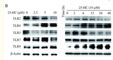
Application: WB    Species: Human    Sample: HepG2 cells

Figure 5 The migration of HepG2 cells induced by 25-HC is partly mediated by the up-regulation of TLR4. (A) HepG2 cells were treated with the indicated concentrations of 25-HC for 24 hours before mRNA was extracted and expressions of TLR2, TLR4, TLR5, TLR7 and TLR9 were examined by RT-qPCR. (B) HepG2 cells were treated with the indicated concentrations of 25-HC for 24 hours or treated with 25-HC at 10 μM for the indicated times before protein was collected to detect the TLR2, TLR4, TLR5, TLR7 and TLR9 expressions by Western blotting. HepG2 cells were transfected with siNC or siTLR4 or siTLR9 for 24 hours, (C) then cells were treated with the indicated concentrations of 25-HC for 24 hours before proteins were collected and the expressions of MMP1, MMP2, MMP3 and MMP9 were determined by Western blotting. (D) Or migratory ability of HepG2 cells after treated with 25-HC for 36 hours was determined by Transwell assay. Results were obtained from 3 independent experiments and are expressed as the means ± SEM. Statistical significance was determined by Student’s t-test. *P<0.05, ***P<0.001.

3). Neuromedin U Induces Pulmonary Group 2 Innate Lymphoid Cell Activation via NMUR1 Pathway During Acute Respiratory Syncytial Virus Infection. Research Square, 2021

Application: WB    Species: Mouse    Sample: lung

Figure 8. Neurons produce NMU in TLR4 and TLR7-dependent manners during RSV infection. E Western blot of TLR4 and TLR7 in DRG neurons after RSV infection.

Restrictive clause


Affinity Biosciences tests all products strictly. Citations are provided as a resource for additional applications that have not been validated by Affinity Biosciences. Please choose the appropriate format for each application and consult Materials and Methods sections for additional details about the use of any product in these publications.

For Research Use Only.
Not for use in diagnostic or therapeutic procedures. Not for resale. Not for distribution without written consent. Affinity Biosciences will not be held responsible for patent infringement or other violations that may occur with the use of our products. Affinity Biosciences, Affinity Biosciences Logo and all other trademarks are the property of Affinity Biosciences LTD.