Product: NGF Antibody
Catalog: DF6061
Description: Rabbit polyclonal antibody to NGF
Application: WB IHC
Reactivity: Human, Mouse, Rat
Prediction: Pig, Bovine, Horse, Sheep, Rabbit, Dog
Mol.Wt.: 27kDa; 27kD(Calculated).
Uniprot: P01138
RRID: AB_2838030

View similar products>>

   Size Price Inventory
 100ul $280 In stock
 200ul $350 In stock

Lead Time: Same day delivery

For pricing and ordering contact:
Local distributors

Product Info

Source:
Rabbit
Application:
WB 1:500-1:2000, IHC 1:50-1:200
*The optimal dilutions should be determined by the end user.
*Tips:

WB: For western blot detection of denatured protein samples. IHC: For immunohistochemical detection of paraffin sections (IHC-p) or frozen sections (IHC-f) of tissue samples. IF/ICC: For immunofluorescence detection of cell samples. ELISA(peptide): For ELISA detection of antigenic peptide.

Reactivity:
Human,Mouse,Rat
Prediction:
Pig(88%), Bovine(88%), Horse(88%), Sheep(88%), Rabbit(100%), Dog(88%)
Clonality:
Polyclonal
Specificity:
NGF Antibody detects endogenous levels of total NGF.
RRID:
AB_2838030
Cite Format: Affinity Biosciences Cat# DF6061, RRID:AB_2838030.
Conjugate:
Unconjugated.
Purification:
The antiserum was purified by peptide affinity chromatography using SulfoLink™ Coupling Resin (Thermo Fisher Scientific).
Storage:
Rabbit IgG in phosphate buffered saline , pH 7.4, 150mM NaCl, 0.02% sodium azide and 50% glycerol. Store at -20 °C. Stable for 12 months from date of receipt.
Alias:

Fold/Unfold

Beta nerve growth factor; Beta NGF; Beta-nerve growth factor; Beta-NGF; HSAN5; MGC161426; MGC161428; Nerve growth factor (beta polypeptide); Nerve growth factor; Nerve growth factor beta; Nerve growth factor beta polypeptide; Nerve growth factor beta subunit; NGF; NGF_HUMAN; NGFB; NID67;

Immunogens

Immunogen:
Uniprot:
Gene(ID):
Description:
Nerve growth factor (NGF) is a small, secreted protein and member of the neurotrophin family of growth factors that promote neuronal cell survival and differentiation (1). Producing cells release NGF that bind and activate TrkA high affinity receptors to mediate NGF-driven signaling. NGF also binds to a low affinity p75 (NTR) receptors, which belong to the death receptor family (2). Although NGF has been classically described as favoring neuron survival and differentiation, nerve growth factor can promote apoptosis in cells that contain p75 (NTR) and lack TrkA. NGF can induce neuron death in a variety of neurodegenerative conditions, including Alzheimer disease (3). Besides its neurotrophic actions, NGF has an effect on non-neuronal cells and may help mediate inflammation, angiogenesis, and stimulate breast cancer cell growth (4-6). NGF signaling is looking increasingly promising as potential drug targets for diseases.
Sequence:
MSMLFYTLITAFLIGIQAEPHSESNVPAGHTIPQAHWTKLQHSLDTALRRARSAPAAAIAARVAGQTRNITVDPRLFKKRRLRSPRVLFSTQPPREAADTQDLDFEVGGAAPFNRTHRSKRSSSHPIFHRGEFSVCDSVSVWVGDKTTATDIKGKEVMVLGEVNINNSVFKQYFFETKCRDPNPVDSGCRGIDSKHWNSYCTTTHTFVKALTMDGKQAAWRFIRIDTACVCVLSRKAVRRA

Predictions

Predictions:

Score>80(red) has high confidence and is suggested to be used for WB detection. *The prediction model is mainly based on the alignment of immunogen sequences, the results are for reference only, not as the basis of quality assurance.

Species
Results
Score
Rabbit
100
Pig
88
Horse
88
Bovine
88
Sheep
88
Dog
88
Zebrafish
75
Chicken
67
Xenopus
63
Model Confidence:
High(score>80) Medium(80>score>50) Low(score<50) No confidence

PTMs - P01138 As Substrate

Site PTM Type Enzyme
S43 O-Glycosylation
T46 O-Glycosylation
S84 Phosphorylation
K155 Acetylation

Research Backgrounds

Function:

Nerve growth factor is important for the development and maintenance of the sympathetic and sensory nervous systems. Extracellular ligand for the NTRK1 and NGFR receptors, activates cellular signaling cascades to regulate neuronal proliferation, differentiation and survival (Probable). The immature NGF precursor (proNGF) functions as ligand for the heterodimeric receptor formed by SORCS2 and NGFR, and activates cellular signaling cascades that lead to inactivation of RAC1 and/or RAC2, reorganization of the actin cytoskeleton and neuronal growth cone collapse. In contrast to mature NGF, the precursor form (proNGF) promotes neuronal apoptosis (in vitro) (By similarity). Inhibits metalloproteinase-dependent proteolysis of platelet glycoprotein VI. Binds lysophosphatidylinositol and lysophosphatidylserine between the two chains of the homodimer. The lipid-bound form promotes histamine relase from mast cells, contrary to the lipid-free form (By similarity).

Subcellular Location:

Secreted. Endosome lumen.
Note: ProNGF is endocytosed after binding to the cell surface receptor formed by SORT1 and NGFR.

Extracellular region or secreted Cytosol Plasma membrane Cytoskeleton Lysosome Endosome Peroxisome ER Golgi apparatus Nucleus Mitochondrion Manual annotation Automatic computational assertionSubcellular location
Subunit Structure:

Homodimer. The homodimer interacts with a single NTRK1 chain. The homodimer interacts with a single NGFR chain. The NGF dimer interacts with a single SORCS2 chain (via extracellular domain) (By similarity). The NGF precursor (proNGF) binds to a receptor complex formed by SORT1 and NGFR, which leads to NGF endocytosis. Both mature NGF and the immature NGF precursor (proNGF) interact with SORCS2 and with the heterodimer formed by SORCS2 and NGFR (via extracellular domains) (By similarity). The NGF precursor (proNGF) has much higher affinity for SORCS2 than mature NGF. The NGF precursor (proNGF) has much higher affinity for SORT1 than mature NGF (By similarity). Interacts with ADAM10 in a divalent cation-dependent manner.

Family&Domains:

Belongs to the NGF-beta family.

Research Fields

· Cellular Processes > Cell growth and death > Apoptosis.   (View pathway)

· Environmental Information Processing > Signal transduction > MAPK signaling pathway.   (View pathway)

· Environmental Information Processing > Signal transduction > Ras signaling pathway.   (View pathway)

· Environmental Information Processing > Signal transduction > Rap1 signaling pathway.   (View pathway)

· Environmental Information Processing > Signal transduction > PI3K-Akt signaling pathway.   (View pathway)

· Organismal Systems > Nervous system > Neurotrophin signaling pathway.   (View pathway)

· Organismal Systems > Sensory system > Inflammatory mediator regulation of TRP channels.   (View pathway)

References

1). Hepcidin deficiency impairs hippocampal neurogenesis and mediates brain atrophy and memory decline in mice. Journal of neuroinflammation, 2024 (PubMed: 38195497) [IF=9.3]

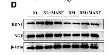
2). MANF Promotes Diabetic Corneal Epithelial Wound Healing and Nerve Regeneration by Attenuating Hyperglycemia-Induced Endoplasmic Reticulum Stress. DIABETES, 2020 (PubMed: 32312869) [IF=7.7]

Application: WB    Species: mice    Sample: corneal epithelial cells

Figure 3—Exogenous MANF promoted corneal nerve regeneration and BDNF and NGF expression. Normal and diabetic corneas pretreated through subconjunctival injection with PBS (as the control) or rhMANF 24 h before, 0 h, and 24 h after removal of the corneal epithelium. At 48 h, corneal epithelial cells were collected for Western blotting. At 5 days postwounding, corneal whole-mount staining was performed to observe nerve regeneration. Corneal sensation was tested before epithelium debridement until 7 days postwounding. To observe nerve regeneration and corneal sensation, another injection was performed at 48 h postwounding. A: Staining for b-tubulin III was performed, and images of the entire cornea were captured as shown in the top panels, with central cornea images shown in the bottom panels. B: Nerve densities of entire corneas (A, top panels) were calculated from the areas staining positive for b-tubulin III using Image J software (n 5 3). C: Corneal sensation is represented with a line chart (n 5 5). The time immediately before epithelium debridement was defined as 0 h. D: Western blot bands observed with or without rhMANF. Two samples/lane represent each condition, three corneas as one sample. E: Western blot analysis of BDNF expression (n 5 4 samples). F: Western blot analysis of NGF expression (n 5 4 samples). *P , 0.05, **P , 0.01, ***P , 0.001 (one-way ANOVA). DM, wounded mice with diabetes mellitus; DM 1 MANF, wounded mice with diabetes mellitus treated with rhMANF; NL, wounded normoglycemic mice treated with PBS; NL 1 MANF, wounded normoglycemic mice treated with rhMANF; n.s., not significant.

3). XQ-1H alleviates cerebral ischemia in mice through inhibition of apoptosis and promotion of neurogenesis in a Wnt/β-catenin signaling dependent way. LIFE SCIENCES, 2019 (PubMed: 31499069) [IF=6.1]

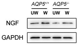
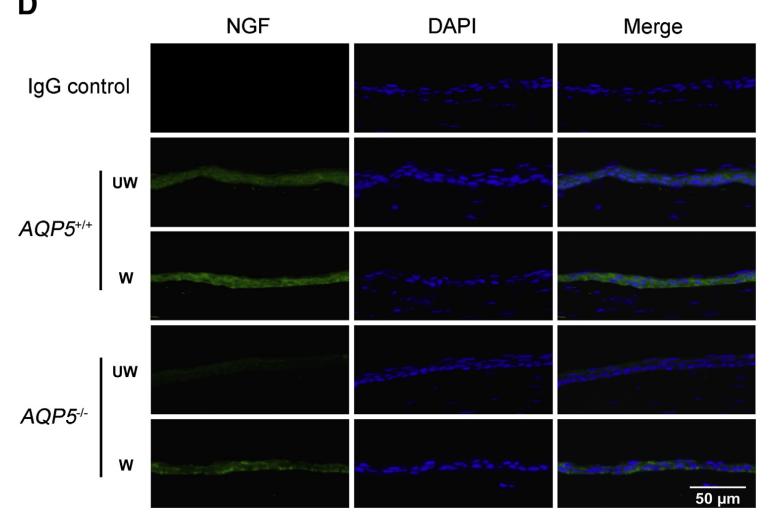
4). AQP5 Facilitates Corneal Epithelial Wound Healing and Nerve Regeneration by Reactivating Akt Signaling Pathway. AMERICAN JOURNAL OF PATHOLOGY, 2021 (PubMed: 34390680) [IF=6.0]

Application: WB    Species: Mice    Sample: corneal epithelium

Figure 3 AQP5 affects corneal nerve regeneration by affecting nerve growth factor (NGF). A: The expression levels of NGF, pigment epithelium-derived factor (PEDF), glial cell-derived neurotrophic factor (GDNF), ciliary neurotrophic factor (CNTF), brain-derived neurotrophic factor (BDNF), and tachykinin 1 (TAC-1) in AQP5þ/þ and AQP5/ mice before and 24 hours after central corneal scraping were evaluated by real-time fluorescence quantitative PCR. B: Western blot analysis was used to confirm the level of NGF protein in the corneal epithelium of AQP5þ/þ and AQP5/ mice before and 24 hours after central corneal scraping. Two corneal epithelia were pooled into one sample. C: Quantification of the intensities of Western blot analysis bands of NGF compared with glyceraldehyde-3-phosphate dehydrogenase (GAPDH). D: Immunofluorescence staining with anti-NGF antibody in AQP5þ/þ and AQP5/ mice before and 24 hours after central corneal scraping. n Z 3 (C). *P < 0.05, **P < 0.01, and ***P < 0.001. Scale bar Z 50 mm (D). UW, unwounded; W, wounded.

Application: IF/ICC    Species: Mice    Sample: corneal epithelium

Figure 3 AQP5 affects corneal nerve regeneration by affecting nerve growth factor (NGF). A: The expression levels of NGF, pigment epithelium-derived factor (PEDF), glial cell-derived neurotrophic factor (GDNF), ciliary neurotrophic factor (CNTF), brain-derived neurotrophic factor (BDNF), and tachykinin 1 (TAC-1) in AQP5þ/þ and AQP5/ mice before and 24 hours after central corneal scraping were evaluated by real-time fluorescence quantitative PCR. B: Western blot analysis was used to confirm the level of NGF protein in the corneal epithelium of AQP5þ/þ and AQP5/ mice before and 24 hours after central corneal scraping. Two corneal epithelia were pooled into one sample. C: Quantification of the intensities of Western blot analysis bands of NGF compared with glyceraldehyde-3-phosphate dehydrogenase (GAPDH). D: Immunofluorescence staining with anti-NGF antibody in AQP5þ/þ and AQP5/ mice before and 24 hours after central corneal scraping. n Z 3 (C). *P < 0.05, **P < 0.01, and ***P < 0.001. Scale bar Z 50 mm (D). UW, unwounded; W, wounded.

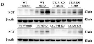
5). CB2R Deficiency Exacerbates Imiquimod-Induced Psoriasiform Dermatitis and Itch Through the Neuro-Immune Pathway. Frontiers in Pharmacology, 2022 (PubMed: 35173615) [IF=5.6]

Application: WB    Species: Mice    Sample:

FIGURE 5 Effects of CB2R on the scratching behavior and expression of nerve fibers in IMQ-induced PsD. (A, B) The behavior results of mice in different groups. ( C) PGP 9.5 stained (red fluorescence) nerve fibers in the skin biopsies from mice, co-stained with DAPI (blue fluorescence) to detect the nucleus. Scale bar represents 100 μm. (D) Protein level of NGF was analyzed by western blotting in the six groups of mice. β-actin served as the loading control. ( E) The mRNA expression levels of NGF was measured by qRT-PCR. Error bars represent mean ± SD. (n = 5 for each group). *p < 0.05, **p < 0.01, and ***p < 0.001 when compared. All the assays were repeated three times with consistent results.

6). Oligo-Porphyran Ameliorates Neurobehavioral Deficits in Parkinsonian Mice by Regulating the PI3K/Akt/Bcl-2 Pathway. Marine Drugs, 2018 (PubMed: 29509717) [IF=5.4]

Application: WB    Species: mouse    Sample: striatum

Figure 6. |Effects of OP on nerve growth factor (NGF) and tropomyosin receptor kinase A (TrkA),expression. After pretreated with MPTP for 7 days, the C57BL/6 mice were administrated with MA or different concentrations of OP for the followed 7 days.(A) Original bands of NGF, pTrkA, TrkA, andβ-actin.

7). Transdifferentiation effects and related mechanisms of nerve growth factor and internal limiting membrane on Müller cells. EXPERIMENTAL EYE RESEARCH, 2019 (PubMed: 30578789) [IF=3.4]

8). Metformin protects against pericyte apoptosis and promotes neurogenesis through suppressing JNK p38 MAPK signalling activation in ischemia/reperfusion injury. Neuroscience Letters, 2022 (PubMed: 35660649) [IF=2.5]

9). Network Pharmacology and Molecular Docking Verify the Mechanism of Qinshi Simiao San in Treating Chronic Prostatitis in the Rat Model. Evidence-based Complementary and Alternative Medicine, 2022 (PubMed: 35069766)

Restrictive clause


Affinity Biosciences tests all products strictly. Citations are provided as a resource for additional applications that have not been validated by Affinity Biosciences. Please choose the appropriate format for each application and consult Materials and Methods sections for additional details about the use of any product in these publications.

For Research Use Only.
Not for use in diagnostic or therapeutic procedures. Not for resale. Not for distribution without written consent. Affinity Biosciences will not be held responsible for patent infringement or other violations that may occur with the use of our products. Affinity Biosciences, Affinity Biosciences Logo and all other trademarks are the property of Affinity Biosciences LTD.