Product: ATF6 Antibody
Catalog: DF6009
Description: Rabbit polyclonal antibody to ATF6
Application: WB IHC IF/ICC
Reactivity: Human, Mouse, Rat
Prediction: Pig, Zebrafish, Horse, Sheep, Rabbit, Dog, Xenopus
Mol.Wt.: 50~75kD(cleaved),90~100kD(full); 75kD(Calculated).
Uniprot: P18850
RRID: AB_2833019

View similar products>>

   Size Price Inventory
 100ul $280 In stock
 200ul $350 In stock

Lead Time: Same day delivery

For pricing and ordering contact:
Local distributors

Product Info

Source:
Rabbit
Application:
WB 1:500-1:2000, IHC 1:50-1:200, IF/ICC 1:100-1:500
*The optimal dilutions should be determined by the end user.
*Tips:

WB: For western blot detection of denatured protein samples. IHC: For immunohistochemical detection of paraffin sections (IHC-p) or frozen sections (IHC-f) of tissue samples. IF/ICC: For immunofluorescence detection of cell samples. ELISA(peptide): For ELISA detection of antigenic peptide.

Reactivity:
Human,Mouse,Rat
Prediction:
Pig(100%), Zebrafish(100%), Horse(100%), Sheep(100%), Rabbit(100%), Dog(100%), Xenopus(100%)
Clonality:
Polyclonal
Specificity:
ATF6 Antibody detects endogenous levels of total ATF6.
RRID:
AB_2833019
Cite Format: Affinity Biosciences Cat# DF6009, RRID:AB_2833019.
Conjugate:
Unconjugated.
Purification:
The antiserum was purified by peptide affinity chromatography using SulfoLink™ Coupling Resin (Thermo Fisher Scientific).
Storage:
Rabbit IgG in phosphate buffered saline , pH 7.4, 150mM NaCl, 0.02% sodium azide and 50% glycerol. Store at -20 °C. Stable for 12 months from date of receipt.
Alias:

Fold/Unfold

Activating transcription factor 6 alpha; Activating transcription factor 6; ATF 6; ATF6 alpha; ATF6; ATF6-alpha; ATF6A; ATF6A_HUMAN; cAMP dependent transcription factor ATF 6 alpha; cAMP-dependent transcription factor ATF-6 alpha; Cyclic AMP dependent transcription factor ATF 6 alpha; DKFZp686P2194; ESTM49; FLJ21663; Processed cyclic AMP dependent transcription factor ATF 6 alpha; Processed cyclic AMP-dependent transcription factor ATF-6 alpha;

Immunogens

Immunogen:
Uniprot:
Gene(ID):
Expression:
P18850 ATF6A_HUMAN:

Ubiquitous.

Description:
This gene encodes a transcription factor that activates target genes for the unfolded protein response (UPR) during endoplasmic reticulum (ER) stress. Although it is a transcription factor, this protein is unusual in that it is synthesized as a transmembrane protein that is embedded in the ER. It functions as an ER stress sensor/transducer, and following ER stress-induced proteolysis, it functions as a nuclear transcription factor via a cis-acting ER stress response element (ERSE) that is present in the promoters of genes encoding ER chaperones. This protein has been identified as a survival factor for quiescent but not proliferative squamous carcinoma cells. There have been conflicting reports about the association of polymorphisms in this gene with diabetes in different populations, but another polymorphism has been associated with increased plasma cholesterol levels. This gene is also thought to be a potential therapeutic target for cystic fibrosis. [provided by RefSeq, Aug 2011]
Sequence:
MGEPAGVAGTMESPFSPGLFHRLDEDWDSALFAELGYFTDTDELQLEAANETYENNFDNLDFDLDLMPWESDIWDINNQICTVKDIKAEPQPLSPASSSYSVSSPRSVDSYSSTQHVPEELDLSSSSQMSPLSLYGENSNSLSSAEPLKEDKPVTGPRNKTENGLTPKKKIQVNSKPSIQPKPLLLPAAPKTQTNSSVPAKTIIIQTVPTLMPLAKQQPIISLQPAPTKGQTVLLSQPTVVQLQAPGVLPSAQPVLAVAGGVTQLPNHVVNVVPAPSANSPVNGKLSVTKPVLQSTMRNVGSDIAVLRRQQRMIKNRESACQSRKKKKEYMLGLEARLKAALSENEQLKKENGTLKRQLDEVVSENQRLKVPSPKRRVVCVMIVLAFIILNYGPMSMLEQDSRRMNPSVSPANQRRHLLGFSAKEAQDTSDGIIQKNSYRYDHSVSNDKALMVLTEEPLLYIPPPPCQPLINTTESLRLNHELRGWVHRHEVERTKSRRMTNNQQKTRILQGALEQGSNSQLMAVQYTETTSSISRNSGSELQVYYASPRSYQDFFEAIRRRGDTFYVVSFRRDHLLLPATTHNKTTRPKMSIVLPAININENVINGQDYEVMMQIDCQVMDTRILHIKSSSVPPYLRDQQRNQTNTFFGSPPAATEATHVVSTIPESLQ

Predictions

Predictions:

Score>80(red) has high confidence and is suggested to be used for WB detection. *The prediction model is mainly based on the alignment of immunogen sequences, the results are for reference only, not as the basis of quality assurance.

Species
Results
Score
Pig
100
Horse
100
Sheep
100
Dog
100
Xenopus
100
Zebrafish
100
Rabbit
100
Bovine
0
Chicken
0
Model Confidence:
High(score>80) Medium(80>score>50) Low(score<50) No confidence

PTMs - P18850 As Substrate

Site PTM Type Enzyme
S16 Phosphorylation
K87 Sumoylation
K87 Ubiquitination
S94 Phosphorylation
Y100 Phosphorylation
S104 Phosphorylation
K152 Sumoylation
T166 Phosphorylation Q16539 (MAPK14)
K176 Acetylation
K176 Sumoylation
K176 Ubiquitination
K182 Sumoylation
K191 Sumoylation
K201 Ubiquitination
K216 Ubiquitination
K290 Sumoylation
K290 Ubiquitination
T296 Phosphorylation
Y330 Phosphorylation
K339 Ubiquitination
K349 Ubiquitination
S373 Phosphorylation
S410 Phosphorylation
K424 Ubiquitination
K436 Ubiquitination
N472 N-Glycosylation
K506 Ubiquitination
T565 Phosphorylation
N584 N-Glycosylation
K629 Ubiquitination
S632 Phosphorylation
N643 N-Glycosylation

Research Backgrounds

Function:

Transmembrane glycoprotein of the endoplasmic reticulum that functions as a transcription activator and initiates the unfolded protein response (UPR) during endoplasmic reticulum stress. Cleaved upon ER stress, the N-terminal processed cyclic AMP-dependent transcription factor ATF-6 alpha translocates to the nucleus where it activates transcription of genes involved in the UPR. Binds DNA on the 5'-CCAC[GA]-3'half of the ER stress response element (ERSE) (5'-CCAAT-N(9)-CCAC[GA]-3') and of ERSE II (5'-ATTGG-N-CCACG-3'). Binding to ERSE requires binding of NF-Y to ERSE. Could also be involved in activation of transcription by the serum response factor. May play a role in foveal development and cone function in the retina.

PTMs:

During unfolded protein response, a fragment of approximately 50 kDa containing the cytoplasmic transcription factor domain is released by proteolysis. The cleavage seems to be performed sequentially by site-1 and site-2 proteases.

N-glycosylated. The glycosylation status may serve as a sensor for ER homeostasis, resulting in ATF6 activation to trigger the unfolded protein response (UPR).

Phosphorylated in vitro by MAPK14/P38MAPK.

Subcellular Location:

Endoplasmic reticulum membrane>Single-pass type II membrane protein.

Nucleus.
Note: Under ER stress the cleaved N-terminal cytoplasmic domain translocates into the nucleus. THBS4 promotes its nuclear shuttling.

Extracellular region or secreted Cytosol Plasma membrane Cytoskeleton Lysosome Endosome Peroxisome ER Golgi apparatus Nucleus Mitochondrion Manual annotation Automatic computational assertionSubcellular location
Tissue Specificity:

Ubiquitous.

Subunit Structure:

Homodimer and heterodimer with ATF6-beta. The dimer interacts with the nuclear transcription factor Y (NF-Y) trimer through direct binding to NF-Y subunit C (NF-YC). Interacts also with the transcription factors GTF2I, YY1 and SRF. Interacts (via lumenal domain) with THBS1 (By similarity). Interacts with THBS4 (via EGF-like 3; calcium-binding domain) which facilitates its processing, activation and nuclear translocation. Interacts with XBP1 isoform 2; the interaction occurs in a ER stress-dependent manner.

Family&Domains:

The basic domain functions as a nuclear localization signal.

The basic leucine-zipper domain is sufficient for association with the NF-Y trimer and binding to ERSE.

Belongs to the bZIP family. ATF subfamily.

Research Fields

· Genetic Information Processing > Folding, sorting and degradation > Protein processing in endoplasmic reticulum.   (View pathway)

· Human Diseases > Neurodegenerative diseases > Alzheimer's disease.

References

1). Branched chain amino acids exacerbate myocardial ischemia/reperfusion vulnerability via enhancing GCN2/ATF6/PPAR-α pathway-dependent fatty acid oxidation. Theranostics, 2020 (PubMed: 32373236) [IF=12.4]

Application: WB    Species: mouse    Sample: Heart

Figure 6. BCAA increase PPAR-α expression in a GCN2/ATF6 pathway-dependent manner. (A) Expression of p-GCN2, GCN2 and ATF6 in the presence of increasing concentrations of BCAA (0, 0.429 mM, 0.858 mM, 1.716 mM, 3.432 mM) by western blotting (n=6). (B) Expression of p-GCN2, GCN2 and ATF6 in the presence of increasing concentrations of BCKA (0, 0.429 mM, 0.858 mM, 1.716 mM, 3.432 mM) by western blotting (n=6). BCKA mixture is composed of αKIC, αKIV and αKMV (weight ratio, αKIC: αKIV: αKMV= 2:1:1). (C) NRVMs were treated with control siRNA and ATF6 siRNA. 48 h after transfection, expression of ATF6 was determined by western blotting (n=4). (D-E) ATF6 siRNA transferred NRVMs were treated with or without BCAA (3.432 mM) (n=6). (D) PPAR-α expression was determined by western blotting. (E) Expression of Acaa2, Acadm, Cd36 and Cpt1b by real-time PCR. (F-G) ATF6 siRNA transferred NRVMs were treated with or without BCKA (3.432 mM) (n=6). (F) PPAR-α expression was determined by western blotting. (G) Expression of Acaa2, Acadm, Cd36 and Cpt1b by real-time PCR. (C) Data were analyzed by Student’s t test. (A-B) and (D-G) Data were analyzed by one-way ANOVA, followed by a Bonferroni post-hoc test. * P<0.05, ** P<0.01. All values are presented as mean ± SEM.

2). Sleep Deprivation Induces Gut Damage via Ferroptosis. Journal of pineal research, 2024 (PubMed: 38975671) [IF=10.3]

3). PPM1H is down-regulated by ATF6 and dephosphorylates p-RPS6KB1 to inhibit progression of hepatocellular carcinoma. Molecular therapy. Nucleic acids, 2023 (PubMed: 37456776) [IF=8.8]

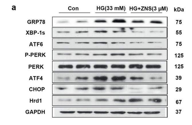
4). Zonisamide, an antiepileptic drug, alleviates diabetic cardiomyopathy by inhibiting endoplasmic reticulum stress. Acta Pharmacologica Sinica, 2020 (PubMed: 32647341) [IF=8.2]

Application: WB    Species: mice    Sample: NRCMs

Fig. 6 Zonisamide alleviates HG-induced cardiac hypertrophy and apoptosis in cultured primary neonatal rat cardiomyocytes (NRCMs) via suppression of activated ER stress. NRCMs were pretreated with 5 mM 4-PBA (an inhibitor of ERS) or 10 ng/mL tunicamycin (Tm, an ERS inducer) for 2 h and then exposed to glucose (33 mM) in the presence or absence of ZNS (3 μM) for 24 h. a–b Representative and quantitative images showing the protein expression of ERS markers, including GRP78, XBP-1s, ATF6, p-PERK, PERK, ATF4, CHOP, and Hrd1. c Immunofluorescence staining of cardiomyocytes with phalloidin (red) and cell nuclei with DAPI (blue), Scale bar = 50 μm. d Quantitative analysis of cell surface area by ImageJ software. e–f Representative Western blotting and analysis of Bax and Bcl-2 expression. g–h Representative and quantitative images of GRP78, ATF6, p-PERK, PERK, ATF4, and CHOP expression. All values are the fold changes normalized to their control group. The results are presented as the means ± SEM (n = 6). *P < 0.05, **P < 0.01 vs. Con; #P < 0.05, ##P < 0.01 vs. HG; $P < 0.05, $$P < 0.01 vs. HG + ZNS.

5). Patulin induces ROS-dependent cardiac cell toxicity by inducing DNA damage and activating endoplasmic reticulum stress apoptotic pathway. Ecotoxicology and environmental safety, 2024 (PubMed: 38061079) [IF=6.8]

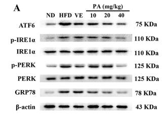
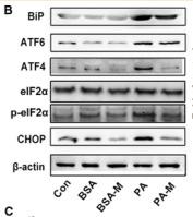
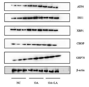
6). Oleic acid protects saturated fatty acid mediated lipotoxicity in hepatocytes and rat of non-alcoholic steatohepatitis. LIFE SCIENCES, 2018 (PubMed: 29709653) [IF=6.1]

7). Network-based identification and mechanism exploration of active ingredients against Alzheimer's disease via targeting endoplasmic reticulum stress from traditional chinese medicine. Computational and structural biotechnology journal, 2024 (PubMed: 38261917) [IF=6.0]

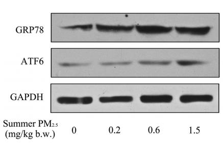
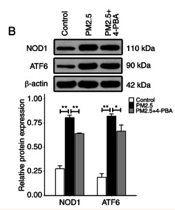
8). The potential immunotoxicity of fine particulate matter based on SD rat spleen. ENVIRONMENTAL SCIENCE AND POLLUTION RESEARCH, 2019 (PubMed: 31218585) [IF=5.8]

Application: WB    Species: rat    Sample: spleen

Fig. 3 | PM2.5 induced ERS in spleen of SD rats. a, b The levels of spleen GRP78 and ATF6 mRNA were measured using qRT-PCR by summer and winter PM2.5 treatment in SD rat. c, dThe levels of spleen GRP78 and ATF6 protein were measured using western blotting in summer PM2.5 treatment in SD rats and quantification of analysis

9). Chrysin ameliorates synovitis and fibrosis of osteoarthritic fibroblast-like synoviocytes in rats through PERK/TXNIP/NLRP3 signaling. Frontiers in Pharmacology, 2023 (PubMed: 37021049) [IF=5.6]

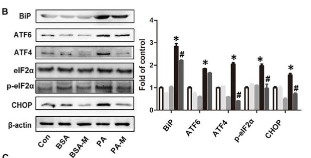
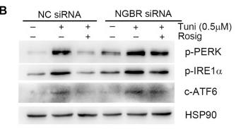
10). Peroxisome Proliferator-Activated Receptor-Gamma Reduces ER Stress and Inflammation via Targeting NGBR Expression. Frontiers in Pharmacology, 2022 (PubMed: 35111067) [IF=5.6]

Application: WB    Species: Human    Sample: HUVEC cells

FIGURE 6 PPARγ reduces tunicamycin-induced ER stress by regulating NGBR. (A–C) HUVEC cells in a six-well plate were transfected with control siRNA (NC siRNA) or NGBR siRNA for 24 h in serum-free medium, followed by switching the cells into complete medium to culture for another 24 h. After treatment with rosiglitazone (10 μM) for 12 h, the cells were treated with tunicamycin (0.5 μg/ml) with or without rosiglitazone for another 12 h. Expression of CHOP, BIP, NGBR p-PERK, p-IRE1α and c-ATF6 protein was determined by Western blot (A, B). Expression of CHOP, BIP and NGBR mRNA was determined by qPCR (C). Values were expressed as means ± SD. *p < 0.05; **p < 0.01; ns, not significant (n = 3). (D) liver total proteins were collected from Figure 4. Expression of BIP and CHOP in mouse liver was determined by Western blot. Values were expressed as means ± SD. *p < 0.05; **p < 0.01; ns, not significant (n = 3).

Load more

Restrictive clause


Affinity Biosciences tests all products strictly. Citations are provided as a resource for additional applications that have not been validated by Affinity Biosciences. Please choose the appropriate format for each application and consult Materials and Methods sections for additional details about the use of any product in these publications.

For Research Use Only.
Not for use in diagnostic or therapeutic procedures. Not for resale. Not for distribution without written consent. Affinity Biosciences will not be held responsible for patent infringement or other violations that may occur with the use of our products. Affinity Biosciences, Affinity Biosciences Logo and all other trademarks are the property of Affinity Biosciences LTD.