Product: TGFBR2 Antibody
Catalog: AF5449
Description: Rabbit polyclonal antibody to TGFBR2
Application: WB IHC
Reactivity: Human, Mouse, Rat
Prediction: Pig, Bovine, Horse, Rabbit, Dog, Chicken, Xenopus
Mol.Wt.: 63 kDa; 65kD(Calculated).
Uniprot: P37173
RRID: AB_2837933

View similar products>>

   Size Price Inventory
 100ul $280 In stock
 200ul $350 In stock

Lead Time: Same day delivery

For pricing and ordering contact:
Local distributors

Product Info

Source:
Rabbit
Application:
WB 1:500-1:2000, IHC 1:50-1:200
*The optimal dilutions should be determined by the end user.
*Tips:

WB: For western blot detection of denatured protein samples. IHC: For immunohistochemical detection of paraffin sections (IHC-p) or frozen sections (IHC-f) of tissue samples. IF/ICC: For immunofluorescence detection of cell samples. ELISA(peptide): For ELISA detection of antigenic peptide.

Reactivity:
Human,Mouse,Rat
Prediction:
Pig(100%), Bovine(100%), Horse(100%), Rabbit(100%), Dog(100%), Chicken(88%), Xenopus(100%)
Clonality:
Polyclonal
Specificity:
TGFBR2 Antibody detects endogenous levels of total TGFBR2.
RRID:
AB_2837933
Cite Format: Affinity Biosciences Cat# AF5449, RRID:AB_2837933.
Conjugate:
Unconjugated.
Purification:
The antiserum was purified by peptide affinity chromatography using SulfoLink™ Coupling Resin (Thermo Fisher Scientific).
Storage:
Rabbit IgG in phosphate buffered saline , pH 7.4, 150mM NaCl, 0.02% sodium azide and 50% glycerol. Store at -20 °C. Stable for 12 months from date of receipt.
Alias:

Fold/Unfold

AAT3; FAA3; LDS1B; LDS2; LDS2B; MFS2; RIIC; TAAD2; TbetaR II; TbetaR-II; TGF beta receptor type 2; TGF beta receptor type II; TGF beta receptor type IIB; TGF beta type II receptor; TGF-beta receptor type II; TGF-beta receptor type-2; TGF-beta type II receptor; TGF-beta-R2; TGFB R2; TGFbeta - RII; TGFbeta RII; Tgfbr2; TGFR-2; TGFR2_HUMAN; Transforming growth factor beta receptor II; Transforming growth factor beta receptor type II; Transforming growth factor beta receptor type IIC; Transforming growth factor, beta receptor II (70/80kDa); transforming growth factor, beta receptor II alpha; transforming growth factor, beta receptor II beta; transforming growth factor, beta receptor II delta; transforming growth factor, beta receptor II epsilon; transforming growth factor, beta receptor II gamma; Transforming growth factor-beta receptor type II;

Immunogens

Immunogen:
Uniprot:
Gene(ID):
Description:
Transmembrane serine/threonine kinase forming with the TGF-beta type I serine/threonine kinase receptor, TGFBR1, the non-promiscuous receptor for the TGF-beta cytokines TGFB1, TGFB2 and TGFB3. Transduces the TGFB1, TGFB2 and TGFB3 signal from the cell surface to the cytoplasm and is thus regulating a plethora of physiological and pathological processes including cell cycle arrest in epithelial and hematopoietic cells
Sequence:
MGRGLLRGLWPLHIVLWTRIASTIPPHVQKSVNNDMIVTDNNGAVKFPQLCKFCDVRFSTCDNQKSCMSNCSITSICEKPQEVCVAVWRKNDENITLETVCHDPKLPYHDFILEDAASPKCIMKEKKKPGETFFMCSCSSDECNDNIIFSEEYNTSNPDLLLVIFQVTGISLLPPLGVAISVIIIFYCYRVNRQQKLSSTWETGKTRKLMEFSEHCAIILEDDRSDISSTCANNINHNTELLPIELDTLVGKGRFAEVYKAKLKQNTSEQFETVAVKIFPYEEYASWKTEKDIFSDINLKHENILQFLTAEERKTELGKQYWLITAFHAKGNLQEYLTRHVISWEDLRKLGSSLARGIAHLHSDHTPCGRPKMPIVHRDLKSSNILVKNDLTCCLCDFGLSLRLDPTLSVDDLANSGQVGTARYMAPEVLESRMNLENVESFKQTDVYSMALVLWEMTSRCNAVGEVKDYEPPFGSKVREHPCVESMKDNVLRDRGRPEIPSFWLNHQGIQMVCETLTECWDHDPEARLTAQCVAERFSELEHLDRLSGRSCSEEKIPEDGSLNTTK

Predictions

Predictions:

Score>80(red) has high confidence and is suggested to be used for WB detection. *The prediction model is mainly based on the alignment of immunogen sequences, the results are for reference only, not as the basis of quality assurance.

Species
Results
Score
Pig
100
Horse
100
Bovine
100
Dog
100
Xenopus
100
Rabbit
100
Chicken
88
Sheep
0
Zebrafish
0
Model Confidence:
High(score>80) Medium(80>score>50) Low(score<50) No confidence

PTMs - P37173 As Substrate

Site PTM Type Enzyme
T39 O-Glycosylation
K205 Ubiquitination
K208 Ubiquitination
S213 Phosphorylation P37173 (TGFBR2)
Y259 Phosphorylation P37173 (TGFBR2)
K260 Ubiquitination
K264 Ubiquitination
Y284 Phosphorylation P12931 (SRC)
K291 Ubiquitination
K300 Ubiquitination
Y336 Phosphorylation P37173 (TGFBR2)
S352 Phosphorylation
K381 Ubiquitination
S409 Phosphorylation P37173 (TGFBR2)
S416 Phosphorylation P37173 (TGFBR2)
Y424 Phosphorylation P37173 (TGFBR2)
T445 Phosphorylation
Y470 Phosphorylation
K477 Ubiquitination
S486 Phosphorylation
K488 Ubiquitination
S548 Phosphorylation
S551 Phosphorylation
S553 Phosphorylation
K556 Ubiquitination
S562 Phosphorylation
T566 Phosphorylation

PTMs - P37173 As Enzyme

Substrate Site Source
P17813 (ENG) S634 Uniprot
P17813 (ENG) S635 Uniprot
P36897 (TGFBR1) S165 Uniprot
P36897-1 (TGFBR1) S172 Uniprot
P36897 (TGFBR1) T176 Uniprot
P36897 (TGFBR1) T185 Uniprot
P36897 (TGFBR1) T186 Uniprot
P36897 (TGFBR1) S187 Uniprot
P36897 (TGFBR1) S189 Uniprot
P36897 (TGFBR1) S191 Uniprot
P37173 (TGFBR2) S213 Uniprot
P37173-2 (TGFBR2) S238 Uniprot
P37173 (TGFBR2) Y259 Uniprot
P37173 (TGFBR2) Y336 Uniprot
P37173-1 (TGFBR2) S409 Uniprot
P37173 (TGFBR2) S416 Uniprot
P37173 (TGFBR2) Y424 Uniprot
P37173-2 (TGFBR2) S434 Uniprot
P37173-2 (TGFBR2) S441 Uniprot
Q03167 (TGFBR3) T843 Uniprot
Q9NPB6 (PARD6A) S345 Uniprot

Research Backgrounds

Function:

Transmembrane serine/threonine kinase forming with the TGF-beta type I serine/threonine kinase receptor, TGFBR1, the non-promiscuous receptor for the TGF-beta cytokines TGFB1, TGFB2 and TGFB3. Transduces the TGFB1, TGFB2 and TGFB3 signal from the cell surface to the cytoplasm and is thus regulating a plethora of physiological and pathological processes including cell cycle arrest in epithelial and hematopoietic cells, control of mesenchymal cell proliferation and differentiation, wound healing, extracellular matrix production, immunosuppression and carcinogenesis. The formation of the receptor complex composed of 2 TGFBR1 and 2 TGFBR2 molecules symmetrically bound to the cytokine dimer results in the phosphorylation and the activation of TGFRB1 by the constitutively active TGFBR2. Activated TGFBR1 phosphorylates SMAD2 which dissociates from the receptor and interacts with SMAD4. The SMAD2-SMAD4 complex is subsequently translocated to the nucleus where it modulates the transcription of the TGF-beta-regulated genes. This constitutes the canonical SMAD-dependent TGF-beta signaling cascade. Also involved in non-canonical, SMAD-independent TGF-beta signaling pathways.

PTMs:

Phosphorylated on a Ser/Thr residue in the cytoplasmic domain.

Subcellular Location:

Cell membrane>Single-pass type I membrane protein. Membrane raft.

Extracellular region or secreted Cytosol Plasma membrane Cytoskeleton Lysosome Endosome Peroxisome ER Golgi apparatus Nucleus Mitochondrion Manual annotation Automatic computational assertionSubcellular location
Subunit Structure:

Homodimer. Heterohexamer; TGFB1, TGFB2 and TGFB3 homodimeric ligands assemble a functional receptor composed of two TGFBR1 and TGFBR2 heterodimers to form a ligand-receptor heterohexamer. The respective affinity of TGFRB1 and TGFRB2 for the ligands may modulate the kinetics of assembly of the receptor and may explain the different biological activities of TGFB1, TGFB2 and TGFB3. Interacts with DAXX. Interacts with TCTEX1D4. Interacts with ZFYVE9; ZFYVE9 recruits SMAD2 and SMAD3 to the TGF-beta receptor. Interacts with and is activated by SCUBE3; this interaction does not affect TGFB1-binding to TGFBR2. Interacts with VPS39; this interaction is independent of the receptor kinase activity and of the presence of TGF-beta. Interacts with CLU.

Family&Domains:

Belongs to the protein kinase superfamily. TKL Ser/Thr protein kinase family. TGFB receptor subfamily.

Research Fields

· Cellular Processes > Transport and catabolism > Endocytosis.   (View pathway)

· Cellular Processes > Cell growth and death > Cellular senescence.   (View pathway)

· Cellular Processes > Cellular community - eukaryotes > Adherens junction.   (View pathway)

· Environmental Information Processing > Signal transduction > MAPK signaling pathway.   (View pathway)

· Environmental Information Processing > Signaling molecules and interaction > Cytokine-cytokine receptor interaction.   (View pathway)

· Environmental Information Processing > Signal transduction > FoxO signaling pathway.   (View pathway)

· Environmental Information Processing > Signal transduction > TGF-beta signaling pathway.   (View pathway)

· Environmental Information Processing > Signal transduction > Hippo signaling pathway.   (View pathway)

· Human Diseases > Infectious diseases: Parasitic > Chagas disease (American trypanosomiasis).

· Human Diseases > Infectious diseases: Viral > HTLV-I infection.

· Human Diseases > Cancers: Overview > Pathways in cancer.   (View pathway)

· Human Diseases > Cancers: Overview > Transcriptional misregulation in cancer.

· Human Diseases > Cancers: Specific types > Colorectal cancer.   (View pathway)

· Human Diseases > Cancers: Specific types > Pancreatic cancer.   (View pathway)

· Human Diseases > Cancers: Specific types > Chronic myeloid leukemia.   (View pathway)

· Human Diseases > Cancers: Specific types > Hepatocellular carcinoma.   (View pathway)

· Human Diseases > Cancers: Specific types > Gastric cancer.   (View pathway)

· Organismal Systems > Development > Osteoclast differentiation.   (View pathway)

· Organismal Systems > Immune system > Th17 cell differentiation.   (View pathway)

· Organismal Systems > Endocrine system > Relaxin signaling pathway.

References

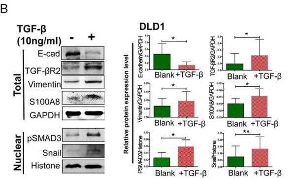
1). S100A8 promotes epithelial‐mesenchymal transition and metastasis under TGF‐β/USF2 axis in colorectal cancer. Cancer Communications, 2021 (PubMed: 33389821) [IF=16.2]

Application: WB    Species: human    Sample: DLD1 cells

FIGURE 6| TGF-β promoted EMT and cell mobility by up-regulating USF2/S100A8. A-B. TGF-β induced EMT and S100A8 expression in SW480 (A) and DLD1 (B) cells.

2). Circular RNA TGFBR2 acts as a ceRNA to suppress nasopharyngeal carcinoma progression by sponging miR-107. CANCER LETTERS, 2021 (PubMed: 33160003) [IF=9.7]

Application: WB    Species: human    Sample: NPC cells

Fig.3.f Relative protein levels in NPC cells, as determined by western blotting.

3). Beauvericin suppresses the proliferation and pulmonary metastasis of osteosarcoma by selectively inhibiting TGFBR2 pathway. International Journal of Biological Sciences, 2023 (PubMed: 37781043) [IF=9.2]

Application: WB    Species: Mouse    Sample: OS cells

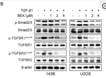
Figure 2 BEA inhibits the TGF-β/Smad2/3 signaling pathway in OS cells. (A) KEGG pathway enrichment analysis in BEA-treated and untreated 143B cells. (B) The effect of BEA on the TGF-β1-induced phosphorylation of Smad2/3, TGFBR1, and TGFBR2 in OS cells was evaluated by Western blotting. (C) Cellular immunofluorescence assay was performed to assess the nuclear translocation of p-Smad2/3 in OS cells treated with BEA. Scale bar: 20 μm. (D) The transcriptional activity of Smad2/3 in OS cells treated as indicated was determined by a Smad2/3 luciferase reporter assay. (E) 143B and U2OS cells were treated with BEA (2 μM) for 24 h and total RNA was harvested and analyzed by qPCR. Quantification of the mRNA levels of COL1A1, COL3A1, COL6A1, COL10A1, MMP2, LOX, LOXL2 in OS cells.

4). Transforming Growth Factor-β3 Chitosan Sponge (TGF-β3/CS) Facilitates Osteogenic Differentiation of Human Periodontal Ligament Stem Cells. INTERNATIONAL JOURNAL OF MOLECULAR SCIENCES, 2019 (PubMed: 31600954) [IF=5.6]

Application: WB    Species: human    Sample: hPDLSCs

Figure 6. |Mechanism of TGF-β3 in osteogenic differentiation of hPDLSCs based on this study. (A)Schematic representation of the mechanism of TGF-β3 in osteogenic differentiation of hPDLSCs. (B)After osteogenic induction of hPDLSCs with various concentrations of TGF-β3 for 7 and 14 d,expression of osteogenic pathway proteins was detected by (B) western blotting and analyzed by grayscale scanning for (C) TGF-βRI, (D) TGF-βRII, (E) t-p38, (F) Pp38, and (G) Runx2 (n = 3). ns means no significant differences, p >0.05 vs. control; *** p < 0.001 vs. control. Abbreviations: TGF-βRI,transforming growth factor-β receptor I; TGF-βRII, transforming growth factor-β receptor II; t-p38,total p38; Pp38, phosphorylated p38; Runx2, runt-related transcription factor 2.

5). Does Chronic Pancreatitis in Growing Pigs Lead to Articular Cartilage Degradation and Alterations in Subchondral Bone?. International journal of molecular sciences, 2024 (PubMed: 38396667) [IF=5.6]

Application: IHC    Species: pig    Sample:

Figure 6 (a) Representative immunohistochemical (IHC) staining for TGF-β1, TGFβR1, TGFβR2, pSMAD2, and pSMAD4 in the articular cartilage of the distal femur from control (CONT) and chronic pancreatitis (CP) pigs. Scale bars represent 50 μm. (b) Bar graphs illustrating the optical density (OD) of expression for the analyzed proteins. Measurements were conducted on two histological samples per animal in at least ten randomly selected areas. Bars indicate mean values ± standard error (n = 5 pigs per group). Statistical significance is denoted as * p < 0.05; ** p < 0.01; *** p < 0.001.

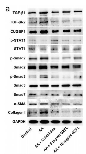
6). Guizhi Fuling pill attenuates liver fibrosis in vitro and in vivo via inhibiting TGF-β1/Smad2/3 and activating IFN-γ/Smad7 signaling pathways. Bioengineered, 2022 (PubMed: 35387552) [IF=4.9]

Application: WB    Species: human    Sample: LX-2 cells

Figure 2.| GZFL inhibits acetaldehyde-induced LX-2 cells activation through suppressing TGF-β1/Smad2/3 signaling and activating IFN-γ/STAT1/Smad7 signaling. LX-2 cells were exposed to acetaldehyde (AA; 400 μM) for 24 h, followed by exposure to colchicine (4 μg/ml) or GZFL (8 or 10 mg/ml) for another 24 h. (a, b) TGF-β1, TGF-βR2, CUGBP1, p-STAT1, p-Smad2, p-Smad3, Smad7,α-SMA and Collagen I expressions in LX-2 cells were detected by western blot assay.

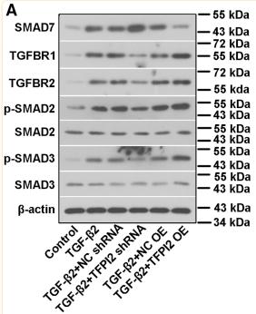
7). TFPI2 suppresses the interaction of TGF-β2 pathway regulators to promote endothelial-mesenchymal transition in diabetic nephropathy. Journal of Biological Chemistry, 2022 (PubMed: 35157852) [IF=4.8]

Application: WB    Species: Human    Sample: hRGECs

Figure 9 TFPI2 promotes TGF-β/Smad signaling activation. Human renal glomerular endothelial cells (hRGECs) were infected with adenovirus encoding shRNA targeting TFPI2 (TFPI2 shRNA) or overexpressing TFPI2 (TFPI2 OE), followed by stimulation of 5 ng/ml TGF-β2 for 48 h. A, the expression of SMAD7, TGFBR1, TGFBR2, SMAD2/3, and phospho-SMAD2/3 (p-SMAD2/3) was determined by Western blot. Semiquantitative analysis of (B) SMAD7, (C) TGFBR1, and (D) TGFBR2, as well as (E and F) the ratio of p-SMAD2/3 to SMAD2/3. G and H, immunofluorescent staining of SMAD2/3 in hRGECs. Yellow arrows indicated nuclear translocation of SMAD2/3. Data are shown as the mean ± SD (n = 3). ∗p < 0.05, ∗∗p < 0.01, ∗∗∗p < 0.001. TGF-β, transforming growth factor beta; TFP12, tissue factor pathway inhibitor 2.

8). Bone marrow mesenchymal stem cell transplantation alleviates radiation-induced myocardial fibrosis through inhibition of the TGF-β1/Smad2/3 signaling pathway in rabbit model. Regenerative Therapy, 2023 (PubMed: 37292187) [IF=4.3]

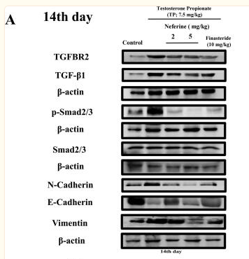
9). Neferine attenuates development of testosterone-induced benign prostatic hyperplasia in mice by regulating androgen and TGF-β/Smad signaling pathways. Saudi Pharmaceutical Journal, 2023 (PubMed: 37293563) [IF=4.1]

Application: WB    Species: Human    Sample: prostate tissues

Fig. 7 Effects of neferine (2, 5 mg/kg; 10–100 μM) and finasteride (10 mg/kg; 10–100 μM) on the expression of TGFBR2, TGF-β1, p-Smad2/3, Smad2/3, N-cadherin, E-cadherin and vimentin in prostate tissues at 14 days (A) and WPMY-1 cells at 24 h (B). (C-D) Data are presented as mean ± SEM. (*p < 0.05 compared with control, #p < 0.05 compared with TP or testosterone group).

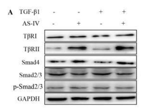
10). Astragaloside IV inhibits cell proliferation in vulvar squamous cell carcinoma through the TGF‐β/Smad signaling pathway. Dermatologic Therapy, 2019 (PubMed: 30536730) [IF=3.6]

Application: WB    Species: human    Sample: SW962 cells

Figure 3. |AS-Ⅳ reverses TGF-β/Smad signaling abnormalities and inhibits TGF-β1-mediated cell proliferation and anti-apoptosis in SW962 cells. SW962 cells were treated with TGF-β1 (10 ng/ml) alone or in combination with AS-Ⅳ (800 μg/ml) for 24 h and 48 h, respectively.Western blot assays evaluated the protein levels of TGF-βRI, TGF-βRII,Smad2/3, p-Smad2/3 and Smad4 (A,B).

Load more

Restrictive clause


Affinity Biosciences tests all products strictly. Citations are provided as a resource for additional applications that have not been validated by Affinity Biosciences. Please choose the appropriate format for each application and consult Materials and Methods sections for additional details about the use of any product in these publications.

For Research Use Only.
Not for use in diagnostic or therapeutic procedures. Not for resale. Not for distribution without written consent. Affinity Biosciences will not be held responsible for patent infringement or other violations that may occur with the use of our products. Affinity Biosciences, Affinity Biosciences Logo and all other trademarks are the property of Affinity Biosciences LTD.