Product: Cyclin D2 Antibody
Catalog: AF5410
Description: Rabbit polyclonal antibody to Cyclin D2
Application: WB IF/ICC
Reactivity: Human, Mouse, Rat
Prediction: Pig, Bovine, Horse, Sheep, Rabbit, Dog, Chicken, Xenopus
Mol.Wt.: 33 kDa; 33kD(Calculated).
Uniprot: P30279
RRID: AB_2837894

View similar products>>

   Size Price Inventory
 100ul $280 In stock
 200ul $350 In stock

Lead Time: Same day delivery

For pricing and ordering contact:
Local distributors

Product Info

Source:
Rabbit
Application:
WB 1:500-1:2000, IF/ICC 1:100-1:500
*The optimal dilutions should be determined by the end user.
*Tips:

WB: For western blot detection of denatured protein samples. IHC: For immunohistochemical detection of paraffin sections (IHC-p) or frozen sections (IHC-f) of tissue samples. IF/ICC: For immunofluorescence detection of cell samples. ELISA(peptide): For ELISA detection of antigenic peptide.

Reactivity:
Human,Mouse,Rat
Prediction:
Pig(100%), Bovine(100%), Horse(100%), Sheep(100%), Rabbit(100%), Dog(100%), Chicken(100%), Xenopus(100%)
Clonality:
Polyclonal
Specificity:
Cyclin D2 Antibody detects endogenous levels of total Cyclin D2.
RRID:
AB_2837894
Cite Format: Affinity Biosciences Cat# AF5410, RRID:AB_2837894.
Conjugate:
Unconjugated.
Purification:
The antiserum was purified by peptide affinity chromatography using SulfoLink™ Coupling Resin (Thermo Fisher Scientific).
Storage:
Rabbit IgG in phosphate buffered saline , pH 7.4, 150mM NaCl, 0.02% sodium azide and 50% glycerol. Store at -20 °C. Stable for 12 months from date of receipt.
Alias:

Fold/Unfold

CCND 2; ccnd2; CCND2_HUMAN; CyclinD2; G1/S specific cyclin D2; G1/S-specific cyclin-D2; KIAK0002; MGC102758; MPPH3;

Immunogens

Immunogen:
Uniprot:
Gene(ID):
Description:
Regulatory component of the cyclin D2-CDK4 (DC) complex that phosphorylates and inhibits members of the retinoblastoma (RB) protein family including RB1 and regulates the cell-cycle during G(1)/S transition. Phosphorylation of RB1 allows dissociation of the transcription factor E2F from the RB/E2F complex and the subsequent transcription of E2F target genes which are responsible for the progression through the G(1) phase.
Sequence:
MELLCHEVDPVRRAVRDRNLLRDDRVLQNLLTIEERYLPQCSYFKCVQKDIQPYMRRMVATWMLEVCEEQKCEEEVFPLAMNYLDRFLAGVPTPKSHLQLLGAVCMFLASKLKETSPLTAEKLCIYTDNSIKPQELLEWELVVLGKLKWNLAAVTPHDFIEHILRKLPQQREKLSLIRKHAQTFIALCATDFKFAMYPPSMIATGSVGAAICGLQQDEEVSSLTCDALTELLAKITNTDVDCLKACQEQIEAVLLNSLQQYRQDQRDGSKSEDELDQASTPTDVRDIDL

Predictions

Predictions:

Score>80(red) has high confidence and is suggested to be used for WB detection. *The prediction model is mainly based on the alignment of immunogen sequences, the results are for reference only, not as the basis of quality assurance.

Species
Results
Score
Pig
100
Horse
100
Bovine
100
Sheep
100
Dog
100
Xenopus
100
Chicken
100
Rabbit
100
Zebrafish
0
Model Confidence:
High(score>80) Medium(80>score>50) Low(score<50) No confidence

PTMs - P30279 As Substrate

Site PTM Type Enzyme
K45 Ubiquitination
K49 Ubiquitination
K113 Ubiquitination
K173 Ubiquitination
K244 Ubiquitination
S269 Phosphorylation
S271 Phosphorylation
S279 Phosphorylation
T280 Phosphorylation Q16539 (MAPK14) , P49841 (GSK3B)
T282 Phosphorylation

PTMs - P30279 As Enzyme

Substrate Site Source
P06400 (RB1) S780 Uniprot

Research Backgrounds

Function:

Regulatory component of the cyclin D2-CDK4 (DC) complex that phosphorylates and inhibits members of the retinoblastoma (RB) protein family including RB1 and regulates the cell-cycle during G(1)/S transition. Phosphorylation of RB1 allows dissociation of the transcription factor E2F from the RB/E2F complex and the subsequent transcription of E2F target genes which are responsible for the progression through the G(1) phase. Hypophosphorylates RB1 in early G(1) phase. Cyclin D-CDK4 complexes are major integrators of various mitogenenic and antimitogenic signals. Also substrate for SMAD3, phosphorylating SMAD3 in a cell-cycle-dependent manner and repressing its transcriptional activity. Component of the ternary complex, cyclin D2/CDK4/CDKN1B, required for nuclear translocation and activity of the cyclin D-CDK4 complex (By similarity).

PTMs:

Polyubiquitinated by the SCF(FBXL2) complex, leading to proteasomal degradation.

Subcellular Location:

Nucleus. Cytoplasm. Membrane.
Note: Cyclin D-CDK4 complexes accumulate at the nuclear membrane and are then translocated into the nucleus through interaction with KIP/CIP family members.

Cytoplasm.

Extracellular region or secreted Cytosol Plasma membrane Cytoskeleton Lysosome Endosome Peroxisome ER Golgi apparatus Nucleus Mitochondrion Manual annotation Automatic computational assertionSubcellular location
Subunit Structure:

Interacts with either CDK4 or CDK6 protein kinase to form a serine/threonine kinase holoenzyme complex. The cyclin subunit imparts substrate specificity to the complex. Component of the ternary complex cyclin D/CDK4/CDKN1B required for nuclear translocation and modulation of CDK4-mediated kinase activity.

Family&Domains:

Belongs to the cyclin family. Cyclin D subfamily.

Research Fields

· Cellular Processes > Cell growth and death > Cell cycle.   (View pathway)

· Cellular Processes > Cell growth and death > p53 signaling pathway.   (View pathway)

· Cellular Processes > Cell growth and death > Cellular senescence.   (View pathway)

· Cellular Processes > Cellular community - eukaryotes > Focal adhesion.   (View pathway)

· Environmental Information Processing > Signal transduction > FoxO signaling pathway.   (View pathway)

· Environmental Information Processing > Signal transduction > PI3K-Akt signaling pathway.   (View pathway)

· Environmental Information Processing > Signal transduction > Wnt signaling pathway.   (View pathway)

· Environmental Information Processing > Signal transduction > Hedgehog signaling pathway.   (View pathway)

· Environmental Information Processing > Signal transduction > Hippo signaling pathway.   (View pathway)

· Environmental Information Processing > Signal transduction > Jak-STAT signaling pathway.   (View pathway)

· Human Diseases > Infectious diseases: Viral > Measles.

· Human Diseases > Infectious diseases: Viral > Human papillomavirus infection.

· Human Diseases > Infectious diseases: Viral > HTLV-I infection.

· Human Diseases > Cancers: Overview > Pathways in cancer.   (View pathway)

· Human Diseases > Cancers: Overview > Transcriptional misregulation in cancer.

· Human Diseases > Cancers: Overview > Viral carcinogenesis.

· Human Diseases > Cancers: Overview > MicroRNAs in cancer.

· Organismal Systems > Endocrine system > Prolactin signaling pathway.   (View pathway)

References

1). Apigenin ameliorates imiquimod-induced psoriasis in C57BL/6J mice by inactivating STAT3 and NF-κB. Food Science and Human Wellness, 2024 [IF=7.0]

Application: WB    Species: Mouse    Sample: skin tissues

Fig. 6. API inhibits IMQ-induced cell proliferation in vivo. A, skin sections were conducted immunofluorescent staining to determine Ki-67 protein expression with quantitation of MFI (n = 5). B, protein expression of cyclinD2, ERK1/2, p-ERK1/2 in skin tissues was determined by Western blot with quantitative analysis of band density (n = 5). C, skin sections were conducted immunofluorescent staining to determine cyclinD2 protein expression with quantitation of MFI (n = 5). The red arrows indicate the positive regions of Ki-67 or cyclinD2. Scale bar: 200 μm; *** P < 0.001 vs. Ctrl group; ### P < 0.001 vs. IMQ treated group.

Application: IF/ICC    Species: Mouse    Sample: skin tissues

Fig. 6. API inhibits IMQ-induced cell proliferation in vivo. A, skin sections were conducted immunofluorescent staining to determine Ki-67 protein expression with quantitation of MFI (n = 5). B, protein expression of cyclinD2, ERK1/2, p-ERK1/2 in skin tissues was determined by Western blot with quantitative analysis of band density (n = 5). C, skin sections were conducted immunofluorescent staining to determine cyclinD2 protein expression with quantitation of MFI (n = 5). The red arrows indicate the positive regions of Ki-67 or cyclinD2. Scale bar: 200 μm; *** P < 0.001 vs. Ctrl group; ### P < 0.001 vs. IMQ treated group.

2). Prostaglandin E2 accelerated recovery of chemotherapy-induced intestinal damage by increasing expression of cyclin D. EXPERIMENTAL CELL RESEARCH, 2020 (PubMed: 31917964) [IF=3.7]

3). Transcription Factor SATB2 Regulates Skeletal Muscle Cell Proliferation and Migration via HDAC4 in Pigs. Genes, 2024 (PubMed: 38254955) [IF=3.5]

4). Regulation of myo-miR-24-3p on the Myogenesis and Fiber Type Transformation of Skeletal Muscle. Genes, 2024 [IF=3.5]

Application: WB    Species: Mouse    Sample:

Figure 2. Effects of miR-24-3p on C2C12 myoblast proliferation. (A–D) The proliferation of C2C12 myoblasts was analyzed after overexpression, and the knockdown of miR-24-3p was determined by a CCK-8 assay and EdU staining. (E,F) qRT-PCR was used to analyze proliferation marker (KI67, PCNA, CDK4, Cyclin D1, and Cyclin E1) expression after miR-24-3p overexpression and knockdown. (G,H) Western blot was used to analyze proliferation marker (KI67, PCNA, and Cyclin D1) expression after miR-24-3p overexpression and knockdown. The data were presented as mean ± S.E.M. and analyzed for statistical differences between groups using unpaired two-tailed t-tests.

5). Long non-coding RNA NEAT1 regulates E2F3 expression by competitively binding to miR-377 in non-small cell lung cancer. Oncology Letters, 2017 (PubMed: 29085511) [IF=2.9]

Application: WB    Species: human    Sample: A549 cells

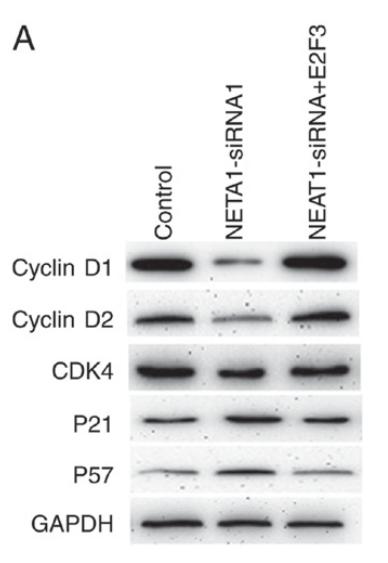
Figure 4.| E2F3 promotes NSCLC proliferation through regulating the cell cycle in NSCLC. The expression of cell cycle‑associated proteins in A549 were detected by western blot (A). E2F3 can rescue the cell growth inhibition induced by decreased NEAT1 (B). P<0.05.

6). Hsa-miR-326 targets CCND1 and inhibits non-small cell lung cancer development. Oncotarget, 2016 (PubMed: 26840018)

Application: WB    Species:    Sample:

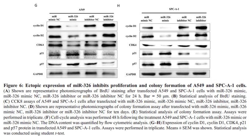
(G–H) Expression of cyclin D1, cyclin D1, CDK4, p21 and p57 protein in transfected A549 and SPC-A-1 cells. Assays were performed in triplicate. Means ± SEM was shown. Statistical analysis was conducted using student t-test.

7). Wnt/PCP-YAP-BIRC2 axis maintains cartilage stem/progenitor cell homeostasis in osteoarthritis. Research Square, 2022

Application: WB    Species: Human    Sample: CSPCs

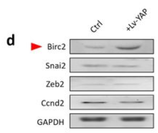
Figure 7. Transcriptomic analysis. a KEGG and GO enrichment analysis of DEGs. b The detailed DEGs that enriched in cell cycle, cellular senescence, autophagy, apoptosis, Hippo signaling pathway, and TNF signaling pathways. c The direct downstream target genes of YAP. d The protein level of Birc2, Snai2, Zenb2, and Ccnd2 in CSPCs.

Restrictive clause


Affinity Biosciences tests all products strictly. Citations are provided as a resource for additional applications that have not been validated by Affinity Biosciences. Please choose the appropriate format for each application and consult Materials and Methods sections for additional details about the use of any product in these publications.

For Research Use Only.
Not for use in diagnostic or therapeutic procedures. Not for resale. Not for distribution without written consent. Affinity Biosciences will not be held responsible for patent infringement or other violations that may occur with the use of our products. Affinity Biosciences, Affinity Biosciences Logo and all other trademarks are the property of Affinity Biosciences LTD.