Product: GRO alpha Antibody
Catalog: AF5403
Description: Rabbit polyclonal antibody to GRO alpha
Application: WB IHC IF/ICC
Cited expt.: WB, IHC, IF/ICC
Reactivity: Human, Mouse, Rat
Mol.Wt.: 11 kDa; 11kD(Calculated).
Uniprot: P09341
RRID: AB_2837887

View similar products>>

   Size Price Inventory
 100ul $280 In stock
 200ul $350 In stock

Lead Time: Same day delivery

For pricing and ordering contact:
Local distributors

Product Info

Source:
Rabbit
Application:
WB 1:500-1:2000, IHC 1:50-1:200, IF/ICC 1:100-1:500
*The optimal dilutions should be determined by the end user.
*Tips:

WB: For western blot detection of denatured protein samples. IHC: For immunohistochemical detection of paraffin sections (IHC-p) or frozen sections (IHC-f) of tissue samples. IF/ICC: For immunofluorescence detection of cell samples. ELISA(peptide): For ELISA detection of antigenic peptide.

Reactivity:
Human,Mouse,Rat
Clonality:
Polyclonal
Specificity:
GRO alpha Antibody detects endogenous levels of total GRO alpha.
RRID:
AB_2837887
Cite Format: Affinity Biosciences Cat# AF5403, RRID:AB_2837887.
Conjugate:
Unconjugated.
Purification:
The antiserum was purified by peptide affinity chromatography using SulfoLink™ Coupling Resin (Thermo Fisher Scientific).
Storage:
Rabbit IgG in phosphate buffered saline , pH 7.4, 150mM NaCl, 0.02% sodium azide and 50% glycerol. Store at -20 °C. Stable for 12 months from date of receipt.
Alias:

Fold/Unfold

C-X-C motif chemokine 1; Chemokine (C-X-C motif) ligand 1 (melanoma growth stimulating activity, alpha); chemokine (C-X-C motif) ligand 1; CINC-1; CXCL1; Cytokine-induced neutrophil chemoattractant 1; Fibroblast secretory protein; Fsp; Gro 1; Gro A; Gro; GRO protein, alpha; GRO-alpha(1-73); GRO-alpha(6-73); Gro1; GRO1 oncogene (melanoma growth stimulating activity, alpha); GRO1 oncogene (melanoma growth-stimulating activity); Gro1 oncogene; GROa; GROA_HUMAN; Growth-regulated alpha protein; KC; KC chemokine, mouse, homolog of; melanoma growth stimulatory activity alpha; Melanoma growth stimulatory activity; Melanoma growth stimulatory activity, alpha; MGSA alpha; MGSA; MGSA-a; N51; NAP-3; NAP3; Neutrophil-activating protein 3; Platelet-derived growth factor-inducible protein KC; Scyb 1; Scyb1; Secretory protein N51; Small inducible cytokine subfamily B, member 1;

Immunogens

Immunogen:

A synthesized peptide derived from human GRO alpha, corresponding to a region within the internal amino acids.

Uniprot:
Gene(ID):
Description:
Has chemotactic activity for neutrophils. May play a role in inflammation and exerts its effects on endothelial cells in an autocrine fashion. In vitro, the processed forms GRO-alpha(4-73), GRO-alpha(5-73) and GRO-alpha(6-73) show a 30-fold higher chemotactic activity.
Sequence:
MARAALSAAPSNPRLLRVALLLLLLVAAGRRAAGASVATELRCQCLQTLQGIHPKNIQSVNVKSPGPHCAQTEVIATLKNGRKACLNPASPIVKKIIEKMLNSDKSN

Research Backgrounds

Function:

Has chemotactic activity for neutrophils. May play a role in inflammation and exerts its effects on endothelial cells in an autocrine fashion. In vitro, the processed forms GRO-alpha(4-73), GRO-alpha(5-73) and GRO-alpha(6-73) show a 30-fold higher chemotactic activity.

PTMs:

N-terminal processed forms GRO-alpha(4-73), GRO-alpha(5-73) and GRO-alpha(6-73) are produced by proteolytic cleavage after secretion from peripheral blood monocytes.

Subcellular Location:

Secreted.

Extracellular region or secreted Cytosol Plasma membrane Cytoskeleton Lysosome Endosome Peroxisome ER Golgi apparatus Nucleus Mitochondrion Manual annotation Automatic computational assertionSubcellular location
Family&Domains:

Belongs to the intercrine alpha (chemokine CxC) family.

Research Fields

· Environmental Information Processing > Signaling molecules and interaction > Cytokine-cytokine receptor interaction.   (View pathway)

· Environmental Information Processing > Signal transduction > TNF signaling pathway.   (View pathway)

· Human Diseases > Infectious diseases: Bacterial > Epithelial cell signaling in Helicobacter pylori infection.

· Human Diseases > Infectious diseases: Bacterial > Salmonella infection.

· Human Diseases > Infectious diseases: Bacterial > Legionellosis.

· Human Diseases > Infectious diseases: Parasitic > Amoebiasis.

· Human Diseases > Immune diseases > Rheumatoid arthritis.

· Organismal Systems > Immune system > Chemokine signaling pathway.   (View pathway)

· Organismal Systems > Immune system > NOD-like receptor signaling pathway.   (View pathway)

· Organismal Systems > Immune system > IL-17 signaling pathway.   (View pathway)

References

1). QDPR deficiency drives immune suppression in pancreatic cancer. Cell metabolism, 2024 (PubMed: 38642552) [IF=27.7]

2). Baohuoside I chemosensitises breast cancer to paclitaxel by suppressing extracellular vesicle/CXCL1 signal released from apoptotic cells. Journal of extracellular vesicles, 2024 (PubMed: 39051750) [IF=16.0]

Application: IF/ICC    Species: Mouse    Sample: Raw264.7 cells

FIGURE 4 BHS inhibits the CXCL1 cargo in EV-Apo and leads to decreased M2 polarization of Raw264.7 macrophages. (a) M2 polarization changes of Raw264.7 macrophages after treatments as indicated for 48 h were detected by an F4/80+/CD206+ population analysis. EVs were used at a concentration of 100 µg/mL. n = 3. (b) EV-Apo and EV-ApoBHS uptake by Raw264.7 macrophages quantified by flow cytometry. EVs were labelled with PKH67 (green). n = 3. (c) EV-Apo and EV-ApoBHS uptake by Raw264.7 macrophages visualized by immunofluorescence labelling. EVs were labelled with PKH67 (green), and Raw264.7 macrophages were labelled with Actin-Red (red) and DAPI (blue). (d) CXCL1 concentrations in EV-Apo and EV-ApoBHS detected by ELISA assay. n = 3. (e) Western blotting analysis of PD-L1 expression in Raw264.7 cells when treated as indicated for 48 h. CXCL1-NA concentration, 5 µg/mL. Recombinant CXCL1 (rCXCL1), 30 ng/mL. n = 3. (f) Immunofluorescence analysis of PD-L1 expression in Raw264.7 cells when treated as indicated for 48 h. n = 3. (g) QPCR analysis of the mRNA expression levels (left) and promoter activity of PD-L1 (right) in Raw264.7 cells when treated as indicated for 48 h. n = 3. **p < 0.01.

3). Chemotherapy-elicited extracellular vesicle CXCL1 from dying cells promotes triple-negative breast cancer metastasis by activating TAM/PD-L1 signaling. Journal of experimental & clinical cancer research : CR, 2024 (PubMed: 38654356) [IF=11.3]

4). XIAOPI formula inhibits the pre-metastatic niche formation in breast cancer via suppressing TAMs/CXCL1 signaling. Cell Communication and Signaling, 2020 (PubMed: 32213179) [IF=8.4]

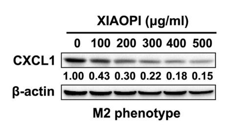
Application: WB    Species: mouse    Sample: M2 phenotype RAW264.7 cells

Fig. 1| XIAOPI formula suppresses the polarization of M2 macrophages and CXCL1 expression.e XIAOPI formula dose-dependently inhibited expression of CXCL1 in M2 phenotype RAW264.7 cells. (The results were obtained from triplicate experiments and were represented as mean values ± SD.,*P < 0.05, **P < 0.01 as compared with control, ##P < 0.01, comparison between three time points of 24 h, 48 h, and 72 h)

5). Aiduqing formula inhibits breast cancer metastasis by suppressing TAM/CXCL1-induced Treg differentiation and infiltration. Cell Communication and Signaling, 2021 (PubMed: 34461944) [IF=8.4]

Application: WB    Species: Mice    Sample: breast tumor tissues

Fig. 2 ADQ inhibits M2 phenotype polarization and CXCL1 secretion by TAMs in vitro and in vivo. a Flow cytometry assay was conducted to investigate the effect of ADQ treatment (0.7 or 1.4 g/kg/day) on TAM infiltration and polarization within the TME of breast tumors. b The expression levels of CD206 (red) and CXCL1 (green) in breast tumor tissues were detected by the tissue immunofluorescence assay. Scale bar = 5 μm. c 40 ng/ml IL-4 and IL-13 were used to induce the transformation of Raw264.7 macrophages into M2-like macrophages (TAMs). Their phenotype validation was conducted by flow cytometry. d The cell viability changes of TAMs when treated with ADQ (20–200 μg/ml) for 12–48 h were detected using the CCK-8 assay. e–f The phenotype changes of Raw264.7-derived TAMs when treated with 20–200 μg/ml ADQ for 24 h were detected by flow cytometry. G–i Raw264.7-derived TAMs were treated with ADQ (20–200 μg/ml) for 24 h. The protein expression, secretion as well as mRNA transcription levels of CXCL1 in Raw264.7-derived TAMs were detected by Western blot, ELISA, and qPCR assays, respectively. j Raw264.7-derived TAMs were treated with ADQ (20–200 μg/Ml) for 24 h. The promoter activity changes of CXCL1 gene in Raw264.7-derived TAMs were detected by the double luciferase reporter gene assay. N = 3. *p < 0.05. **p < 0.01

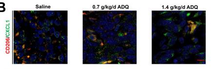
Application: IF/ICC    Species: Mice    Sample: breast tumor tissues

Fig. 2 ADQ inhibits M2 phenotype polarization and CXCL1 secretion by TAMs in vitro and in vivo. a Flow cytometry assay was conducted to investigate the effect of ADQ treatment (0.7 or 1.4 g/kg/day) on TAM infiltration and polarization within the TME of breast tumors. b The expression levels of CD206 (red) and CXCL1 (green) in breast tumor tissues were detected by the tissue immunofluorescence assay. Scale bar = 5 μm. c 40 ng/ml IL-4 and IL-13 were used to induce the transformation of Raw264.7 macrophages into M2-like macrophages (TAMs). Their phenotype validation was conducted by flow cytometry. d The cell viability changes of TAMs when treated with ADQ (20–200 μg/ml) for 12–48 h were detected using the CCK-8 assay. e–f The phenotype changes of Raw264.7-derived TAMs when treated with 20–200 μg/ml ADQ for 24 h were detected by flow cytometry. G–i Raw264.7-derived TAMs were treated with ADQ (20–200 μg/ml) for 24 h. The protein expression, secretion as well as mRNA transcription levels of CXCL1 in Raw264.7-derived TAMs were detected by Western blot, ELISA, and qPCR assays, respectively. j Raw264.7-derived TAMs were treated with ADQ (20–200 μg/Ml) for 24 h. The promoter activity changes of CXCL1 gene in Raw264.7-derived TAMs were detected by the double luciferase reporter gene assay. N = 3. *p < 0.05. **p < 0.01

6). Tumor-associated macrophages/C-X-C motif chemokine ligand 1 promotes breast cancer autophagy-mediated chemoresistance via IGF1R/STAT3/HMGB1 signaling. Cell death & disease, 2024 (PubMed: 39394189) [IF=8.1]

Application: IF/ICC    Species: Mouse    Sample: breast cancer cells

Fig. 2: TAMs/CXCL1 signaling promoted autophagy-mediated chemoresistance in breast cancer cells. A Cell counting assay was conducted to examine CXCL1 (20 ng/mL) and its combination with several chemotherapeutic drugs (Paclitaxel at 50 nM, Epirubicin at 1 μg/mL, Doxorubicin at 1 μg/mL, and 5-FU 5 μg/mL) on cell growth of MDA-MB-231 and MCF-7 cells after 48 h treatment. B Western blotting assay was applied to detect the expressions of ABCG2, SQSTM1/p62 and LC3 levels in the presence of CXCL1 ranging from 0 to 50 ng/mL after 24 h treatment. C Cell counting assay was used to detect the effect of CXCL1 neutralizing antibodies (20 ng/mL) on the chemosensitivity of MDA-MB-231 and MCF-7 in response to TAMs-CM. D Western blotting assay was performed on MDA-MB-231 and MCF-7 cells to determine the effects of TAMs-CM containing a CXCL1 neutralizing antibody (20 ng/mL) on ABCG2, SQSTM1/p62 and LC3 expression levels. E The mRFP-GFP-LC3 reporter assay was carried out to verify autophagic alternations in the presence of either CXCL1 (10–20 ng/mL) or TAMs-CM containing CXCL1 neutralizing antibodies (20 ng/mL) (n = 5, scale bars indicate 20 μm). Values are presented as Mean ± SD, n = 3 unless otherwise indicated, *P 

7). Blocking the CXCL1-CXCR2 axis enhances the effects of doxorubicin in HCC by remodelling the tumour microenvironment via the NF-κB/IL-1β/CXCL1 signalling pathway. Cell Death Discovery, 2023 (PubMed: 37037815) [IF=7.0]

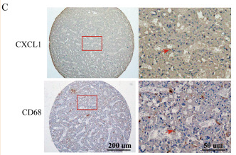
Application: IHC    Species: Human    Sample:

Fig. 1 High CXCL1 expression predicts a poor prognosis in HCC, and is positively related to macrophage enrichment. A Overall survival plot of CXCL1 expression in HCC patients was analysed through the GEPIA database (n = 364). B The expression of CXCL1 in tumour tissues and adjacent nontumor tissues was measured by IHC. Arrows indicate positive staining. Scale bar, 100 μm (up) and 50 μm (down). C–E CD68 and CXCL1 staining and representative images of the same tissue. D CXCL1, CD68, and CD206 staining in the same tissues. Arrows indicate positive staining. Scale bar, 200 μm. E The relationship between CXCL1 expression and the CD206+/CD68+ cell ratio. Data are expressed as the mean ± SEM. **P 

8). Total flavonoids from sea buckthorn ameliorates lipopolysaccharide/cigarette smoke-induced airway inflammation. PHYTOTHERAPY RESEARCH, 2019 (PubMed: 31209984) [IF=6.1]

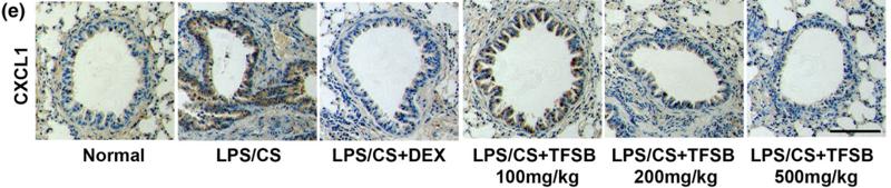
Application: IHC    Species: mouse    Sample: lung

FIGURE 7 |Total flavonoids from sea buckthorn fruit (TFSB) treatment suppressed the expression of cytokines, chemokines, and MUC5AC in bronchial tissues in lipopolysaccharide/cigarette smoke extract (LPS/CS)‐exposed mice. After LPS/CS exposure, mice were treated with TFSB (100, 200, and 500 mg/kg) or dexamethasone (0.25 mg/kg). After treatment of TFSB, lung tissues were used to detect the expression of (a) IL‐1β,(b) IL‐6, (c) COX2, (d) MUC5AC, and (e) CXCL1 through immunohistochemical method. (f) The quantitative results of the expression of IL‐1β, IL‐6,COX2, MUC5AC, and CXCL1 were shown, respectively. Bar = 50 μm. *p < .05, **p < .01, ***p < .001, ****p < .0001, versus LPS/CS alone, n = 4

9). XIAOPI formula inhibits breast cancer stem cells via suppressing TAMs/CXCL1 pathway. Frontiers in Pharmacology, 2019 (PubMed: 31803057) [IF=5.6]

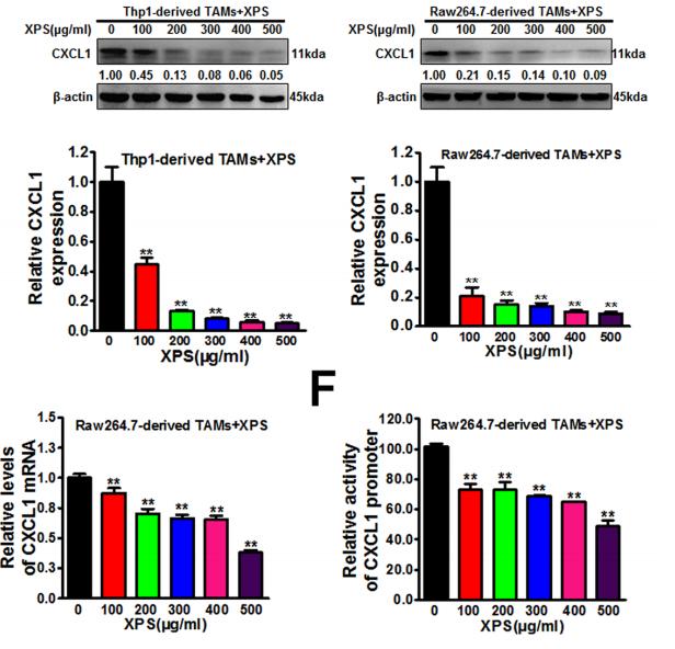
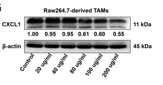
Application: WB    Species: human and mouse    Sample: TAMs

FIGURE 3 | XIAOPI formula (XPS) inhibits M2 phenotype polarization, C-X-C motif chemokine ligand 1 (CXCL1) expression and secretion of tumor-associated macrophages (TAMs).(D–E) Western blot and QPCR results further verified that XIAOPI formula treatment for 48 h could dramatically inhibit CXCL1 protein expression levels (D) and CXCL1 mRNA transcription levels (E) in both human and mouse TAMs. (F) Double luciferase reporter gene assay suggested that XPS treatment for 48 h could suppress the promoter activity of CXCL1 gene in Raw264.7-derived TAMs. All values are presented as the mean ± SD, n = 3, **P < 0.05.

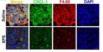
Application: IF/ICC    Species: mouse    Sample: macrophages

FIGURE 5 | XIAOPI formula (XPS) suppresses breast tumor growth and breast cancer stem cells (CSCs) activity in vivo. (D–E) XPS administration significantly decreased the infiltration degree of macrophages as well as their M2 phenotype polarization and C-X-C motif chemokine ligand 1 (CXCL1) expression in the 4T1-Luc xenografts in vivo. n = 3.

10). The Role of Autophagy in the Innate Immune Response to Fungal Keratitis Caused by Aspergillus fumigatus Infection. INVESTIGATIVE OPHTHALMOLOGY & VISUAL SCIENCE, 2020 (PubMed: 32084267) [IF=5.0]

Application: WB    Species: mouse    Sample: corneas

FIGURE 4.| Effect of autophagy on PMN recruitment and morphology. (A-C) The mRNA levels of CXCL-1 in corneas of C57BL/6 mice after 3-MA, CQ, or rapamycin treatment at 3 days p.i.; (D-F) The protein level of CXCL-1 in corneas of C57BL/6 mice after 3-MA, CQ, or rapamycin treatment at 3 days p.

Load more

Restrictive clause


Affinity Biosciences tests all products strictly. Citations are provided as a resource for additional applications that have not been validated by Affinity Biosciences. Please choose the appropriate format for each application and consult Materials and Methods sections for additional details about the use of any product in these publications.

For Research Use Only.
Not for use in diagnostic or therapeutic procedures. Not for resale. Not for distribution without written consent. Affinity Biosciences will not be held responsible for patent infringement or other violations that may occur with the use of our products. Affinity Biosciences, Affinity Biosciences Logo and all other trademarks are the property of Affinity Biosciences LTD.