
, diluted 1/600 was used as secondary antibody.")
 detected by western blotting. GAPDH used as a housekeeping control. Nonirradiated kidney tissues (CTL) used as controls. Expression of LC3-I and LC3-II quantitated by ImageJ software and ratio of LC3-II and LC3-I expression presented (right panel). *p < 0.05 vs CTL; ***p < 0.001 vs CTL. CTL nonirradiated renal tissues, GAPDH glyceraldehyde 3-phosphate dehydrogenase, IHC immunohistochemistry, pmTOR pshosphorylated mammalian target of rapamycin, TBI total body irradiation")
 by immunofluorescence (200×) in different groups was displayed with DAPI, LC3, and MERGE, respectively. Relative fluorescence intensity of LC3")
BECN1 expression was assessed using immunohistochemistry and photographed at 9100 and 9200 magnifications. (B) Representative western blotting results of BECN1, LC3-I, LC3-II, and b‑actin in the corpus cavernosum of the three groups. Expression levels of BECN1 (C) and LC3-II (D), with b‑actin as the loading control, in the corpus cavernosum of the three groups are presented as bar graphs")
 The expression of the autophagy markers P62 and LC3 were determined by western blot analysis in PC12 cells; (B) PC12 cells morphology changed and (C) neurite-bearing cells counted after treatment with M. smegmatis culture supernatant (Sup), the autophagy inducer rapamycin (1µM, Rap), the autophagy inhibitor 3-MA (10 mM, 3-MA), Sup + Rap, and Sup + 3-MA. (20×) for 48 h. Quantitative results were presented as means ± S.D. for 3 replicates. *P < 0.05, **P < 0.01, ***P < 0.001 vs. control group, n = 3.")
 The expression and quantification of HMGB1, RAGE, LC3, and Beclin1 in the A498 cell line were analyzed by Western blot analysis.")
 or p62 siRNA (B),cells were then pre-treated with Andro (20 µM) for 1 h followed by stimulation with Aβ (10 µM)for an additional 24 h, Beclin-1, LC3A/BII, Nrf2, and p21 protein expression levels were evaluated by Western blot analysis. # p < 0.05 versus the Andro + Aβ1–42 group were considered statistically significant differences.")
 Mitochondria dynamics in control and BHPF-exposed oocytes. HSP60, red; α-Tubulin, green; DNA, blue. Arrow, mitochondria aggregation. Scale bar, 10 μm. (B) Relative mtDNA copy numbers in control and BHPF-exposed oocytes. (C) Immunoblot analysis to assess the effects of BHPF on the expression of PINK1, Beclin1, LC3B, TOMM20,and TIMM17A proteins.")


 HUVECs were pretreated for 2h with 10 mM 3-MA, 20μM Z-VAD-FMK, or 10
mM NAC, and then treated with 5μg/ml HMME and light dose of 2 J/cm2
. Cell viability
was detected by CCK8 assay 24h after HMME-PDT.
** p<0.01. (B) Autophagosome
formation was observed in HUVECs 6h after HMME-PDT. Scale bar= 25μm. (C) The
number of early autophagosomes (yellow puncta) per cell was calculated, and more
than 10 cells were quantified for each condition.
** p<0.01. (D) TUNEL staining of
HUVECs was performed 24h after HMME-PDT. Scale bar=100μm. (E) Apoptosis ratio
Journal Pre-proof
was calculated according to the TUNEL staining of HUVECs.
** p<0.01. (F) Western
blotting analysis was used to measure the expression levels of Cytochrome C, Cleaved
Caspase 3, PARP, Cleaved PARP, Beclin 1, LC3, and β-actin in HUVECs 24h after
HMME-PDT")
 The effect of PA on LC3-II and p62 protein levels in hippocampus were in-vestigated by western blot analysis.")

; mitochondrial function markers(B); BDNF signaling-related proteins (C); and representative confocal microscopy images of BDNF staining (green, magnification: ×40,scale Bar 50 μm) (D: a, 8 M–SED; b, 26 M–SED;c, 18 M–MICT; d, 8 M–MICT); and serum BDNF levels (E); and correlation analysis (F).")
. Bar = 50 µm. *P < 0.05 compared to the sham group. ##P < 0.01 compared to the 5/6 Nx model group")

.Bar = 50 µm. *P < 0.05 compared to the sham group. ##P < 0.01 compared to the 5/6 Nx model group. C-E, Representative immune blots of key proteins involved in muscle atrophy and proteins associated with the AMPK/SKP2/CARM1 signalling pathway")
.Corresponding images and quantifcations were shown.")
 and FTO knockdown A2780 cells (B). Western blot analysis of ATG-5 and corresponding band grayscale values in FTO
overexpression SKOV-3 cells (C) and FTO knockdown A2780 cells (D). Western blot analysis of P62 and corresponding band grayscale values in FTO overexpression
SKOV-3 cells (E) and FTO knockdown A2780 cells (F). Western blot analysis of p-AKT and corresponding band grayscale values in FTO overexpression SKOV-3 cells
(H) and FTO knockdown A2780 cells (I). Data are shown as Mean ± SD (n = 3). *P < 0.05, **P < 0.01, ***P < 0.001, ****P < 0.0001. OE: overexpression;
si: small interference RNA.")
 and the ratio of LC3‐II/I and
the relative expression of Beclin‐1 normalized
by GAPDH (B) (*P < .05 vs control group, #
P < .05 vs hyperoxia group). GAPDH,
glyceraldehyde 3‐phosphate dehydrogenase;
RF/6A, rhesus macaque choroid‐retinal
endothelial cells")
 Primary skeletal muscle cells were isolated from the mice of different groups including control, DM, DM+Ex, DM+GLPG0974 and DM+Ex+GLPG0974. The expression of autophagy-related proteins p62, LC3, Beclin1, p-mTOR and mTOR in different groups was analyzed by western blot; (B) quantitative analysis of the ratio of LC3II/LC3I, p-mTOR, Beclin 1 and p62 in the left panel (A); (C) primary skeletal muscle cells were isolated from wild type mice. The insulin resistance skeletal muscle cells were established by the addition of palmitate (PA). Autophagy flux was blocked by the administration of chloroquine. The p-IRSTyr612, IRS, p-AKTSer473 and AKT expression in different treatment groups were determined by western blot; (D) glucose uptake capacity of cells in each group measured by fluorescently labeled 2-deoxyglucose (2-NBDG). * p < 0.05. ** p < 0.01.")
 Dual-Luciferase reporter assay was conducted to verify the targeted relationship between miR-19b-3p and 3’-UTR of PTEN in HEK293T cells. Western blot for (B) PTEN, (C) p-Akt, (D)p-mTOR, (E) LC3II/I, (F) p62 and (G) Beclin-1 in BMSC-derived osteoblasts after transduction.")
 MDC staining was conducted to detect autophagy in Du145 and VCaP cells
after 1 μm GW9662 and 50 μm Hispidulin treatment for 48 h. The relative protein levels of (b) LC3II/I, (c) p-mTOR, (d) p-S6K1, and (e) PPARγ in Du145 and VCaP cells
after 1 μm GW9662 and 50 μm Hispidulin treatment for 48 h. (f) Electrophoretic mobility shift assay for PPARγ in Du145 and VCaP cells after 1 μm GW9662 and 50
μm Hispidulin treatment for 48 h. Data were represented as mean ± SD at least 3 independent experiments and analyzed by one-way analysis of variance (ANOVA)
followed by Tukey’s multiple comparison test. *
P < .05 vs blank or indicated group.")
 RAW264.7 cells were infected with lentivirus carrying SDC4 overexpression
plasmid or sh-SDC4, and induced by RANKL. Thereafter, the protein levels of LC3 and beclin-1 were detected by western blot. (C) RAW264.7 cells infected with
lentivirus carrying SDC4 overexpression plasmid were treated with RANKL and 3-MA. Thereafter, the osteoclast differentiation was assessed by TRAP staining. Scale
bar = 200 μm. (D) The mRNA levels of c-Fos, NFATc1 and cathepsin K were detected by qRT-PCR. * p < 0.05, ** p < 0.01, *** p < 0.001.")
 The average Basso‐Beattie‐Bresnahan (BBB) scores at 0‐, 1‐, 4‐, 7‐, 14‐ and 21‐day post‐SCI. (B) RT‐PCR was used to detect miR‐19b‐3p expression in the femurs at day 1 and day 21 post‐SCI surgery. Western blot for (C) LC3II/I, (D) p62 and (E) Beclin‐1 in the femurs at day 1 and day 21 post‐SCI surgery. Data were presented as means ± SD, and the statistical difference between two groups was determined by Student's t test. # P < .05 vs. Con. All the in vivo experiments were independently repeated for six times (n = 6)")

 Representative images of Beclin1, LC3 and P62 (immunohistochemical staining; magnification, ×400). Quantitative analysis of (B) Beclin1, (C) LC3 and (D) P62 were normalized to the normal control group. (E) Protein expression levels of Beclin1, LC3 and P62 in the liver tissues were detected via western blotting. Semi-quantitative analysis of (F) Beclin1, (G) LC3 and (H) P62 was normalised to β-actin. Data are presented as the mean ± SD, n=3. *P<0.05, ***P<0.001 vs. control group; #P<0.05, ##P<0.01, ###P<0.001 vs. model group. Con, Control; Mod, Model; AS-IV, Astragaloside IV (80 mg/kg); Met, Metformin (200 mg/kg).")
 Representative images of Beclin1, LC3 and P62 (immunohistochemical staining; magnification, ×400). Quantitative analysis of (B) Beclin1, (C) LC3 and (D) P62 were normalized to the normal control group. (E) Protein expression levels of Beclin1, LC3 and P62 in the liver tissues were detected via western blotting. Semi-quantitative analysis of (F) Beclin1, (G) LC3 and (H) P62 was normalised to β-actin. Data are presented as the mean ± SD, n=3. *P<0.05, ***P<0.001 vs. control group; #P<0.05, ##P<0.01, ###P<0.001 vs. model group. Con, Control; Mod, Model; AS-IV, Astragaloside IV (80 mg/kg); Met, Metformin (200 mg/kg).")
2D3 repairs the injured autophagy in H/R-treated HTR-8 cells (A) Representative transmission electron microscopy micrographs of autophagosomes (black arrow) and autolysosomes (red arrow) in each group (Scale bar, 1 μm). (B) Representative images of autophagosomes (yellow; GFP+ RFP+
puncta) and autolysosomes (red; GFP− RFP+ puncta) in each group (Scale bar, 20 μm). (C, D) The autophagy relevant proteins (P62, Beclin1, and LC3II/I) in each
group were detected by Western blot analysis. This experiment was repeated three times with similar results. Data are presented as the mean value ± standard
deviation. Statistical significance are determined by one-way ANOVA followed by Tukey’s post hoc test. ***P < 0.001, ****P < 0.0001 versus Control; ###P < 0.001
versus H/R. H/R, hypoxia/reoxygenation; RFP, red fluorescent protein; GFP, green fluorescent protein; LC3, light chain 3; GAPDH, glyceraldehyde-3-phosphate
dehydrogenase.")
, LC3- II (b), Beclin1 (c), determined by western blotting. Autophagosomes analysis, determined by TEM (d) (The red points to the autophagosome, N indicates the nucleus, Scale Bar=2 μm), values indicate mean ± SD. (n = 6–7) *P < 0.05, compared with the CON group, #P < 0.05, compared with the IR group.")
 Expression levels of beclin 1, LC3 II, and p62 proteins in the LV of mice. n = 6, ∗P < 0.05, ∗∗P < 0.01 vs. control group; #P < 0.05, ##P < 0.01 vs. Ang II group. (b) Representative images of the ultrastructure of H9c2 cells showing the changes of autophagy under a transmission electron microscope. Scale bar in (i), 2 μm and magnification, ×10000; in (ii), 1 μm, ×20000. Yellow arrow refers to the autophagosome, which contains dense electronic content such as autolysosomes or mitochondria. (c) Expression of beclin 1 and LC3 mRNA in H9c2 cells. (d, e) Expression of beclin 1, LC3II, and P62 proteins in H9c2 cells. In cell experiments, (1) control, (2) Ang II, (3) NC, (4) NC + Ang II, (5) miR-128 antag, (6) miR-128 antag + Ang II, (7) Ang II + CQ (chloroquine), and (8) miR-128 antag + Ang II + CQ. n = 3 independent batches, repeated 2 ~ 3 wells/each batch. Data are expressed as mean ± SD. Statistical significance was assessed by using one-way ANOVA followed by LSD. ∗P < 0.05, ∗∗P < 0.01 vs. control group; #P < 0.05, ##P < 0.01 vs. NC + Ang II group; ∆P < 0.05, P < 0.01 vs. Ang II group; ▴P < 0.05 vs. Ang II + CQ group.")
 activity and autophagic fux in CD4+T cells. (A–E) Protein levels of phospho (p)-mTOR,p-P70 S6 kinase (S6K), LC3II/I and p62/sequestome 1 (SQSTM1) in CD4+T cells were quantifed by western blotting in wild-type (WT) mice, mTOR knockout via Cre expression under the control of lymphocyte-specifc protein tyrosine kinase (Lck) (Lck-mTOR) cecal ligation and puncture (CLP) mice, and tuberous sclerosis complex 1(TSC1) knockout (Lck-TSC1)+CLP mice. The amount of each protein level was normalized to that of β-actin. Means±standard deviations (SDs) of four mice per group are shown. **P<0.01, ***P<0.001,****P<0.0001.")
. B, Detection of the autophagy-related proteins LC3II/I, Beclin1, AKT, PTEN, mTOR and STX3 by Western blotting.")
 Expression levels of beclin 1, LC3 II, and p62 proteins in the LV of mice. n = 6, ∗P < 0:05, ∗∗P < 0:01 vs. control group; #P < 0:05, ##P < 0:01 vs. Ang II group.")
 To confrm that the efect of baicalin is mediated by regulating autophagy, 3-MA was used with baicalin treatment, and the levels of the autophagy-related proteins LC3II/I, Beclin1, P62, p-AMPK, AMPK, and mTOR were measured by Western blotting.")

. *P < 0.05, **P < 0.01, vs. Sham group; #P < 0.05, ##P < 0.01, vs. ROSC group; †P < 0.05, ††P < 0.01, vs. BK group (one-way analysis of variance followed by Tukey’s post hoc test). BK: Bradykinin; CP: compound C; IOD: integrated optical density; LC3: microtubule-associated protein 1 light chain 3; NBR1: neighbor of breast cancer 1 gene; p-AMPK: phosphorylated adenosine-monophosphate activated protein kinase; p-mTOR: p-mechanistic target of rapamycin; Ra: rapamycin; ROSC: restoration of spontaneous circulation; S100β: S100 calcium-binding protein B; Sham: sham operation.")
 By IHC staining, the number of Beclin 1+ and LC3II+ cells was significantly reduced in patients with eCRSwNP or noeCRSwNP, compared with control tissues. p62 staining revealed no significant differences among the groups. (c, d) Western blot analysis showed that the levels of Beclin 1 protein in nasal polyps were downregulated in patients with eCRSwNP or noeCRSwNP, compared with control tissues. p62 protein levels increased, whereas LC3II/LC3I levels were significantly reduced, only in the nasal polyps of patients with eCRSwNP, compared with control tissues. (e) TEM showed that autophagosomes formed less frequently in the nasal polyps of patients with eCRSwNP or noeCRSwNP, compared with control tissues. Black arrow: autophagosome. ∗P < 0.05, ∗∗P < 0.001.")
 By IHC staining, the number of Beclin 1+ and LC3II+ cells was significantly reduced in patients with eCRSwNP or noeCRSwNP, compared with control tissues. p62 staining revealed no significant differences among the groups. (c, d) Western blot analysis showed that the levels of Beclin 1 protein in nasal polyps were downregulated in patients with eCRSwNP or noeCRSwNP, compared with control tissues. p62 protein levels increased, whereas LC3II/LC3I levels were significantly reduced, only in the nasal polyps of patients with eCRSwNP, compared with control tissues. (e) TEM showed that autophagosomes formed less frequently in the nasal polyps of patients with eCRSwNP or noeCRSwNP, compared with control tissues. Black arrow: autophagosome. ∗P < 0.05, ∗∗P < 0.001.")


 Representative protein blots of p62, LC3, and Beclin 1. (b) Quantitative analysis of p62, LC3, and Beclin 1 proteins. (c) Representative protein blots of PI3K/AKT pathway-related proteins. (d) Quantitative analysis of PI3K pathway-related targets in each group. ∗P < 0.05 and ∗∗P < 0.01 vs. sham group. ▲P < 0.05 and ▲▲P < 0.01 vs. IR group.")

 Representative images shown by TEM. The red arrow indicates representative mitochondria in WT and YAP1f/f mice lungs treated by CLP or not (n = 3 mice/group). (B) Fluorescence analysis showed representative images of ferritin colocalization (green) with tissue expression of LC3 (red) and quantification fluorescence intensity of LC3. Nuclei were counterstained by DAPI (blue). Light pink fluorescence represented the colocalization of ferritin and LC3. (C–F) Immunoblotting detection of the expression contents of GPX4 (D), SLC7A11 (E), and SFXN1 (F) in mice with indicated treatment. (G–J) Immunoblotting determination of the expression levels of LC3II/LC3I (H), NCOA4 (I), and FTH1 (J) in mice with equivalent treatment. Data are expressed as mean ± SD. n = 6; *p < 0.05, **p < 0.01, ***p < 0.001.")
 Representative images shown by TEM. The red arrow indicates representative mitochondria in WT and YAP1f/f mice lungs treated by CLP or not (n = 3 mice/group). (B) Fluorescence analysis showed representative images of ferritin colocalization (green) with tissue expression of LC3 (red) and quantification fluorescence intensity of LC3. Nuclei were counterstained by DAPI (blue). Light pink fluorescence represented the colocalization of ferritin and LC3. (C–F) Immunoblotting detection of the expression contents of GPX4 (D), SLC7A11 (E), and SFXN1 (F) in mice with indicated treatment. (G–J) Immunoblotting determination of the expression levels of LC3II/LC3I (H), NCOA4 (I), and FTH1 (J) in mice with equivalent treatment. Data are expressed as mean ± SD. n = 6; *p < 0.05, **p < 0.01, ***p < 0.001.")
 Protein levels of beclin-1, p62, and LC3II as measured by western blotting. (B–D) Relative density values showing P62, beclin1, LC3- II/LC3- I expression. (E,F) Representative images of TEM and immunohistochemical staining for P62. The white arrows indicate autophagosomes and autolysosomes, the yellow arrowheads indicate abnormal mitochondria. Data are expressed as *p < 0.05, **p < 0.01, compared with sham group; #p < 0.05, ##p < 0.01, compared with model group.")
 Immunofluorescence staining of LC3 and F4/80 in the lung tissues of WT and Nrf2−/− mice before or after CLP treatment. (C,D) Western blot analyses of autophagy-associated proteins including LC3 I, LC3 II, Beclin1, and p62 in lung tissues of each group. ** p < 0.01, *** p < 0.001. (E) Transmission electron microscopy assay detected the numbers of double-membrane autophagosomes and autolysosomes (red arrows in the pictures) in lung tissues of each group.")
 Immunofluorescence staining of LC3 and F4/80 in the lung tissues of WT and Nrf2−/− mice before or after CLP treatment. (C,D) Western blot analyses of autophagy-associated proteins including LC3 I, LC3 II, Beclin1, and p62 in lung tissues of each group. ** p < 0.01, *** p < 0.001. (E) Transmission electron microscopy assay detected the numbers of double-membrane autophagosomes and autolysosomes (red arrows in the pictures) in lung tissues of each group.")
 treated VSMCs. The relative protein expressions in Ang II-treated VSMCs with presence of C/EBPα overexpression or knockdown were analyzed by Western blot. GAPDH was used as an internal control. Data are presented as means ± SEM. (a, b) Evaluation of the influence of C/EBPα on the expression of α-SMA, SM-MHC, and OPN proteins. (c, d) Evaluation of the influence of C/EBPα on the expression of PIK3C2A protein. (e, f) Evaluation of the influence of C/EBPα on the expression of Beclin-1, p62, LC3II, and LC3I proteins. (g, h) Evaluation of the influence of C/EBPα on the expression of MMP-2 and MMP-9 proteins. (i) Intracellular ROS was measured by flow cytometry using DCFH-DA probe with presence of C/EBPα overexpression or knockdown. Control: control group; model: treated with 1 μM Ang II; vector: treated with 1 μM Ang II in vector transfected VSMCs; pcC/EBPα: treated with 1 μM Ang II in C/EBPα-overexpressed VSMCs; siNC: treated with 1 μM Ang II in siRNA NC-transfected VSMCs; siC/EBPα: treated with 1 μM Ang II in C/EBPα-knockdown VSMCs. ∗P < 0.05 vs. control,")
 Protein levels of PINK1, Beclin1, p62, and LC3II as measured by western blotting. (b–f) Relative density values showing PINK1, Beclin1, LC3-II, LC3-II/LC3-I, and p62 expression. (g) Representative images of immunohistochemical staining (400×) for p62. ∗P < 0.05, ∗∗P < 0.01, compared with the NC group; #P < 0.05, ##P < 0.01, compared with BLM group.")
. C-E: Immunofluorescence assay of the expressions of Beclin1, LC3B, and p62 (White arrows indicate positive expression of the autophagy proteins, scale bars=10 μm).")
 LC3B, (A to D) p62, and (A to D) Beclin-1 were determined using western blotting. Data are expressed as the mean ± SD. n = 5 for each group. *P < 0.05, **P < 0.01, and ***P < 0.001 versus Control group. VPA: valproic acid; PFC: prefrontal cortex; HC: hippocampus; CB: cerebellum.")
. TGF-β1 (10 ng/mL) applied to primary HTM cells for 48–72 h, increased relative mRNA expression of (A) ATG-5 and (B) LC3-II. It also increased levels of (C) ATG-5 proteins and lipidation of LC3 II, (D,E) Densitometry analysis of bands. The results represent the averages of three separate tests conducted. The error bars represent the standard deviation.")

 and CQ (autophagy inhibitor) intervention. (a) LC3 immunofluorescence staining of VSMCs. Scale bar: 100 μm. (b, n = 8) Body weight of mice at sacrifice. (c) α-SMA and P62 immunofluorescence staining of aortic root sections. (d) ORO, (e) Nile Red, (f) HE, and (g) Sirius Red staining of aortic root sections. n = 3–4 per group. Scale bar: 200 μm (100 μm for local magnification). Data were presented as mean ± standard deviation in one-way ANOVA, with Dunnett post hoc tests. ns: not significant")
-a. CT showed multiple hepatic hypointensive foci and splenomegaly (A)-b and revealed portal hypertension and lateral branch circulation formation as well (A)-c. Portal anastomotic stenosis was present after liver transplantation (A)-d. HE staining showed the distorted architecture of liver lobules and the morphology of pseudo-lobules (B)-a ×40, hepatic cells with finely granular cytoplasm and clear cell membrane, along with obvious vesicular steatosis in the portal area (B)-b ×200, and lymphocytic infiltration in the portal area (B)-c 100. PAS staining identified various sizes of vacuoles in partial hepatocytes (B)-d ×200. Immunohistochemistry on the liver showed hepatocytes positive for ATP6AP2 (C)-a ×100, LC3A/B (C)-b ×100 and mTOR (C)-c ×100, without obvious differences in expression compared to the normal control.")
. Western blotting showed alterations of the proteins (B). ATP6AP2 was increased without significant difference (C), while the expression of FLAG confirmed the elevated level in the mutant (D). LC3A/B was upregulated without significant difference (E). But mTOR signaling was significantly upregulated (F). ns represents no statistical significance, * represents p < 0.05, and *** represents p < 0.001.")
. *P < 0.05, **P < 0.01, vs. Sham group; #P < 0.05, ##P < 0.01, vs. ROSC group; †P < 0.05, ††P < 0.01, vs. BK group (one-way analysis of variance followed by Tukey’s post hoc test). BK: Bradykinin; CP: compound C; IOD: integrated optical density; LC3: microtubule-associated protein 1 light chain 3; NBR1: neighbor of breast cancer 1 gene; p-AMPK: phosphorylated adenosine-monophosphate activated protein kinase; p-mTOR: p-mechanistic target of rapamycin; Ra: rapamycin; ROSC: restoration of spontaneous circulation; S100β: S100 calcium-binding protein B; Sham: sham operation.")
 Representative immunofluorescence images (A, original magnification 400×) and statistical analysis (B) of LC3 fluorescence intensity. LC3 fluorescence intensity (green Alexa Fluor® 488) was decreased in neurons (marked by NeuN, red, Alexa Fluor® 647) in the model group compared with that in the sham group; BA and methylprednisolone treatment enhanced LC3 fluorescence intensity (green, Alexa Fluor® 488). Scale bars: 100 μm. (C, D) Representative immunofluorescence images (C, original magnification 400×) and statistical analysis of P62 fluorescence intensity (D). Scale bars: 100 μm. P62 fluorescence intensity (green, Alexa Fluor® 488) in neurons (marked by NeuN, red, Alexa Fluor® 647) was stronger in the model group than in the sham group; BA and methylprednisolone treatment decreased P62 fluorescence intensity (green Alexa Fluor® 488). Data are expressed as mean ± SD (n = 6). **P < 0.01, vs. sham group; ##P < 0.01, vs. model group (one-way analysis of variance followed by Tukey's post hoc test). BA: Biochanin A; DAPI: 4,6-diamino-2-phenylindole; LC3: microtube-associated protein 1 light-chain 3; P62: sequestosome-1; PC: positive control; SCI: spinal cord injury.")

. According to the integrity of the lung tissues and the degree of inflammatory cell aggregation, the degree of lung damage was scored, being proportional to the score. C-H. The NLRP3 inflammasome and autophagy-related protein levels of the lung were measured by Western blot (n=3). The levels of ASC, pro-caspase 1, and NLRP3 protein in the lungs of CLP rats were reduced by Rap treatment, and the Beclin-1 and ratio of LC3Ⅱ to Ⅰ levels were increased by Rap treatment, while 3-MA treatment had the contrary effect. Data are presented as the mean ± standard deviation. ##P")
 BECLIN1, (C,D) LC3 were observed at the protein level and photographed under a microscope at 200× magnification. Scale bars represent 100 μm. typical areas were magnified at 400×. Scale bar represents 50 μm. Compared with Sham group: *p")
 Western blotting of LC3, Beclin 1, ATG5, and p62 levels in chondrocytes stimulated by IL-1β or/and transfected with miR-103-3p inhibitor. (F-G) Immunofluorescence staining of LC3 in chondrocytes. (H-L) Western blotting of LC3, Beclin 1, ATG5, and p62 protein levels in chondrocytes stimulated by IL-1β or/and transfected with miR-103-3p mimic. (M-N) Flow cytometry evaluated apoptosis in IL-1β-stimulated chondrocytes treated with miR-103-3p mimic or 3-MA. (O-S) Western blotting of Bax, Bcl-2, MMP13, and collagen II protein levels in chondrocytes. N = 3. ***p")
 HK-2 cells treated with solvent or albumin (100 mg/mL) and combined with miRNA antagomir NC or miR-664a-5p antagomir. After transfection with NC siRNA or HIPK2 siRNA for 72 h, intracellular Calpain1, GSα shear, LC3II/I ratio and p62 protein content were detected by Western Blot, using GAPDH as reference (left panel). The statistical results of grey value of WB strip are shown on the right. (B) HK-2 cells were infected with RFP-GFP-LC3 adenovirus and treated with solvent or albumin (100 mg/mL) and combined with miRNA antagomir NC or miR-664a-5p antagomir in the presence of NC siRNA or HIPK2 siRNA. After 72 h, the changes of autophagy flux in each group were observed under laser confocal microscope. The white arrow indicates autophagic lysosome that has fused with lysosome (GFP−RFP+ punctate aggregate) and the blue arrow indicates autophagosome that has not fused with lysosome (GFP+RFP+ punctate aggregate). Scale = 20 μm. (C-E) HK-2 cells were treated with solvent or albumin (100 mg/mL), and treated with miR-664a-5p antagomir or HIPK2 overexpression or Calpain inhibitor SJA6017 combined with autophagy inhibitor chloroquine meanwhile for 72 h. (C) LC3II/I ratio and p62 protein content in each group were detected by Western blot, using GAPDH as the internal reference. (D) CCK-8 kit was used to detect the ability of HK-2 cell growth. (E) Flow cytometry with Annexin V-FITC/PI dye was used to detect cell apoptosis in each group. Data were expressed as Mean ± SD. One-way analysis of variance was used for statistical analysis between groups, with ** indicating p")
 HK-2 cells treated with solvent or albumin (100 mg/mL) and combined with miRNA antagomir NC or miR-664a-5p antagomir. After transfection with NC siRNA or HIPK2 siRNA for 72 h, intracellular Calpain1, GSα shear, LC3II/I ratio and p62 protein content were detected by Western Blot, using GAPDH as reference (left panel). The statistical results of grey value of WB strip are shown on the right. (B) HK-2 cells were infected with RFP-GFP-LC3 adenovirus and treated with solvent or albumin (100 mg/mL) and combined with miRNA antagomir NC or miR-664a-5p antagomir in the presence of NC siRNA or HIPK2 siRNA. After 72 h, the changes of autophagy flux in each group were observed under laser confocal microscope. The white arrow indicates autophagic lysosome that has fused with lysosome (GFP−RFP+ punctate aggregate) and the blue arrow indicates autophagosome that has not fused with lysosome (GFP+RFP+ punctate aggregate). Scale = 20 μm. (C-E) HK-2 cells were treated with solvent or albumin (100 mg/mL), and treated with miR-664a-5p antagomir or HIPK2 overexpression or Calpain inhibitor SJA6017 combined with autophagy inhibitor chloroquine meanwhile for 72 h. (C) LC3II/I ratio and p62 protein content in each group were detected by Western blot, using GAPDH as the internal reference. (D) CCK-8 kit was used to detect the ability of HK-2 cell growth. (E) Flow cytometry with Annexin V-FITC/PI dye was used to detect cell apoptosis in each group. Data were expressed as Mean ± SD. One-way analysis of variance was used for statistical analysis between groups, with ** indicating p")
. The red arrow indicates a double membrane autophagosome. Scale bar, 1 μm. B The mRNA expression levels of Beclin1 and LC3 were detected by RT-qPCR. C The protein expression levels of Beclin1, p62, and LC3I/II were detected by western blot. D The LC3 puncta was detected by immunofluorescence staining (IF). Scale bar, 10 μm. Data represented mean ± SD (n = 3 biological replicates). *P")
")

. A. The CD4+ T cells in spleens of CLP rats by flow cytometry. B. The CD8+ T cells in spleens of CLP rats by flow cytometry. C. Bands of proteins; D-H. The statistical analysis results of NLRP3, Sirt1, Pro-IL-1β, Beclin-1, and LC3 Ⅱ/LC3 Ⅰ proteins bands by Western blot. Sham means the group underwent sham operation; CLP meats the group underwent CLP operation; HNK means the group was injected intraperitoneally with HNK 24h after the operation; And all rats were injected intraperitoneally with HNK (5 mg/kg twice) or PBS in the same volume. HNK: honokiol; CLP: cecal ligation and puncture; PBS: Phosphate buffer saline; NLRP3: NOD-like receptor thermal protein domain associated protein 3; Sirt1: Sirtuin1; ProIL-1β: Pro-Interleukin-1β; LC3: MAP1LC3.")
 and renal fibroblasts (B); (C) Ad-mCherry-GFP-LC3B virus was used to trace autophagy with a scale of 20 μm; (D) After protein extraction from renal fibroblasts, LC3 and p62 levels were detected by Western blot; (E) CCK-8 was used to detect the cell viability; (F) Western blot was used to detected α-SMA and Collagen I. (A) **p < 0.01 VS Control, ##p < 0.01 VS Ang II, ^^p < 0.01 VS Ang II + Low SJZT; (B) **p < 0.01 VS Control, ##p < 0.01 VS Ang II + Con-serum; (D–F) **p < 0.01 VS Con-serum, ##p < 0.01 VS Ang II + Con-serum, ^^p < 0.01 VS Ang II + SJZT-serum.")
 on cellular autophagy. A and B, The formation of autophagic vesicles in different groups was detected by electron microscopy, and statistical analysis was done, The results of one-way analysis of variance (ANOVA) were F (DFn, DFd) = F (3, 20) = 2628, P < 0.0001; C, immunoblotting was performed to detect the protein levels of Beclin-1 and LC3A/B in liver tissues. Data are mean ± standard deviation. Ischemia-reperfusion vs. Sham. *** P < 0.001; IR + Sch B vs. IR, ### P < 0.001; IR + Rap + Sch B vs. IR + Sch B, $$$ P < 0.001.")
 (A). Comparison of expression levels of cGAS, STING, PINK1, Parkin, and LC3-II/I proteins in the four groups (x ± s, n = 3). #p")
 Beclin1, (B) LC3B, (C) p62 (scale bars, 10 μm; red arrows indicate positive expression). (D) Histological analysis of tissues from WKY rats and SHRs. (E) Western blotting assay was conducted to determine LC3-II/LC3-I, Beclin1, and p62 expression. (F) Statistical analysis of autophagy-related proteins. ⁎P < 0.05 vs. WKY-CN; #P < 0.05 vs. SHR-CN. WKY, Wistar-Kyoto; SHRs, spontaneously hypertensive rats; WKY-CN, WKY control group; SHR-CN, SHR control group.")
, nuclear receptor coactivator 4 (NCOA4), microtubule-associated protein light chain 3 (LC3), sequestosome 1 (SQSTM1/p62), and ferritin heavy chain 1 (FTH1) in normal and high glucose groups; H: Co-localization of ferritinophagy-related proteins observed by immunofluorescence double staining in human kidney-2 cells (scale bars = 50 μm); I-O: Western blot and quantitative reverse transcription-polymerase chain reaction analyses of ATG7, NCOA4, LC3, SQSTM1/p62, and FTH1 in the four groups; P-V: Western blot analysis of ATG7, glutathione peroxidase 4, solute carrier family 7 member 11, LC3, SQSTM1/p62, and FTH1 after NCOA4 knockdown. NG: Normal group; HG: High glucose; ATG7: Autophagy related 7; NCOA4: Nuclear receptor coactivator 4; LC3: Microtubule-associated protein light chain 3; SQSTM1/p62: Sequestosome 1; FTH1: Ferritin heavy chain 1; Fer-1: Ferrostatin-1; KKBS:: Kunkui Baoshen decoction; GPX4: Glutathione peroxidase 4; SLC7A11: Solute carrier family 7 member 11; GAPDH: Glyceraldehyde 3-phosphate dehydrogenase; si-NC: Negative control small interfering RNA; si-NCOA4: Nuclear receptor coactivator 4-targeting small interfering RNA. aP < 0.05 vs normal group, bP < 0.01 vs normal group, dP < 0.0001 vs normal group; eP < 0.05 vs high glucose group, fP < 0.01 vs high glucose group, gP < 0.001 vs high glucose group, hP < 0.0001 vs high glucose group; iP < 0.05 vs high glucose + ferrostatin-1 group, jP < 0.01 vs high glucose + ferrostatin-1 group, kP < 0.001 vs high glucose + ferrostatin-1 group, lP < 0.0001 vs high glucose + ferrostatin-1 group; mP < 0.05 vs negative control small interfering RNA group; nP < 0.05 vs nuclear receptor coactivator 4-targeting small interfering RNA group, and oP < 0.01 vs nuclear receptor coactivator 4-targeting small interfering RNA group.")
 The protein levels of LC3-I/II and p62 were measured by western blot. (B) Fluorescence images of cells expressing mCherry-GFP-LC3B. (C) TEM images of cells, with red arrows indicating the autophagosome. Data shown in the bar chart are presented as mean ± SD, inter-group statistical analyses were performed by one-way ANOVA. Ns represent p > 0.05, and ** represent p")
| Product: | LC3A/B Antibody |
| Catalog: | AF5402 |
| Description: | Rabbit polyclonal antibody to LC3A/B |
| Application: | WB IHC IF/ICC |
| Cited expt.: | WB, IHC, IF/ICC |
| Reactivity: | Human, Mouse, Rat |
| Prediction: | Pig, Zebrafish, Bovine, Sheep, Dog, Xenopus |
| Mol.Wt.: | 14kDa,16 kDa(Observed); 14kD,15kD(Calculated). |
| Uniprot: | Q9H492 | Q9GZQ8 |
| RRID: | AB_2837886 |
Control Products

Related Downloads
Protocols
Product Info
*The optimal dilutions should be determined by the end user. For optimal experimental results, antibody reuse is not recommended.
*Tips:
WB: For western blot detection of denatured protein samples. IHC: For immunohistochemical detection of paraffin sections (IHC-p) or frozen sections (IHC-f) of tissue samples. IF/ICC: For immunofluorescence detection of cell samples. ELISA(peptide): For ELISA detection of antigenic peptide.
Cite Format: Affinity Biosciences Cat# AF5402, RRID:AB_2837886.
Fold/Unfold
ATG8E; Autophagy-related protein LC3 A; Autophagy-related ubiquitin-like modifier LC3 A; LC3; LC3A; MAP1 light chain 3 like protein 1; MAP1 light chain 3-like protein 1; MAP1A/1B light chain 3 A; MAP1A/MAP1B LC3 A; MAP1A/MAP1B light chain 3 A; MAP1ALC3; MAP1BLC3; Map1lc3a; Microtubule associated proteins 1A/1B light chain 3; Microtubule-associated protein 1 light chain 3 alpha; Microtubule-associated proteins 1A and 1B, light chain 3; Microtubule-associated proteins 1A/1B light chain 3A; MLP3A_HUMAN; ATG8F; Autophagy-related protein LC3 B; Autophagy-related ubiquitin-like modifier LC3 B; LC3B; LC3II; MAP1 light chain 3 like protein 2; MAP1 light chain 3-like protein 2; MAP1A/1BLC3; MAP1A/MAP1B LC3 B; MAP1A/MAP1B light chain 3 B; MAP1ALC3; MAP1LC3B a; Map1lc3b; Microtubule associated protein 1 light chain 3 beta; Microtubule-associated protein 1 light chain 3 beta; Microtubule-associated proteins 1A/1B light chain 3B; MLP3B_HUMAN;
Immunogens
A synthesized peptide derived from human LC3B, corresponding to a region within N-terminal amino acids.
Most abundant in heart, brain, liver, skeletal muscle and testis but absent in thymus and peripheral blood leukocytes.
Q9GZQ8 MLP3B_HUMAN:Most abundant in heart, brain, skeletal muscle and testis. Little expression observed in liver.
- Q9H492 MLP3A_HUMAN:
- Protein BLAST With
- NCBI/
- ExPASy/
- Uniprot
MPSDRPFKQRRSFADRCKEVQQIRDQHPSKIPVIIERYKGEKQLPVLDKTKFLVPDHVNMSELVKIIRRRLQLNPTQAFFLLVNQHSMVSVSTPIADIYEQEKDEDGFLYMVYASQETFGF
- Q9GZQ8 MLP3B_HUMAN:
- Protein BLAST With
- NCBI/
- ExPASy/
- Uniprot
MPSEKTFKQRRTFEQRVEDVRLIREQHPTKIPVIIERYKGEKQLPVLDKTKFLVPDHVNMSELIKIIRRRLQLNANQAFFLLVNGHSMVSVSTPISEVYESEKDEDGFLYMVYASQETFGMKLSV
Predictions
Score>80(red) has high confidence and is suggested to be used for WB detection. *The prediction model is mainly based on the alignment of immunogen sequences, the results are for reference only, not as the basis of quality assurance.
High(score>80) Medium(80>score>50) Low(score<50) No confidence
Research Backgrounds
Ubiquitin-like modifier involved in formation of autophagosomal vacuoles (autophagosomes). Whereas LC3s are involved in elongation of the phagophore membrane, the GABARAP/GATE-16 subfamily is essential for a later stage in autophagosome maturation. Through its interaction with the reticulophagy receptor TEX264, paticipates in the remodeling of subdomains of the endoplasmic reticulum into autophagosomes upon nutrient stress, which then fuse with lysosomes for endoplasmic reticulum turnover.
The precursor molecule is cleaved by ATG4B to form the cytosolic form, LC3-I. This is activated by APG7L/ATG7, transferred to ATG3 and conjugated to phospholipid to form the membrane-bound form, LC3-II.
The Legionella effector RavZ is a deconjugating enzyme that produces an ATG8 product that would be resistant to reconjugation by the host machinery due to the cleavage of the reactive C-terminal glycine.
Phosphorylation at Ser-12 by PKA inhibits conjugation to phosphatidylethanolamine (PE). Interaction with MAPK15 reduces the inhibitory phosphorylation and increases autophagy activity.
Cytoplasm>Cytoskeleton. Endomembrane system>Lipid-anchor. Cytoplasmic vesicle>Autophagosome membrane>Lipid-anchor. Cytoplasmic vesicle>Autophagosome.
Note: LC3-II binds to the autophagic membranes.
Most abundant in heart, brain, liver, skeletal muscle and testis but absent in thymus and peripheral blood leukocytes.
Belongs to the ATG8 family.
Ubiquitin-like modifier involved in formation of autophagosomal vacuoles (autophagosomes). Plays a role in mitophagy which contributes to regulate mitochondrial quantity and quality by eliminating the mitochondria to a basal level to fulfill cellular energy requirements and preventing excess ROS production. Whereas LC3s are involved in elongation of the phagophore membrane, the GABARAP/GATE-16 subfamily is essential for a later stage in autophagosome maturation. Promotes primary ciliogenesis by removing OFD1 from centriolar satellites via the autophagic pathway. Through its interaction with the reticulophagy receptor TEX264, paticipates in the remodeling of subdomains of the endoplasmic reticulum into autophagosomes upon nutrient stress, which then fuse with lysosomes for endoplasmic reticulum turnover.
The precursor molecule is cleaved by ATG4B to form the cytosolic form, LC3-I. This is activated by APG7L/ATG7, transferred to ATG3 and conjugated to phospholipid to form the membrane-bound form, LC3-II.
The Legionella effector RavZ is a deconjugating enzyme that produces an ATG8 product that would be resistant to reconjugation by the host machinery due to the cleavage of the reactive C-terminal glycine.
Phosphorylation at Thr-12 by PKA inhibits conjugation to phosphatidylethanolamine (PE) (By similarity). Interaction with MAPK15 reduces the inhibitory phosphorylation and increases autophagy activity.
Cytoplasm>Cytoskeleton. Endomembrane system>Lipid-anchor. Cytoplasmic vesicle>Autophagosome membrane>Lipid-anchor. Cytoplasmic vesicle>Autophagosome.
Note: LC3-II binds to the autophagic membranes. Localizes also to discrete punctae along the ciliary axoneme (By similarity).
Most abundant in heart, brain, skeletal muscle and testis. Little expression observed in liver.
Belongs to the ATG8 family.
Research Fields
· Cellular Processes > Cell growth and death > Ferroptosis. (View pathway)
References

Application: WB Species: human Sample: A549 cells

Application: WB Species: Human Sample: NPC cells
Application: WB Species: Pig Sample: colonic tissues

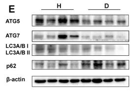
Application: WB Species: Mouse Sample:

Application: WB Species: Mouse Sample: normoxic and hypoxic BV2 cells
Restrictive clause
Affinity Biosciences tests all products strictly. Citations are provided as a resource for additional applications that have not been validated by Affinity Biosciences. Please choose the appropriate format for each application and consult Materials and Methods sections for additional details about the use of any product in these publications.
For Research Use Only.
Not for use in diagnostic or therapeutic procedures. Not for resale. Not for distribution without written consent. Affinity Biosciences will not be held responsible for patent infringement or other violations that may occur with the use of our products. Affinity Biosciences, Affinity Biosciences Logo and all other trademarks are the property of Affinity Biosciences LTD.